Under the lens of a microscope lies a world of wonder, and one of the fascinating creatures to observe is the humble worm. These ancient beings have been roaming the earth for hundreds of millions of years, evolving into the diverse forms we see today.
In 2018, an article published in National Geographic unveiled the discovery of a 500-million-year-old fossil in British Columbia, which belonged to a bristle worm. This ancient organism possessed numerous tiny hairs for swimming and a pair of tube-like palps on its head, serving as sensory organs. Though this specific creature no longer exists today, it is believed that modern leeches and earthworms are among its descendants.
The father of evolutionary thought, Charles Darwin, found himself captivated by earthworms, dedicating considerable time to study their lives and behaviors. His profound research allowed him to unravel the secrets of how these fascinating creatures behave, their preferred food choices, and intricate anatomical features, among many other characteristics.
Through their meandering paths in the soil and their feeding habits, Darwin also shed light on the vital role of earthworms in agriculture. These unassuming creatures play a significant part in soil aeration and nutrient cycling, ultimately benefiting the growth of plants and crops.
As a fascinating historical side note, the early years of microscopy presented some curious misconceptions. Sperm cells, when first observed, were mistaken for parasitic worms by some scientists and philosophers, aptly referred to as “spermatic worms.”
In conclusion, the worm’s journey through time is a tale of resilience and adaptability. From the ancient bristle worm to the diverse earthworms and leeches of today, these microscopic marvels continue to play essential roles in ecosystems and agriculture, reminding us of the wonders that unfold when we explore the hidden realms under the lens of a microscope.
What is Worm?
Worms, those enigmatic invertebrate creatures, inhabit diverse corners of our planet, each group displaying unique traits that intrigue and fascinate. They can be broadly categorized into three major groups: flatworms, roundworms, and segmented worms. While their appearances may differ, worms are universally characterized by an elongated and slender body that boasts bilateral symmetry.
One of the most notable features of worms is their lack of limbs. Instead, they employ a set of muscles to gracefully creep across various surfaces. Some species, however, possess hair-like structures that aid them in their movements, adding an extra touch of intrigue to their locomotion.
These versatile organisms have adapted to a multitude of habitats around the world. Earthworms, for instance, thrive in moist terrestrial environments, burrowing through soil and decomposing organic matter, playing a vital role in maintaining soil health. On the other hand, Polychaetes, a type of marine worm, are abundant in oceanic habitats, boasting an astounding diversity that enchants marine enthusiasts. Fascinatingly, tube worms like Riftia pachyptila form a symbiotic relationship with sulfur bacteria, inhabiting the mysterious depths of the sea floor.
But worms aren’t just content to reside in diverse habitats; some have adopted more intimate living arrangements. A few species of worms have chosen the life of a parasite, residing within plants or animals, showcasing the marvels of co-evolution and adaptation.
Intriguingly, worms possess several common characteristics that define their biological makeup. They boast a body cavity known as a coelom, which provides essential structural support and room for organ development. A majority of worm species feature a digestive system to extract nutrients from their environments, and their bodies are organized using primary germ layers, indicating their developmental origin.
Let us briefly explore some well-known representatives of the worm world:
- Earthworms: Familiar garden dwellers, diligently aerating the soil and breaking down organic matter, contributing to the ecological balance.
- Leeches: These blood-sucking worms have a historical reputation in medicine, with their use in bloodletting for medical treatment in the past.
- Polychaete Worms: Dancing gracefully in ocean waters, these marine marvels showcase an incredible array of shapes, colors, and behaviors.
- Pseudobiceros filgor: A striking flatworm, mesmerizing with its vibrant hues and intriguing behavior.
- Yellow Papillae Flatworm: Another captivating flatworm species that adds beauty to the diversity of invertebrate life.
- Hookworms: A group of parasitic worms that can be found residing in the intestines of various animals, including humans.
In conclusion, worms encompass a fascinating array of invertebrate life. From the mesmerizing marine world of Polychaetes to the industrious earthworms diligently working underground, each species provides a window into the wonders of nature’s diverse and intricate designs. As we delve deeper into the world of worms, we unveil the captivating stories of adaptation, evolution, and coexistence that enrich our understanding of life on Earth.
Objectives Of this Experiment
The primary goal of this enthralling experiment is to provide students with the opportunity to observe and compare members from each group of worms: the flatworms, roundworms, and segmented worms. Through this comparative study, students will gain valuable insights into the similarities and differences among these intriguing creatures.
Upon completion of the activities, students will have achieved the following objectives:
- Proficiency in Microscopy: Through hands-on experience, students will learn how to skillfully use a microscope to observe and examine specimens. This essential skill will open up a world of microscopic wonders, empowering them to explore various organisms and objects with greater detail.
- Specimen Preparation: Understanding the correct methods for preparing specimens is crucial in microscopic studies. This experiment will teach students the proper techniques for preparing worm specimens, ensuring accurate and clear observations.
- Comparative Morphology: Armed with newfound microscopy skills, students will delve into the morphology of the three distinct groups of worms. By comparing and contrasting the physical characteristics of flatworms, roundworms, and segmented worms, they will discern the unique features that distinguish each group.
To aid their investigation, it is recommended that students start their exploration by using a magnifying glass. While offering lower magnification power than a microscope, the magnifying glass serves as a helpful preliminary tool. By magnifying the image several times, students can acquire a general understanding of the worm specimen’s overall morphology.
In conclusion, the objectives of this captivating experiment extend beyond the mere observation of worms under a microscope. Students will embark on a journey of scientific discovery, honing their microscopy skills, perfecting specimen preparation techniques, and unraveling the fascinating diversity of worms. As they delve into the microscopic world, they will acquire a deeper appreciation for the intricacies of life and the wonders that await exploration.
Materials Required
To embark on this captivating worm observation experiment, gather the following materials:
- Magnifying Glass: A typical magnifying glass, boasting a magnification power of approximately 1× to 2×, serves as an initial tool for obtaining a general view of the worm specimens.
- Petri Dish: A shallow, transparent Petri dish provides a suitable container for holding and examining the worms under the microscope.
- Pair of Tweezers: Utilize a pair of tweezers for precise handling and manipulation of the delicate worm specimens during the observation process.
- Specimens: Select three types of worms to compare and contrast their features. These specimens include:a. Earthworm (Segmented Worm): Easily found in gardens and loose soil, earthworms provide valuable insights into the world of segmented worms.b. Planarian Flatworm: Found in shallow water bodies such as ponds and rivers, these flatworms showcase unique characteristics for comparison.c. Eelworm (Roundworm): These worms can be found infecting various crops, like tomatoes, and they represent the intriguing realm of roundworms.
- Pair of Gloves (Nitrile Rubber): To ensure safe and hygienic handling of the worm specimens, wear a pair of nitrile rubber gloves.
- Container: Have a container ready to hold the worm specimens before transferring them to the Petri dish for observation.
- Sieve: Use a sieve to sift through the soil or water to carefully extract the worm specimens without causing harm.
- Mini Garden Shovel: If searching for earthworms in the garden, a mini garden shovel will aid in gently excavating the soil and locating the specimens.
The selection of these three specific types of worms is strategic. They are easily obtainable in their respective habitats, making the process of collection simpler. Additionally, these worms are safe to handle, ensuring a secure and ethical learning experience for students. Moreover, by including representatives from each of the three major groups of worms (segmented worms, flatworms, and roundworms), the comparison process becomes more straightforward and insightful, offering a comprehensive understanding of the diversity within the worm world.
With these materials in hand, students can dive into the world of worms, exploring their unique characteristics and unraveling the wonders of the animal kingdom.
Procedure – Sample Collection
To begin this exciting worm observation experiment, follow these steps to collect the specimens for your study:
- Optional: Wear Gloves
- While the worms used in this experiment are generally harmless, you may choose to wear a pair of gloves for added protection during the collection process.
- Collecting Earthworms:
- Using a mini garden shovel, gently dig around your garden or in loose, moist soils to find one or several earthworms.
- With a pair of tweezers, gently pick up the earthworms and place them in a Petri dish. To prevent their escape, you can partially cover the Petri dish.
- Collecting Planarian Flatworms:
- Use a container to draw water from a shallow pond or river to collect Planarian flatworms.
- Take a closer look at the water in the container, possibly using a magnifying glass, to determine if you have successfully collected the flatworms. These worms are small, ranging from 3 to 15mm in length and may appear black, gray, or brown depending on the species.
- Once you are sure you have Planarian flatworms in your container, sieve the water to easily collect the worms. Keep the specimens in a separate Petri dish, ensuring each type of worm is placed in different dishes. To preserve the worms, you can add a small amount of water to the Petri dish.
- Collecting Eelworms (Roundworms):
- Collecting eelworms may prove challenging as they are barely visible to the naked eye, measuring only 0.4-1.2 mm in size. However, you can find them by identifying an infected crop, such as tomatoes, rhubarb, or potatoes.
- Gently break off the infected part of the crop, such as leaves, into tiny bits.
- Place the leaves in a glass container of water and let them sit for about 30 minutes.
- After 30 minutes, you will observe a cluster of eelworms moving at the bottom of the container.
- Carefully pour off the water, being cautious not to lose the mass of eelworms.
- You can either keep the eelworms in the container or gently transfer them to a Petri dish with a small amount of water.
- Observe Using Magnifying Glass:
- Once you have collected the entire specimen in each Petri dish, you can use a magnifying glass to carefully observe the worms. This can be done while the worms are still in the Petri dish.
By following this precise procedure for collecting worm specimens, you are well-prepared to embark on the fascinating observation and comparison of earthworms, Planarian flatworms, and eelworms. Enjoy the wonder of exploring these intriguing creatures under the lens of a magnifying glass and microscope.
Observation
Under the magnifying glass, each type of worm reveals its unique characteristics, providing a fascinating glimpse into their intricate world.
Earthworm:
- As you observe the earthworm under the magnifying glass, you will notice distinct segments running along the entire length of its body. These segments allow the earthworm to maneuver and twist gracefully.
- In good lighting conditions, you may be able to glimpse some internal organs, such as vessels, within the worm. However, in certain species with a darker epidermis, these internal structures may be difficult to discern.
- At the anterior part of the earthworm’s body, you’ll observe a narrow and pointed head, which is smaller compared to the other segments. Additionally, near the middle part of the body, a distinct segment appears swollen and lighter in color; this is known as the clitellum, which plays a vital role in the worm’s reproduction.
- Through the magnifying glass, you can also spot setae, hair-like structures present on each segment, aiding in the worm’s movement. The body of the earthworm appears cylindrical in shape, contributing to its efficient burrowing capabilities.
Planarian Flatworms:
- Planarian flatworms, while possessing an elongated body, are significantly smaller in size, ranging from 3 to 15mm. Their body shape is distinctly flattened, reminiscent of a leaf-like structure.
- On closer inspection, you may notice eyespots at the head region, providing the worm with a simple form of vision. The posterior end, or tail, appears more pointed compared to the broader anterior part.
- A closer examination may also reveal the presence of a pharynx located in the central part of the body, which plays a crucial role in the planarian’s feeding process.
Eelworms:
- Eelworms, unlike earthworms and planarian flatworms, are tiny creatures. Their diminutive size makes it challenging to discern different body parts under the magnifying glass. However, you can observe them moving about collectively, forming a group.
In conclusion, the observation of these diverse worms under a magnifying glass allows us to appreciate the intricate details of their bodies. From the segmented and cylindrical form of the earthworm to the flattened shape and eyespots of planarian flatworms, and the enigmatic collective movement of eelworms, each species offers a unique insight into the wonders of the microscopic world. This observation process fuels our curiosity and fascination with the diverse life forms that thrive in the hidden realms of the natural world.
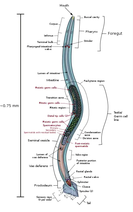
Earthworm under a Microscope
Through the use of a microscope, you will be able to observe both the external and internal (via dissection) anatomy of the earthworm.
Materials Required
To embark on a captivating journey of observing earthworms under a microscope, gather the following materials:
- Earthworm Specimen:
- Obtain a live or preserved earthworm specimen for examination. You can find earthworms in gardens or moist soils, or you may choose to use a preserved specimen if live ones are not readily available.
- Alcohol Solution:
- Prepare an alcohol solution for the purpose of preserving the earthworm specimen, if needed. The alcohol solution will help maintain the integrity of the specimen and prevent decomposition.
- Microscope:
- A high-quality microscope is essential for detailed observation of the earthworm’s anatomical features. Ensure that the microscope is in good working condition to provide clear and sharp images.
- Glass Slide:
- Place the earthworm specimen on a glass slide for observation under the microscope. The glass slide provides a stable surface and allows you to adjust the positioning of the specimen for better visibility.
- Water:
- Use water to create a suitable environment for the earthworm specimen on the glass slide. Adding a small amount of water helps keep the specimen moist during the observation process.
- A Pair of Tweezers:
- Utilize a pair of tweezers to carefully handle the earthworm specimen. Tweezers allow for precise positioning of the specimen on the glass slide without causing any damage.
With these materials at hand, you are well-prepared to embark on the microscopic exploration of the fascinating earthworm. The combination of a high-quality microscope, glass slide, water, and a pair of tweezers ensures a smooth and insightful observation experience. Whether you are using a live or preserved earthworm specimen, this endeavor promises to unveil the hidden wonders of the earthworm’s anatomy and behavior under the lens of a microscope.
Procedure for Preparation
To ensure a successful and clear observation of the earthworm under a microscope, follow these precise steps for preparation:
- Prepare Alcohol Solution:
- In a clean Petri dish, create an alcohol solution by mixing 1 part alcohol with 9 parts water. The alcohol solution will serve to preserve and immobilize the earthworm for microscopy.
- Placing Earthworm in Alcohol Solution:
- Using a pair of tweezers, carefully pick one of the earthworms and place it gently into the alcohol solution. This step is crucial as it serves to euthanize the earthworm, making it suitable for detailed microscopic examination.
- Transfer Earthworm to Water:
- After the earthworm has been in the alcohol solution for an appropriate amount of time to ensure its immobility, pick the earthworm once more using the tweezers. Transfer the earthworm into a clean Petri dish filled with water. This step is aimed at washing off any dirt or mucus that may be present on the earthworm’s epidermis.
By following these precise steps for preparation, you ensure that the earthworm is suitably preserved and cleansed for detailed microscopic observation. The use of the alcohol solution effectively immobilizes the earthworm, making it easier to position for examination. Subsequently, washing the earthworm in water removes any external impurities, providing a clear and unobstructed view of its intricate anatomy under the microscope. With this preparation complete, you are now ready to embark on the fascinating journey of exploring the hidden wonders of the earthworm’s anatomy and behavior through the lens of a microscope.
Stereo Microscopy of Earthworm
To explore the external anatomy of an earthworm in detail, a stereomicroscope proves to be an invaluable tool. Follow this step-by-step procedure to embark on an exciting journey of observation:
Procedure:
- Prepare the Specimen:
- Place the earthworm gently in a clean Petri dish. Ensure that the worm is positioned in a way that allows you to observe its body from various angles.
- Set the Magnification:
- Begin by turning the revolving turret of the stereomicroscope to set the lowest magnification objective in place. This initial magnification provides a broader view of the worm’s external features.
- Position the Petri Dish:
- Carefully place the Petri dish containing the earthworm specimen on the stage of the stereomicroscope.
- Focus the Image:
- While looking through the eyepiece, gently turn the focus knob of the stereomicroscope to bring the image of the earthworm into sharp focus. Adjust the focus until the specimen’s details become clear and easily observable.
- Adjust the Light Intensity:
- For optimal visibility, you can adjust the intensity of the light by manipulating the condenser. This will allow you to control the illumination and enhance the clarity of the earthworm’s external features.
- Explore the Anatomy:
- Once the image is in focus, start your observation by identifying the anterior end of the earthworm. From there, carefully move the stage to investigate the external anatomy of the rest of the body.
- As you explore, take note of the different body segments, setae (hair-like structures), and any other notable external characteristics.
- Increase Magnification:
- For a more detailed examination, adjust the stereomicroscope to a higher magnification setting. This will enable you to get a closer look at specific regions of interest on the earthworm’s body.
- Record Your Observations:
- As you make your observations, take detailed notes or use a camera attachment to capture images of the earthworm’s external features.
- Note: The same procedure can be repeated with a live earthworm to observe its movement and the motion of its body and mouthparts.
By following this procedure, you will be able to unlock the hidden wonders of the earthworm’s external anatomy through the lens of a stereomicroscope. This exploration will deepen your understanding of the remarkable adaptations and intricate structures that contribute to the earthworm’s survival and success in its environment.
Observation
Under higher magnification power, the stereomicroscope offers a remarkable view of the earthworm’s external anatomy, revealing intriguing details and features:
- Prostomium:
- At the anterior part of the earthworm, a fleshy bump known as the prostomium becomes clearly visible. This specialized structure surrounds the mouth area of the worm, playing a vital role in the earthworm’s feeding processes.
- Anus:
- On the opposite end of the earthworm’s body, at the posterior part, the anus can be observed. This opening serves as the exit point for waste elimination.
- Septum:
- Under the microscope, you can discern a septum that acts as a partition, separating the tiny body segments of the earthworm. This characteristic feature aids in the earthworm’s efficient and coordinated movement.
- Dorsal and Ventral Surfaces:
- Notable differences between the dorsal and ventral surfaces of the earthworm become apparent. The ventral surface, which appears darker, is flatter compared to the rounded dorsal surface.
- Setae and Pores:
- The microscope unveils the presence of setae, which are tiny hair-like bristles that project from each body segment of the earthworm. These setae contribute to the worm’s locomotion and gripping abilities.
- Additionally, you will observe pores on each body segment of the earthworm. These pores play a role in the worm’s respiration and excretion processes.
- Genital Pores:
- Alongside the regular pores on the body segments, larger pores known as genital pores can be easily seen near the anterior part of the earthworm. These genital pores are crucial for the worm’s reproductive activities.
Through the lens of the stereomicroscope, the earthworm’s external anatomy comes to life, revealing a myriad of intricate features that contribute to its survival and functionality. The observation of the prostomium, anus, septum, setae, pores, and genital pores provides a deeper understanding of the earthworm’s adaptation to its environment. This exploration ignites our curiosity and appreciation for the wonders of nature, as we uncover the hidden marvels of even the smallest creatures on Earth.
Dissection of Earthworm
To gain a deeper understanding of the internal anatomy of the earthworm, a dissection is a valuable and informative technique. By carefully exposing the inner structures, we can unravel the complexities of this fascinating creature. Here’s what you’ll need and the steps to follow:
Requirements/Materials:
- Dissecting Pins: These are used to hold the specimen in place during the dissection, ensuring stability and precision.
- Specimen: Obtain a fresh or preserved earthworm specimen for the dissection. Choose one that is intact and of suitable size for examination.
- Dissecting Knife/Blade or Dissecting Scissors: A sharp and precise dissecting tool is essential for making clean and accurate incisions during the dissection process.
- Microscope: After the dissection, you can use a microscope to further observe and study the internal structures of the earthworm in greater detail.
- Dissecting Tray: This serves as a clean and organized workspace for conducting the dissection.
- Pair of Forceps: Forceps are handy tools for gently holding and maneuvering delicate structures during the dissection.
Procedure for Dissection of Earthworm
To explore the internal anatomy of the earthworm through dissection, follow these precise steps for a thorough and enlightening examination:
- Preparation and Positioning:
- Place the earthworm specimen on the dissecting tray with its dorsal side facing up. Position it in a way that provides a clear and stable view for the dissection process.
- Securing the Specimen:
- Use the dissecting pins to secure each end of the earthworm in place on the dissecting tray. This ensures that the specimen remains steady and immobile during the dissection.
- Creating an Incision:
- With the dissecting blade, carefully make a small opening just below the clitellum, a swollen band located near the middle part of the earthworm’s body. This opening will serve as the starting point for the dissection.
- Cutting the Epidermis:
- Utilize the forceps to gently lift the skin around the incision. This allows you to insert the dissecting scissors and cut the epidermis along a straight line, moving both towards the head and towards the posterior end of the earthworm.
- Revealing the Internal Anatomy:
- Using the forceps, gently pull apart the epidermis along the incision lines. Be meticulous in this step to ensure the internal anatomy of the worm is clearly visible.
- Securely pin the opened epidermis to the dissecting tray using multiple pins, enabling a well-exposed view of the earthworm’s internal structures.
- Observation under the Microscope:
- As you proceed with the dissection, take moments to observe the specimen under the microscope to explore the intricate details of its internal anatomy.
By following this precise procedure for the dissection of the earthworm, you will gain an in-depth understanding of its internal structures and systems. From the digestive system and nervous system to the reproductive and circulatory systems, this hands-on approach offers invaluable insights into the complex organization of this fascinating creature. Through patient exploration and careful dissection, you will uncover the hidden marvels of the earthworm’s internal anatomy, deepening your appreciation for the wonders of the natural world.
Observation of Earthworm’s Internal Anatomy: Unveiling the Intriguing Structures
Under the microscope, a whole new world of the earthworm’s internal anatomy awaits, offering a detailed view of its fascinating structures and systems:
- Aortic Arches (Heart):
- The microscope reveals five dark loops encircling the esophagus, serving as the earthworm’s heart, also known as the aortic arches. These loops play a vital role in pumping blood and distributing nutrients throughout the body.
- Pharynx:
- Located at the anterior part of the earthworm, inside the mouth, the pharynx is a distinctive organ. Composed of several muscles, it appears as a lightly colored structure involved in the ingestion and processing of food.
- Reproductive Organs:
- Near the heart, you will observe the reproductive organs of the earthworm. These organs may appear as lightly colored tissues or tiny white structures, reflecting their essential function in the worm’s reproductive processes.
- Gizzard:
- Positioned just behind the crop, the gizzard serves as a food storage chamber in the earthworm. Through the microscope, you can identify this specialized structure that aids in breaking down and grinding the ingested food.
- Intestine:
- The microscope allows you to follow the path of the intestine, a long tube extending from the gizzard towards the posterior part of the body. The intestine plays a crucial role in nutrient absorption and waste elimination.
- Central Nerve Cord:
- By carefully moving the intestine aside, you will uncover the central nerve cord, located on the dorsal part of the earthworm’s body. This nerve cord appears as a long white cord and is essential for transmitting nerve signals throughout the worm’s body, coordinating its various functions.
Through meticulous observation of the earthworm’s internal anatomy under the microscope, you gain valuable insights into its intricate structures and systems. The identification of the aortic arches as the heart, the pharynx as the organ for food intake, the reproductive organs for reproduction, the gizzard for food storage, the intestine for nutrient absorption, and the central nerve cord for nerve signal transmission deepens our understanding of this remarkable creature’s physiology. This observation journey fosters curiosity and appreciation for the complexity and efficiency of the earthworm’s internal organization, exemplifying the wonders of nature’s design.
Planarian Flatworm under a Microscope
Materials Required
To conduct detailed and precise microscopic observation, gather the following essential materials:
- Microscope Glass Slides:
- Obtain clean and transparent microscope glass slides. These slides serve as the platform on which you place the specimen for observation.
- Microscope Coverslip:
- Secure microscope coverslips, which are small, thin, and transparent pieces of glass or plastic. They are used to cover the specimen on the glass slide, protecting it and preventing distortion during observation.
- Agar:
- Agar is a gelatinous substance used to create a solid medium for the examination of living specimens under the microscope. It provides a stable and controlled environment for observation.
- Compound Microscope:
- A compound microscope is an essential tool for magnifying and observing specimens at high magnification. It consists of multiple lenses, allowing for detailed examination of minute structures.
- Vaseline:
- Vaseline, also known as petroleum jelly, is used to create a seal around the edges of the coverslip. This seal prevents air bubbles from forming and ensures a clear and undistorted view of the specimen.
- Water:
- Water is often used to create a wet mount, where a drop of water is placed on the glass slide to suspend and view specimens. It provides a hydrated environment for live specimens and allows for easier examination.
By gathering these materials, you are well-equipped to embark on a comprehensive microscopic observation of various specimens. The microscope glass slides, coverslips, agar, and water facilitate the preparation and mounting of specimens for examination. With a high-quality compound microscope at your disposal, you can explore the intricate details and hidden wonders of the microscopic world. The addition of Vaseline ensures optimal viewing conditions, creating a secure and clear observation experience. By utilizing these materials, you can delve into the fascinating realm of microscopy, unlocking the mysteries of the smallest organisms and structures that make up our world.
Preparation of Temporary Slides
To observe live worms under the microscope, the technique of preparing temporary slides with agar is employed. This method ensures a controlled and stable environment for the worms, allowing for detailed and real-time examination. Follow these steps for the preparation of temporary slides:
- Applying Hot Agar:
- Begin by adding a drop of hot agar solution (4 to 5 percent agar) on a clean glass slide. The agar used for this technique consists of Oxoid Agar Technical and cupric sulphate, providing a suitable medium for the live worms.
- Flattening the Agar:
- Use another clean glass slide to gently and evenly flatten the hot agar drop. This ensures a smooth and consistent surface for the subsequent steps.
- Allowing Agar to Set:
- Once the agar has set, carefully remove the glass slide used to flatten the agar, leaving behind a stable agar bed for the live worms.
- Adding Water:
- Using a dropper, add a small drop of water onto the surface of the agar. This provides a hydrated environment for the live worms and allows them to remain active during observation.
- Introducing the Flatworm:
- Carefully place the flatworm on the drop of water on the agar bed. Position the worm in a way that facilitates optimal observation of its structures and behavior.
- Covering with a Coverslip:
- Place a clean coverslip over the flatworm, gently lowering it onto the water drop and agar bed. The coverslip protects the specimen and provides a clear viewing window for the microscope.
- Preventing Movement:
- To prevent the flatworm from moving during observation, a small amount of vaseline can be applied to the edge of the coverslip. This helps create a seal and keeps the specimen in place without causing harm.
By following these steps, you can create temporary slides that provide a suitable habitat for live worms, facilitating detailed examination under the microscope. This technique allows for the observation of the worms’ natural behavior and physiological processes in a controlled environment. The temporary slides with agar offer an invaluable opportunity to explore the dynamic world of live organisms, deepening our understanding of their biology and enhancing our appreciation for the wonders of life.
Microscopy of Temporary Slides
Once the temporary slide has been meticulously prepared, the captivating world of the specimen awaits under the microscope. The following steps outline the process of observing the specimen on the temporary slide:
- Setting the Lowest Power Objective:
- Before mounting the slide, rotate the turret of the microscope to set the lowest power objective (usually 4x or 10x) in place. This initial setting allows for a wide view of the specimen, aiding in locating and focusing on the desired area.
- Placing the Slide on the Stage:
- Carefully position the temporary slide on the stage of the microscope. Secure it in place with the clips to prevent any movement during observation.
- Focusing the Image:
- Peer through the eyepiece and slowly turn the focus knob to bring the image into sharp focus. Be patient and gentle with the adjustments to achieve a clear view of the specimen.
- Exploring Different Parts of the Specimen:
- Utilize the stage adjustment knob to gently move the stage in different directions. This maneuverability allows you to explore various regions of the specimen, unraveling its hidden details.
- Switching to Higher Magnification:
- For a closer examination of specific structures or intricate features, switch to a higher magnification objective (e.g., 40x or 100x). This enhances the level of detail, revealing the microscopic intricacies of the specimen.
- Lowering the Stage and Slide Removal:
- After completing the observation, lower the stage carefully. Then, remove the temporary slide from the stage, ensuring utmost care to avoid damage to both the slide and the microscope.
Microscopy of temporary slides offers a captivating journey into the microscopic world. With precision and patience, the diverse and intricate details of the specimen come to life under the lens of the microscope. From the lowest power objective, providing a broad view, to the higher magnification, uncovering minute structures, each step of the observation process adds to our understanding and appreciation of the wonders that lie beyond the naked eye. The exploration of temporary slides brings to light the remarkable diversity and complexity of life’s building blocks, fostering curiosity and inspiring further discoveries in the fascinating realm of microscopy.
Observation
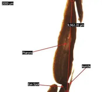
Upon placing the worm specimen under the microscope, a fascinating array of features comes into view, offering insights into its unique anatomy and sensory capabilities:
- Flattened Body Resembling an Elongated Flat-Leaf:
- Through the microscope lens, the body of the worm appears flattened, resembling an elongated flat-leaf. This distinctive feature is characteristic of certain worm species, providing them with an efficient body structure for movement and survival.
- Eyespots at the Head (Anterior Part) of the Worm:
- Notably, the microscope reveals the presence of eyespots located at the head part, also known as the anterior part of the worm. These pigmented spots are sensitive to sunlight, functioning much like eyes. They play a crucial role in detecting light and shadows, aiding the worm in its orientation and behavior.
- Pointed Auricles on the Sides of the Head:
- Depending on the specific species, the head of the worm may exhibit pointed auricles on the sides. These auricles serve as sensory structures involved in smell and touch. They enhance the worm’s ability to interact with its environment and respond to various stimuli.
- Pharynx in the Midsection:
- Further observation through the microscope might reveal the presence of the pharynx in the midsection of the worm. The pharynx is a significant organ involved in feeding and digestion. Its location and structure enable efficient intake and processing of food for nourishment.
The microscopic observation of the worm provides a detailed glimpse into its specialized adaptations and sensory abilities. The flattened body shape, eyespots, auricles, and pharynx each contribute to the worm’s survival and successful navigation in its habitat. This exploration of the worm’s anatomy fosters appreciation for the diverse adaptations and functionalities found in nature’s myriad of organisms. The microscope serves as a powerful tool, granting us access to the intricacies of the natural world and offering a deeper understanding of the wonders that lie beyond the naked eye.
Roundworms (round worms) under the microscope
Culture
Materials Requiremed
To culture vinegar eels, a type of nematode commonly found in vinegar, you will need the following materials:
- Cider Vinegar:
- Cider vinegar serves as the primary medium for the vinegar eels’ culture. It provides the necessary nutrients and environment for their growth and reproduction.
- Vinegar Eels (Turbatrix aceti):
- The vinegar eels themselves are required to initiate the culture. These microscopic nematodes naturally occur in vinegar and will form the foundation of your culture.
- Glass Culture Dish:
- A clean and clear glass culture dish is essential to contain and observe the vinegar eels’ growth. The dish provides a suitable space for their development and easy visualization.
- Pipette:
- A pipette is necessary for transferring the vinegar eels into the culture dish. It allows for precise and controlled dispensing of the eels into the vinegar medium.
- Apple:
- An apple slice or a small piece of apple is used as a food source for the vinegar eels. As the apple decomposes, it releases nutrients that sustain the eels during the culturing process.
By gathering these materials, you can establish a successful culture of vinegar eels. The combination of cider vinegar, vinegar eels, a glass culture dish, a pipette, and an apple creates a suitable and controlled environment for the eels to thrive. As they grow and reproduce, you can observe the fascinating life cycle of these microscopic organisms, deepening your understanding of their biology and ecology. Culturing vinegar eels offers a unique opportunity to explore the hidden world of microorganisms and appreciate the intricate balance of life in the smallest corners of our natural world.
Procedure
Culturing vinegar eels provides a fascinating opportunity to observe these tiny nematodes and their life cycle. Follow these steps to establish and maintain the culture:
- Prepare the Culture Dish:
- Using a pipette, carefully add about 200ml of cider vinegar into a clean glass culture dish. The vinegar will serve as the medium for the eels’ culture.
- Add Apple Slices:
- Cut a few slices of apple and add them into the glass dish containing the vinegar. The apple slices will serve as a food source for the vinegar eels as they decompose, providing essential nutrients for their growth and survival.
- Introduce the Vinegar Eels:
- Using the pipette, introduce the vinegar eels (Turbatrix aceti) into the culture dish containing the vinegar and apple slices. These microscopic nematodes will naturally occur in the cider vinegar.
- Cover the Culture Dish:
- Cover the culture dish loosely to slow down evaporation and maintain a stable environment for the vinegar eels. The loose cover allows for air exchange and prevents excessive moisture loss.
- Aerate the Culture:
- Regularly aerate the culture using a clean pipette. Gently force air into the culture media to provide oxygen and maintain optimal conditions for the eels’ growth and activity.
- Observation:
- To observe the vinegar eels, place the culture dish on a dark paper under a stereoscope. The stereoscope will magnify the eels and allow for easy viewing of their movement and behavior.
By following this procedure, you can successfully culture vinegar eels and witness their intriguing life cycle. The apple slices provide nourishment, and the vinegar medium offers a suitable environment for their growth and reproduction. Regular aeration ensures a well-oxygenated culture, supporting the eels’ thriving population. As you observe the vinegar eels under the stereoscope, you will gain valuable insights into the hidden world of these microscopic organisms, deepening your understanding of their ecology and biology. Culturing vinegar eels is an exciting scientific endeavor that fosters curiosity and appreciation for the diversity of life at the microscopic level.
Microscopy
Materials Requiremed
To conduct a detailed microscopic observation of eelworms, gather the following essential materials:
- Specimen (Eelworms):
- Obtain live eelworms (nematodes) as the primary subject of your observation. These tiny, worm-like organisms are commonly found in water environments and soil.
- Compound Microscope:
- A compound microscope is an essential tool for magnifying and examining the eelworms. This type of microscope has multiple lenses that allow for high-resolution imaging.
- Dropper:
- A dropper, also known as a pipette, is used to handle and transfer the eelworms onto the microscope slide. It enables precise and controlled placement of the specimens.
- Microscope Glass Slide:
- The microscope glass slide serves as the platform on which the eelworms will be mounted for observation. It provides a flat and transparent surface to facilitate microscopy.
By assembling these materials, you can embark on a fascinating journey into the microscopic world of eelworms. The compound microscope will magnify the eelworms, revealing intricate details of their anatomy and behavior. With the dropper, you can carefully position the specimens on the microscope slide, allowing for close examination under the lens. The combination of live eelworms and the microscope glass slide sets the stage for an enlightening exploration of these fascinating organisms. Through microscopic observation, you can unlock the hidden wonders of the microscopic realm and gain a deeper appreciation for the diversity of life that exists at this scale.
Procedure
Observing eelworms under a microscope allows for a closer look at their intricate structures and behaviors. Follow these steps to conduct the observation:
- Collecting Eelworms:
- Using a dropper, carefully collect a few live eelworms from the liquid culture at the bottom of the Petri dish. Gently suction the eelworms into the dropper, ensuring that you handle them with care.
- Preparing Microscope Slides:
- Place about 2 drops of the liquid containing the eelworms onto several clean glass slides. This ensures that each slide has an adequate number of eelworms for observation.
- Mounting the Slides:
- Once the liquid drops are on the slides, place each slide on the microscope stage, ensuring it is secure and level. Position the slide such that the eelworms are within the field of view when the microscope is turned on.
- Initial Observation with Low Magnification:
- Set the microscope to its lowest magnification objective (e.g., 4x or 10x) and look through the eyepiece. Gently turn the focus knob to bring the image into focus. Observe the eelworms under low magnification first to get an overview of their general appearance and movement.
- Switching to Higher Magnification:
- After obtaining a basic view, switch to higher magnification objectives (e.g., 40x or 100x) to observe the eelworms in more detail. Adjust the focus and lighting as necessary to obtain clear and crisp images.
- Record Observations:
- As you observe the eelworms, take notes of their characteristics, movement patterns, and any interesting behaviors or structures you observe. You can also capture images or videos using a camera attachment on the microscope.
- Proper Handling and Disposal:
- After completing the observation, handle the eelworms with care and return them to the liquid culture in the Petri dish. Ensure proper disposal of any waste materials and clean the microscope and slides as needed.
Microscopic observation of eelworms provides a unique opportunity to explore the intricate world of these tiny organisms. By following this procedure, you can gain valuable insights into their biology and behavior, deepening your understanding of their role in the ecosystem. The microscopic realm holds countless wonders waiting to be discovered, and the study of eelworms offers a glimpse into the hidden diversity and complexity of life at the microscale.
Observation
Upon peering through the microscope lens, a captivating world of eelworms comes into view, offering fascinating insights into their anatomy and features:
- Transparent Epidermis:
- The microscope reveals that the eelworm’s body possesses a transparent epidermis, allowing for a clear view of its internal structures. This transparency facilitates detailed observation of the worm’s inner workings.
- Cylindrical Body Shape:
- Contrasting with the flatworm, the eelworm exhibits a cylindrical or tube-like body shape. This elongated form enables efficient movement through various environments, making it well-adapted to its habitat.
- Pointed Posterior:
- Upon closer examination, the posterior end of the eelworm appears pointed. This tail-like structure aids in locomotion and helps the worm navigate its surroundings.
- Anterior Mouth Part:
- Under higher magnification, the anterior end of the eelworm may reveal a distinct mouth part. This specialized structure serves as the entry point for ingesting food and nutrients.
- Intestine Extending from Esophageal Bulb:
- A more detailed observation using increased magnification may uncover an intestine extending from the esophageal bulb towards the posterior part of the eelworm’s body. This digestive organ plays a crucial role in nutrient absorption and waste elimination.
The microscopic observation of eelworms provides a fascinating glimpse into their unique body plan and biological adaptations. The transparent epidermis offers a privileged view of their internal workings, allowing scientists and enthusiasts alike to delve into the intricacies of these tiny organisms. The eelworm’s cylindrical shape, pointed posterior, mouth part, and intestinal structure exemplify the elegance of nature’s design, showcasing the diversity and efficiency of life at the microscopic scale. As researchers explore and study these creatures, they gain valuable insights into the complexities of life’s building blocks and the remarkable adaptations that enable these minuscule organisms to thrive in their diverse habitats. The observation of eelworms under the microscope serves as a testament to the wonders that lie beyond the naked eye, revealing the hidden marvels of the microcosm.
A Worm Under a Microscope Pictures
Oligochaetes: Segmented Wonders of the Annelid World
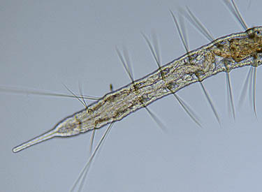
Oligochaetes, a remarkable class of annelids, stand as captivating examples of segmented worms with unique features that set them apart in the microscopic realm. These fascinating organisms possess groups of bristles, known as setae, along their bodies, showcasing a diversity of shapes and sizes that serve essential functions in their lives. The most renowned member of the Oligochaete class is the earthworm, but these intriguing creatures can also be found dwelling in ponds, where their setae become more visible and distinct.
- Setae and Muscular Grip:
- Oligochaetes exhibit a set of bristles, or setae, along their bodies, serving as an integral part of their locomotion and survival. In most pond-dwelling Oligochaetes, the setae are easily visible, presenting a range of lengths and shapes. Some may be long and slender, while others are small and hooked. These bristles operate with the aid of numerous muscles, providing the worms with an extra grip as they wriggle along surfaces or move through soil.
- Earthworms and Transparency:
- While earthworms, a well-known Oligochaete, have relatively tiny and less visible setae, most pond-dwelling Oligochaetes offer a more transparent appearance, allowing observers to easily discern their unique setae and other inner features.
- Well-Developed Digestive and Nervous Systems:
- Oligochaetes boast well-developed digestive and nervous systems, which contribute to their efficient feeding and ability to respond to their environment. These systems work in harmony, ensuring proper digestion and effective control of their movements.
- Visible Inner Anatomy:
- The transparency of Oligochaetes grants observers a privileged glimpse into their inner anatomy. This remarkable feature allows scientists and enthusiasts alike to study and understand the intricate structures and functions of their internal organs and systems.
Oligochaetes, with their segmented bodies and distinctive setae, hold a unique place in the annelid world. From the earthworm’s more inconspicuous setae to the prominent and transparent features of pond-dwelling Oligochaetes, these organisms present an opportunity for researchers and nature enthusiasts to explore the wonders of their anatomy and behavior. Their muscular grip, well-developed systems, and transparent bodies make Oligochaetes an intriguing subject of scientific study, adding to our knowledge of the diverse and captivating life forms that inhabit the world around us.
Nematodes or Roundworms: Versatile and Fascinating Organisms
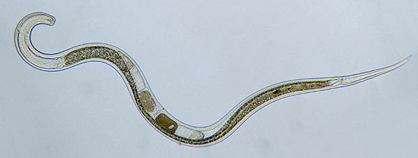
Nematodes, also known as roundworms, are intriguing creatures that share some relationship with rotifers. Characterized by their distinct S-shaped, often highly active movements, these cylindrical organisms possess a tough and slightly flexible skin, known as the cuticle. Nematodes exhibit incredible adaptability, inhabiting a wide range of environments, from ponds to soils, and even as parasites within other organisms. Their ecological significance is profound, playing vital roles in the breakdown of organic matter in soil and the ecology of pond ecosystems.
- Cylindrical Body and S-Shaped Movement:
- The cylindrical shape of nematodes, coupled with their characteristic S-shaped movements, makes them easily distinguishable in the microscopic world. These agile organisms actively navigate their environments, exhibiting impressive locomotion.
- Habitat Diversity:
- Nematodes are not confined to specific habitats; instead, they are highly adaptable to various environments. While some thrive in pond ecosystems, others are well-suited for life as soil dwellers or even as parasitic inhabitants in other organisms.
- Ecological Role:
- In the intricate web of ecological interactions, nematodes play a significant role in the breakdown of organic matter in soil and contribute to nutrient cycling. Moreover, they are crucial components of pond ecosystems, shaping the balance and functioning of these aquatic environments.
- Ideal Subjects for Scientific Study:
- Nematodes, particularly the Caenorhabditis elegans species, have proven to be ideal subjects for scientific research. Scientists have been able to meticulously observe and describe the complete developmental process of this 1 mm long nematode. This groundbreaking study provided invaluable insights into the formation of a multicellular body with specialized organs, all originating from a single cell.
- Compact Cell Structure:
- One of the striking features of nematodes, exemplified by C. elegans, is their limited number of cells. C. elegans, for instance, possesses a mere 959 nuclei, which is remarkably low compared to more complex organisms. This compact cell structure offers a unique platform for studying developmental processes.
- Rapid Development:
- Nematodes exhibit an impressive developmental pace, with C. elegans completing its development in just 3.5 days. This swift life cycle provides researchers with the opportunity to study and understand various aspects of cellular development in a relatively short time frame.
Nematodes, or roundworms, stand as remarkable examples of adaptability and versatility in the microscopic world. Their diverse ecological roles, coupled with their suitability for scientific study, make them invaluable subjects for researchers and enthusiasts alike. As scientists continue to explore and uncover the intricacies of nematodes, their significance in understanding developmental processes and ecological dynamics continues to grow, leaving an indelible mark on the world of scientific discovery.
Flatworms: Simple Yet Fascinating Organisms
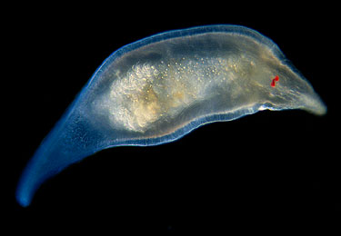
Flatworms, scientifically known as Platyhelminthes, are captivating creatures renowned for their simplicity and unique characteristics. A microscopic observation of these intriguing organisms unveils a range of distinctive features that set them apart in the animal kingdom.
- Ciliated Exterior:
- When viewed under a microscope, one cannot miss the covering of tiny hair-like cilia that adorn the flatworm’s entire body. These cilia play a crucial role in propelling the organism forward, facilitating movement in a manner akin to ciliates, but on a larger scale.
- Defined Head Region and Eyes:
- Flatworms possess a relatively well-defined head region, which may include one or more pairs of simple eyes known as ocelli. These eyes allow the flatworms to detect light and their surroundings, aiding in their navigation through their environment.
- Simple Nervous System:
- In terms of neurological complexity, flatworms boast a relatively simple nervous system. This basic yet effective system enables them to respond to stimuli and coordinate their movements.
- Branched Gut:
- The gut, or intestine, of flatworms is designed as a dead-end sac that branches out. This branching arrangement ensures that food reaches all parts of their body, facilitating digestion and nutrient absorption.
- Mouth Positioning:
- The mouth opening of flatworms is situated on their underside, often near the center of their body. This unique location allows for efficient feeding and the intake of essential nutrients.
- Turbellarian Flatworms in Ponds:
- The flatworms commonly encountered in ponds belong to the group known as Turbellarian flatworms. These freshwater inhabitants are fascinating to study and offer insights into the diversity of life in aquatic environments.
- Parasitic Adaptations:
- While Turbellarian flatworms thrive in ponds, other flatworms, such as flukes and tapeworms, have evolved to live as parasites inside other animals. These parasitic adaptations have allowed them to occupy diverse ecological niches, showcasing their incredible versatility.
Flatworms present an engaging opportunity for researchers and enthusiasts to explore the wonders of simplicity in the animal kingdom. Their ciliated exterior, defined head region, and simple nervous system demonstrate how efficient adaptations can arise even in seemingly simple organisms. Whether thriving in freshwater ponds or inhabiting as parasites, flatworms leave a lasting impression on those who delve into their remarkable world. As scientists continue to study and understand these fascinating creatures, they unveil the intriguing interplay between simplicity and adaptation, providing valuable insights into the vast diversity of life on Earth.
Midge Larva: An Aquatic Insect Larva with Worm-like Features

In the enchanting world of aquatic ecosystems, some organisms may initially be mistaken for worms due to their slender and elongated bodies. Among them, the midge larva stands out as a notable example. Although these larvae share a worm-like appearance, they possess distinct characteristics that set them apart from true worms. Midge larvae belong to a diverse group of aquatic insect larvae and exhibit fascinating adaptations for life in water.
- Slender and Elongated Bodies:
- Midge larvae, like true worms, exhibit slender and elongated bodies that allow them to glide gracefully through aquatic environments. This streamlined shape aids in efficient movement and navigation.
- Segmented Body:
- Upon closer inspection, one can easily differentiate midge larvae from worms by the presence of distinct body segments. Unlike worms, midge larvae’s bodies are divided into segments, contributing to their unique morphology.
- Head and Appendages:
- Another key distinguishing feature is the presence of a well-defined head in midge larvae. Additionally, they possess feet-like appendages, which provide stability and control during their aquatic escapades.
- Aquatic Adaptations:
- Midge larvae are perfectly adapted for life in water. Their elongated bodies and segmented structure allow them to navigate through the aquatic environment with ease, searching for food and evading predators.
- Ecological Role:
- As important members of the aquatic food web, midge larvae play a vital role in nutrient cycling. They serve as a valuable food source for various aquatic predators, contributing to the overall balance and biodiversity of their ecosystems.
- Habitat and Distribution:
- Midge larvae are commonly found in freshwater environments, such as ponds, lakes, streams, and even slow-moving rivers. Their widespread distribution allows them to thrive in various aquatic habitats across the globe.
- Development Stages:
- Midge larvae undergo several developmental stages before transforming into adult midges. These stages, which include pupation, are essential for the larvae’s growth and eventual metamorphosis.
In the realm of aquatic insect larvae, midge larvae stand out with their worm-like appearance but distinct segmented bodies and presence of a head. As these remarkable organisms glide through the water, they serve as a vital component of aquatic ecosystems, contributing to the intricate balance and functionality of these habitats. Observers exploring the wonders of aquatic environments will find fascination in the lives and adaptations of midge larvae, gaining a deeper understanding of the diversity and complexity of life beneath the water’s surface.
Hydra: The Fascinating Freshwater Hydroid
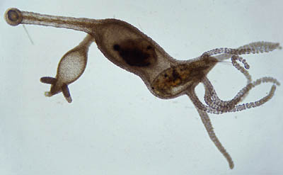
Hydra, a captivating freshwater hydroid related to jellyfish, possesses an intriguing and adaptable body structure. Despite its worm-like appearance, hydras stand out with their long and extendable bodies, adorned with tentacles that set them apart in the aquatic world. This unique organism never fails to amaze with its peculiar abilities and feeding strategies.
- Worm-like Yet Tentacled:
- Hydra’s body may resemble that of a worm, but its distinguishing feature lies in the long, delicate tentacles that surround its mouth opening. These tentacles play a crucial role in capturing prey and defending against potential threats.
- Ingenious Tricks:
- One of Hydra’s most astonishing tricks is its ability to turn itself inside out through its own mouth opening. This remarkable feat showcases the hydra’s remarkable flexibility and adaptability.
- Ferocious Predator:
- Despite its modest size, Hydra is a ferocious predator, capable of capturing relatively large prey like waterfleas and copepods. It uses its tentacles, equipped with specialized stinging cells known as nematocysts, akin to tiny harpoons, to paralyze its prey effectively.
- Enchanting Habitat:
- Hydras can typically be found adorning the leaves of water plants in freshwater environments. These habitats provide the ideal conditions for hydras to thrive and carry out their remarkable feeding and survival strategies.
- Size and Appearance:
- A fully extended hydra can reach a length of approximately 2 centimeters, making it a relatively small organism in the water world. Its elegant tentacles and elongated body make it a fascinating sight to behold.
- Easy Discovery:
- To witness the magic of a hydra, one can easily set up a simple experiment by placing some duckweed and a bit of water in a jar. Given the right conditions, there is a good chance of encountering hydras in this miniature aquatic ecosystem.
In the realm of freshwater organisms, Hydra stands as a captivating marvel with its worm-like body and impressive tentacles. Its ability to perform remarkable tricks and capture prey with its nematocyst-equipped tentacles showcases the incredible diversity and adaptability of life in water environments. As observers delve into the aquatic world and explore the wonders of hydra, they gain a deeper appreciation for the unique adaptations and survival strategies that enable these intriguing creatures to flourish in their natural habitats.
FAQ
What type of worm can be observed under a microscope?
Various types of worms can be observed under a microscope, including earthworms, flatworms, roundworms, and segmented worms.
How do I collect a worm specimen for microscopic observation?
For earthworms, gently dig around moist soil and use tweezers to pick them up. For flatworms, draw water from a shallow pond or river and sieve to collect them. Eelworms can be found in infected crops and collected using water and a glass container.
What equipment do I need to observe a worm under a microscope?
The basic equipment includes a microscope, glass slides, and a dropper or pipette to handle the worm specimen.
Can I observe the internal anatomy of a worm under a microscope?
Yes, by carefully dissecting the worm and preparing temporary slides, you can observe its internal structures such as the digestive system, reproductive organs, and nervous system.
How do I prepare temporary slides for microscopic observation?
Add a drop of hot agar on a glass slide, flatten it, and let it set. Place a drop of water on the agar, introduce the worm specimen, and cover with a coverslip.
What can I observe under a microscope when looking at a worm specimen?
Under the microscope, you can observe the worm’s body segments, setae (hair-like structures), the clitellum (involved in reproduction), and other characteristic features based on the type of worm.
Can I observe the movement of a worm under the microscope?
Yes, by using a stereomicroscope and placing a dark paper under the Petri dish, you can observe the worm’s movement and behavior.
How can I compare different types of worms under a microscope?
You can collect specimens of different worms (e.g., earthworms, flatworms, and eelworms) and observe them separately under the microscope to compare their morphological differences.
What are some interesting historical facts about worm microscopy?
Early scientists once considered sperm cells to be parasitic worms and referred to them as “spermatic worms.”
How can I prepare myself for worm observation under a microscope?
Familiarize yourself with the microscope’s operation and how to prepare slides. Follow proper handling and safety procedures when collecting, observing, and disposing of worm specimens.