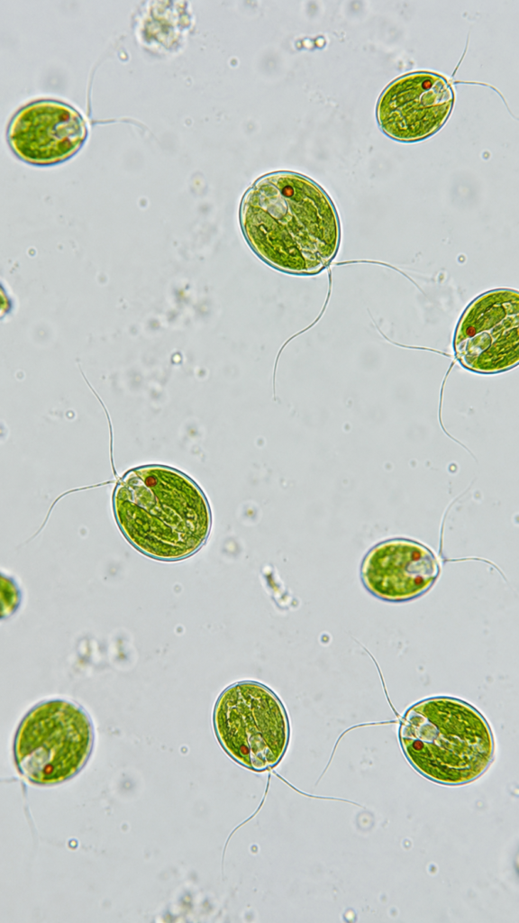
BNO-IMG-080910-01
- Magnification
- 400x
- Image dimensions
- 941 × 1672 px
- Caption
- Volvox Colony Under Microscope

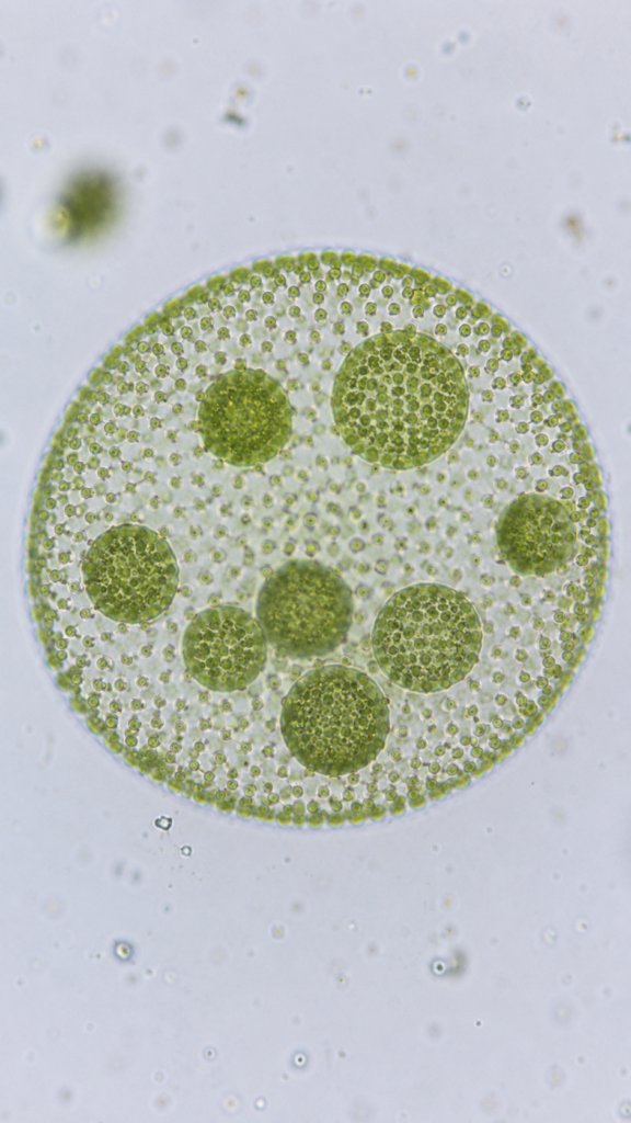


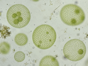
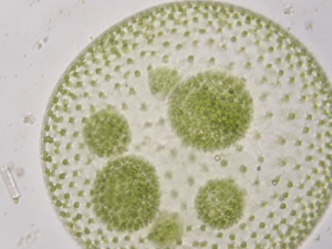
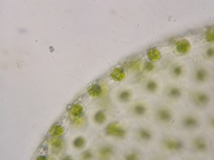
Volvox is a freshwater colonial green alga that forms distinctive spherical colonies containing many chlorophyll-rich cells. In this unstained live wet mount, brightfield microscopy preserves its natural green coloration. At 100×, entire colonies and occasional internal daughter colonies are readily visible. At 400×, the spherical colony surface and numerous small peripheral cells become much clearer. At 1000× using an oil-immersion objective, only a tiny section of the colony is visible, allowing closer observation of individual somatic cells and their green pigmentation, with fine flagella sometimes appearing faintly at favorable focus.
| Field | Value |
|---|---|
| Specimen/common name | Volvox |
| Scientific name | Volvox spp. — genus-level identification; no species is assumed from the supplied name. Volvox is a recognized genus of freshwater green algae in the family Volvocaceae. |
| Stain | None — unstained; natural green chlorophyll pigmentation provides brightfield contrast |
| Microscopy method | Brightfield |
| Tissue/cell type | Spherical colonial green alga composed of numerous peripheral somatic cells; reproductive colonies may contain developing daughter colonies |
| Preparation method | Freshwater live wet mount under a coverslip; no chemical stain. Wet mounting is appropriate for living aquatic microorganisms and preserves natural pigmentation. |
| 100× expected appearance | One or several nearly spherical green colonies visible in a wide field. Colony outline and, in reproductive specimens, rounded daughter colonies within the parent colony may be evident. Individual peripheral cells remain mostly fine green points. Volvox colonies can contain hundreds to thousands of cells. |
| 400× expected appearance | A colony fills much of the field or extends beyond it. The hollow spherical organization is clearer, with numerous small green somatic cells distributed around the peripheral layer. Developing daughter colonies may show stronger green density internally. |
| 1000× expected appearance | Only a very small portion of the colony surface is visible. Individual green somatic cells become the main subject; natural chloroplast pigmentation and cell boundaries are more apparent. Very fine flagella may be faintly detectable near optimally focused peripheral cells, but no ultrastructure should be shown. |
Spherical colony outline, peripheral green somatic cells, natural chloroplast pigmentation, developing daughter colonies when present, and faint flagella at high magnification when favorably focused
Sourav Pan. (2026). Volvox Under Microscope – 100×, 400× and 1000× [BNO-IMG-080910]. Biology Notes Online. https://biologynotesonline.com/microscopic-image/volvox-colony-under-microscope/
This microscopic image is an original work created by Sourav Pan for Biology Notes Online and published on June 21, 2026.
Scientific information associated with this image is provided for educational and student reference. Microscopy details such as specimen, stain, microscopy technique and magnification are shown where available.
The image may be cited using the citation information on this page. For copyright and reuse conditions, see Image Licensing.
If you notice an identification, labeling or scientific accuracy issue, please report it so the record can be reviewed.
 Sourav Pan