BNO-IMG-080933-01
- Magnification
- 400x
- Image dimensions
- 941 × 1672 px
- Caption
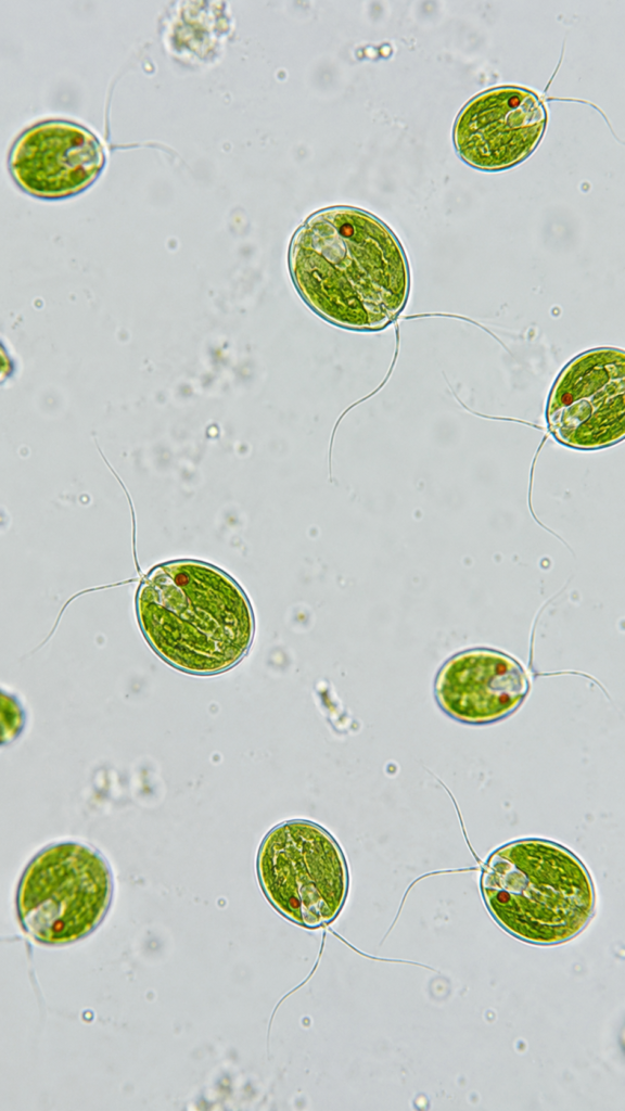
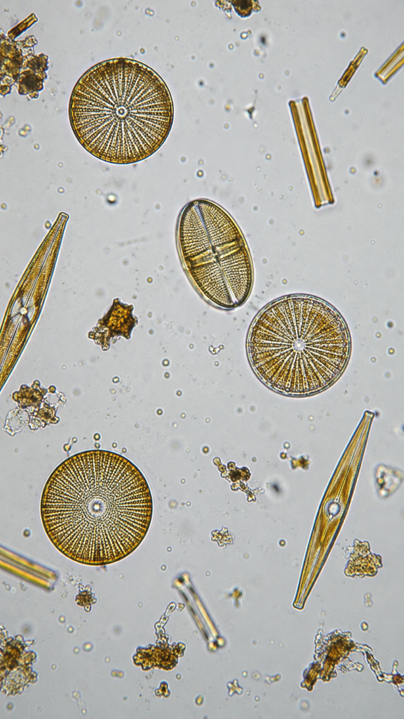
- Chlamydomonas Under Microscope

Chlamydomonas Under Microscope
Microscopic view of Chlamydomonas, single green oval algal cells with two flagella, visible cup-shaped chloroplast and red eyespot, wet mount, brightfield microscope, 400x magnification
Sourav Pan. (2026). Chlamydomonas Under Microscope [BNO-IMG-080933]. Biology Notes Online. https://biologynotesonline.com/microscopic-image/chlamydomonas-under-microscope/
This microscopic image is an original work created by Sourav Pan for Biology Notes Online and published on June 22, 2026.
Scientific information associated with this image is provided for educational and student reference. Microscopy details such as specimen, stain, microscopy technique and magnification are shown where available.
The image may be cited using the citation information on this page. For copyright and reuse conditions, see Image Licensing.
If you notice an identification, labeling or scientific accuracy issue, please report it so the record can be reviewed.
 Sourav Pan