BNO-IMG-080888-01
- Magnification
- 400x
- Image dimensions
- 941 × 1672 px
- Caption
- Paramecium Under Microscope

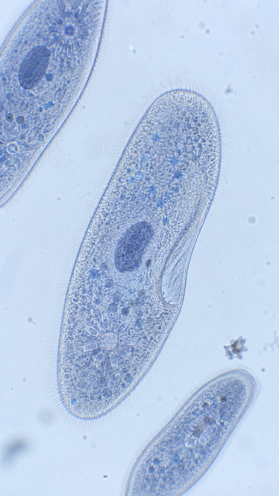

This high-resolution microscopic image captures a live Paramecium specimen observed under light microscopy. This visual reference enables biology students and microscopists to identify characteristic ciliate morphology, including the outer pellicle, dense surface cilia, anterior oral groove, cytostome, contractile vacuoles, and macronucleus.
🔬 For complete laboratory protocols, staining methods, and structural anatomy, read our detailed microscopy guide: Paramecium Under Microscope – Structure, Observation & Identification.
Paramecium Under Microscope
Microscopic view of Paramecium, slipper-shaped protozoan with visible cilia around the body, oral groove, contractile vacuole and nucleus, stained lightly with methylene blue, brightfield microscope, 400x magnification
Sourav Pan. (2026). Paramecium Under the Microscope – Microscopic Specimen Image & Identification [BNO-IMG-080888]. Biology Notes Online. https://biologynotesonline.com/microscopic-image/paramecium-under-microscope/
This microscopic image is an original work created by Sourav Pan for Biology Notes Online and published on June 21, 2026.
Scientific information associated with this image is provided for educational and student reference. Microscopy details such as specimen, stain, microscopy technique and magnification are shown where available.
The image may be cited using the citation information on this page. For copyright and reuse conditions, see Image Licensing.
If you notice an identification, labeling or scientific accuracy issue, please report it so the record can be reviewed.
 Sourav Pan