
Bronchus Section Under Microscope

Image Gallery
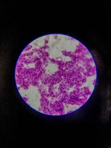
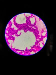
Description
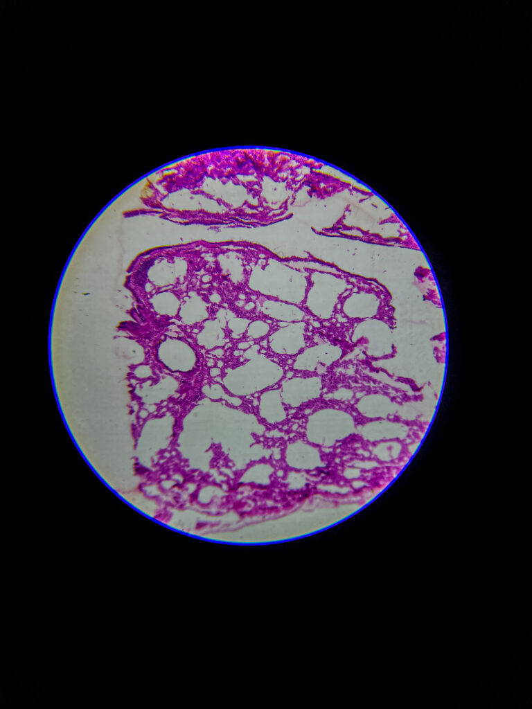
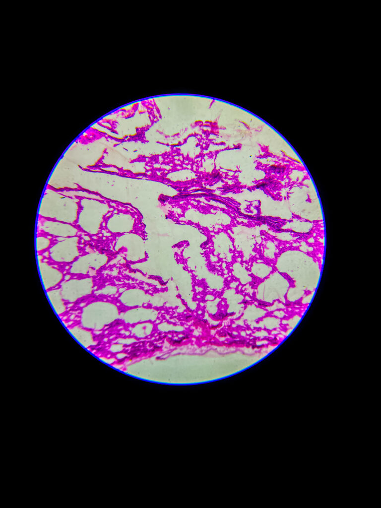
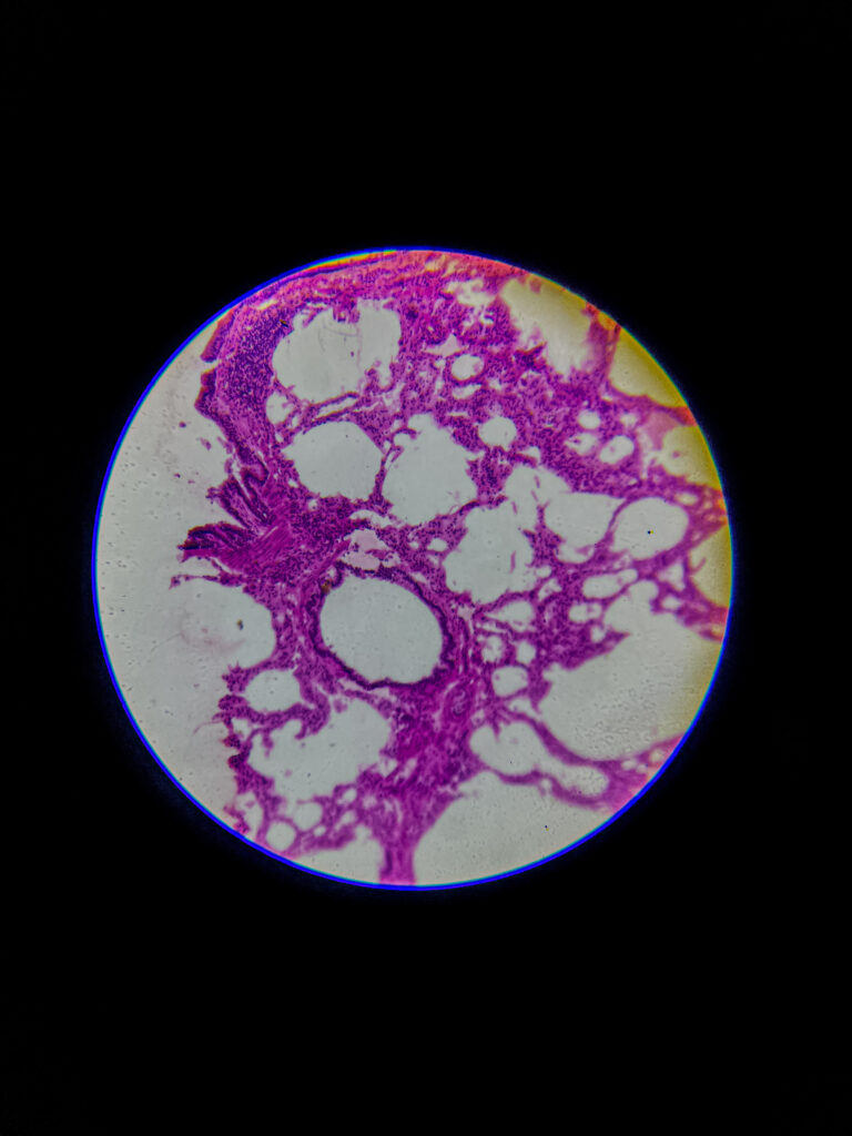
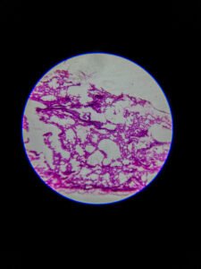
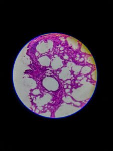
At 40× magnification
Overview of bronchus within lung tissue: visible hyaline cartilage plates surrounded by alveoli
Lumen lined with pseudostratified ciliated columnar epithelium; goblet cells and seromucous glands present in submucosa
Smooth muscle band seen between cartilage and mucosal layer
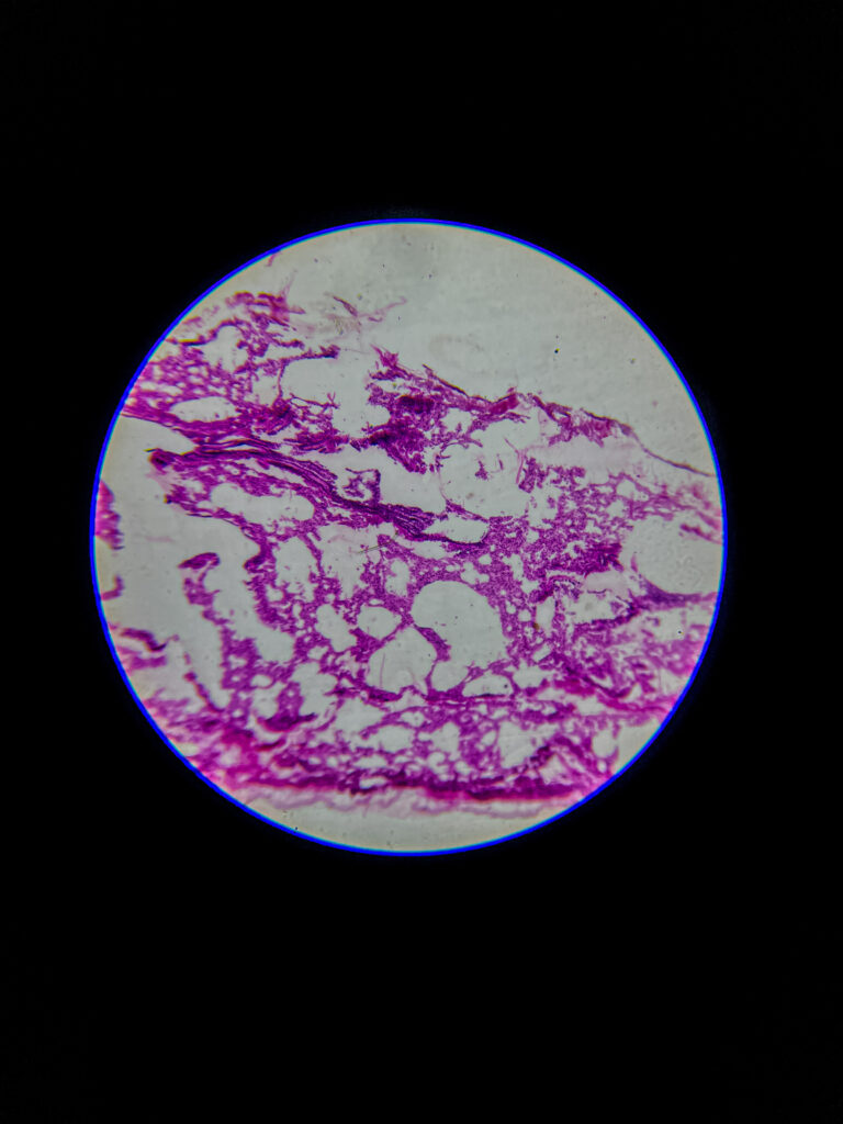
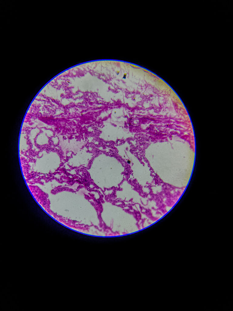
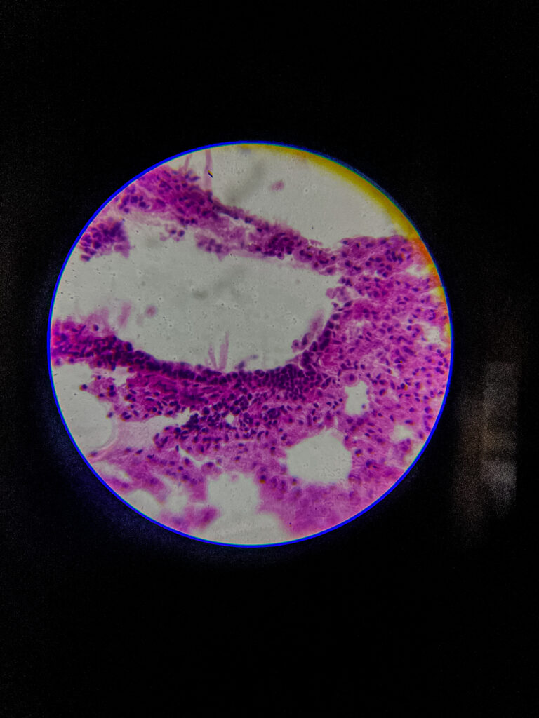
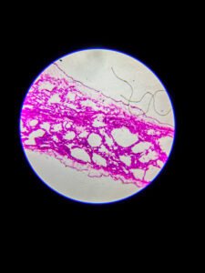
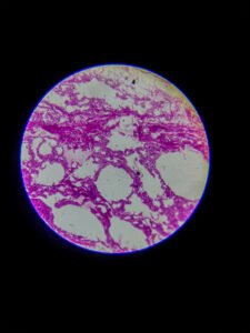
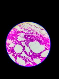
At 100× magnification
Epithelial details clearer: cilia atop columnar cells, goblet cells apparent
Mucosal folds begin to appear, connective tissue in lamina propria visible
Surrounding structures such as alveoli, pulmonary vessels, submucosal glands become discernible
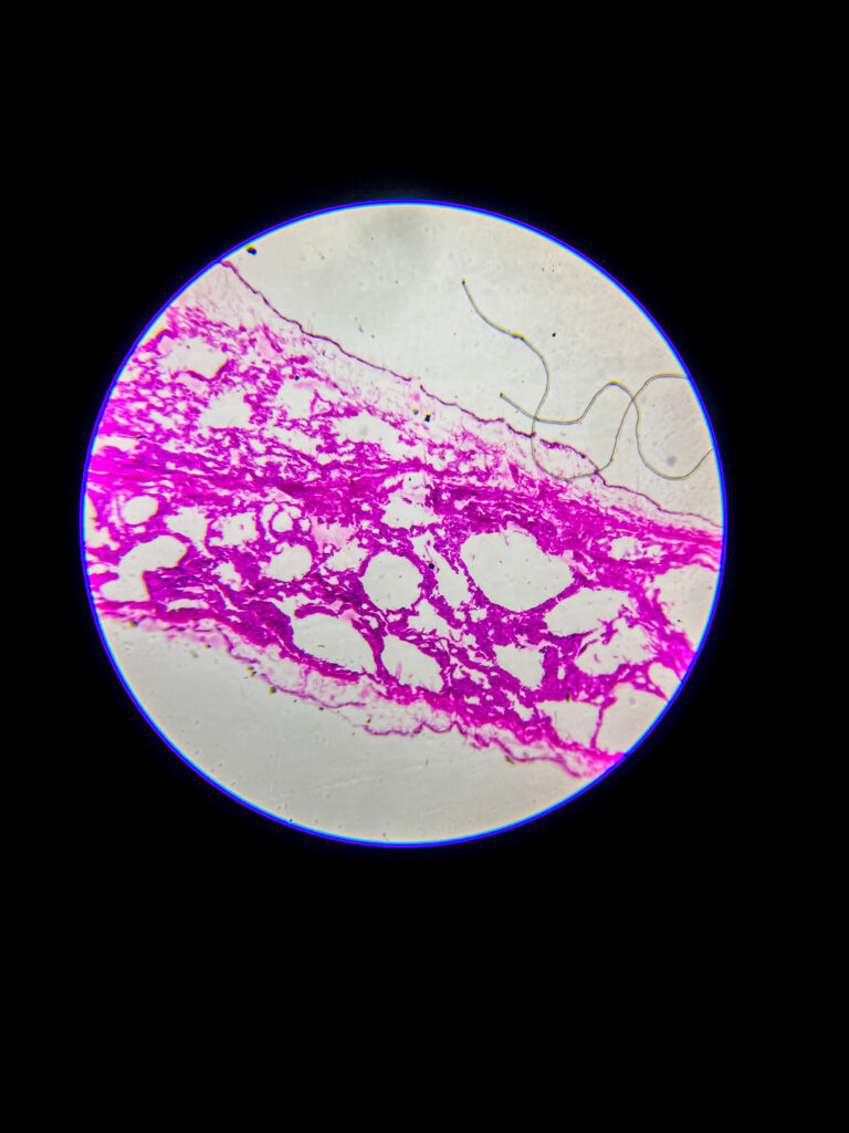
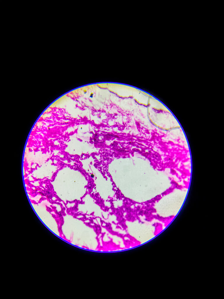
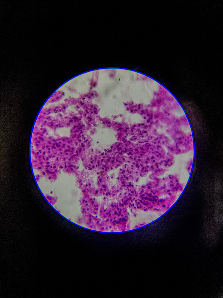
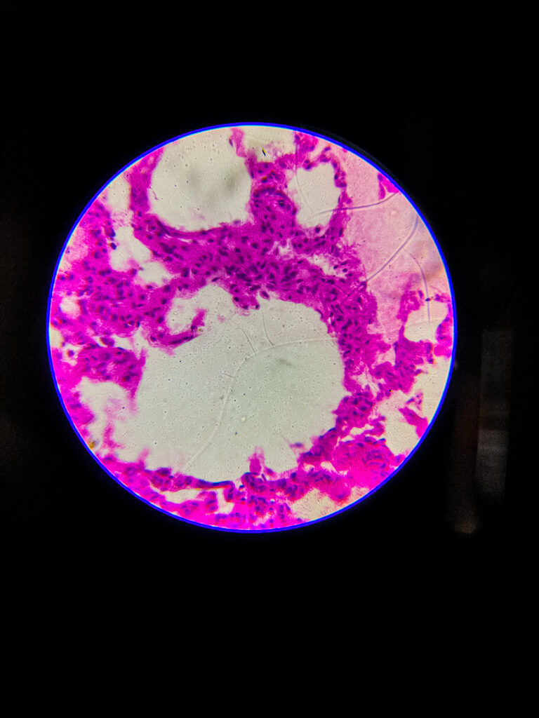
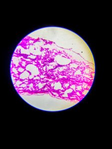
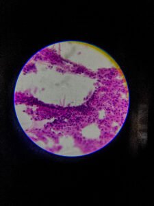
At 400× magnification
High-detail view of respiratory epithelium: individual cilia, goblet cells, basement membrane seen
Smooth muscle ring and seromucous glands visible in submucosa
Cartilage chondrocytes within lacunae clearly identifiable; adventitia and surrounding alveoli evident
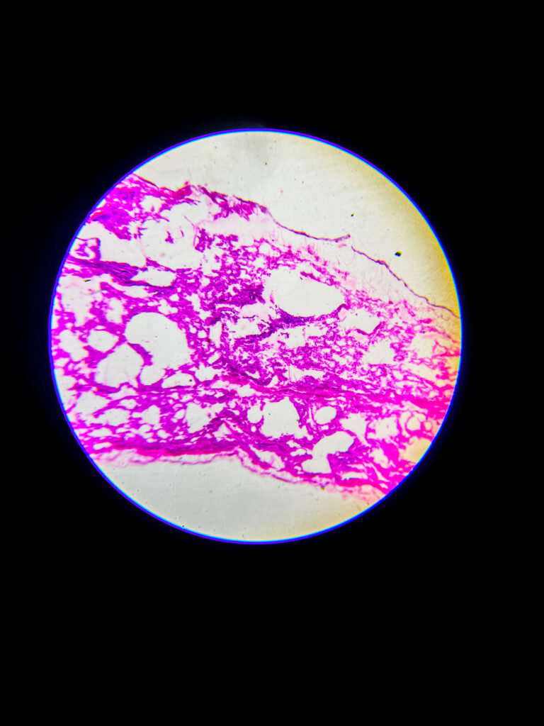
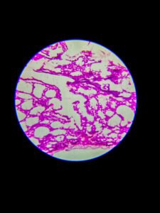
At 1000× magnification (oil immersion)
Focus on cell ultrastructure: ciliary axonemes may be observed, goblet cell mucin granules, basal cells in epithelium
Details of epithelial cell junctions and basement membrane become distinguishable
Smooth muscle cell nuclei and fine connective tissue fibers can be resolved clearly
Functional significance at each magnification
40× provides architectural context—cartilage support, airway branching, relationship to alveoli
100× reveals key tissue components—epithelium type, mucosal structure, glands, vessels
400× allows cellular morphology analysis—cell types, tissue organization, pathological changes
1000× enables study of subcellular features—cilia structure, secretory granules, cell junctions
Summary of histologic layers observed
Epithelium: pseudostratified ciliated columnar with goblet cells
Lamina propria: loose connective tissue with vessels, immune cells
Smooth muscle: circular layer regulating airway diameter
Submucosa: seromucous glands in larger bronchi
Cartilage: plates of hyaline cartilage external to muscle
Adventitia: connective tissue blending into lung parenchyma
Equipment
Compound Microscope
Magnification
40x, 100x, 400x
Staining Technique
Unknow
Image credit: Biology Notes Online / Sourav Pan
Copyright: © 2025 Sourav Pan. All Rights Reserved.
Reuse: Permission may be required. See Image Licensing.
Sourav Pan. (2025). Bronchus Section Under Microscope [BNO-IMG-075899]. Biology Notes Online. https://biologynotesonline.com/community-image/bronchus-section-under-microscope/
About This Microscopic Image
This microscopic image is an original work created by Sourav Pan for Biology Notes Online and published on June 30, 2025.
Scientific information associated with this image is provided for educational and student reference. Microscopy details such as specimen, stain, microscopy technique and magnification are shown where available.
- Image Record ID: BNO-IMG-075899
- Creator: Sourav Pan
- Published: June 30, 2025
The image may be cited using the citation information on this page. For copyright and reuse conditions, see Image Licensing.
If you notice an identification, labeling or scientific accuracy issue, please report it so the record can be reviewed.