hyaline cartilage under microscope
Image ID: BNO-IMG-077500-01
hyaline cartilage under microscope
Image ID: BNO-IMG-077500-02
hyaline cartilage under microscope-3
hyaline cartilage under microscope-3 Image ID: BNO-IMG-077500-03
hyaline cartilage under microscope
hyaline cartilage under microscope Image ID: BNO-IMG-077500-04
hyaline cartilage under microscope
hyaline cartilage under microscope Image ID: BNO-IMG-077500-05
hyaline cartilage under microscope
hyaline cartilage under microscope Image ID: BNO-IMG-077500-06
hyaline cartilage under microscope
hyaline cartilage under microscope Image ID: BNO-IMG-077500-07
hyaline cartilage under microscope
Image ID: BNO-IMG-077500-08
hyaline cartilage under microscope
hyaline cartilage under microscope Image ID: BNO-IMG-077500-09
hyaline cartilage under microscope
hyaline cartilage under microscope Image ID: BNO-IMG-077500-10
hyaline cartilage under microscope
hyaline cartilage under microscope Image ID: BNO-IMG-077500-11

Hyaline Cartilage Under Microscope

Avatar for Sourav Pan Sourav Pan
Uploaded on July 29, 2025

Description

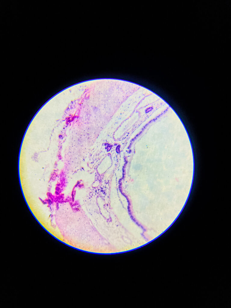
Hyaline cartilage is a type of connective tissue, it consists of a dense network of collagen fibers embedded in a firm, gel-like matrix, mostly it’s made up of type II collagen, it provides smooth surfaces for joint movement, flexibility, and support, it is most common cartilage type in body, that’s found in nose, trachea, larynx, ends of long bones, costal cartilages, and embryonic skeleton.

it’s avascular, so nutrients must diffuse through matrix, no nerves, and no blood vessels, chondrocytes present inside lacunae, surrounded by perichondrium except at articular surfaces.

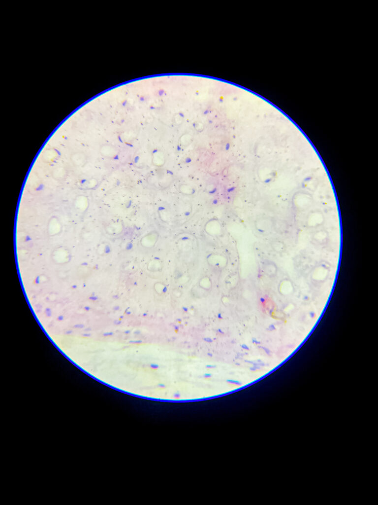
Microscopic appearance – under microscope, hyaline cartilage appears glassy, homogeneous, pale bluish or pinkish if stained, matrix looks smooth and translucent, that’s why called ‘hyaline’ (means glass-like), it’s difficult to distinguish individual collagen fibers because they are very fine and dispersed.

  • chondrocytes are visible as round or oval cells, often clustered in groups called isogenous groups, each chondrocyte lies in a small cavity called lacuna, cytoplasm basophilic due to presence of RNA.
  • peripheral region shows more elongated or flattened chondrocytes, central region contains round chondrocytes.
  • perichondrium, a dense connective tissue layer, can be seen around the cartilage except at articular surfaces.
  • no blood vessels or nerves present inside the tissue, that’s why repair is slow.

Staining features – with H&E stain, matrix looks smooth and light purple, chondrocyte nuclei dark blue, lacunae white or empty spaces, sometimes cytoplasmic shrinkage causes a clear halo around cells.

Summary of key features under microscope

  • smooth, glassy, homogeneous extracellular matrix
  • chondrocytes inside lacunae, sometimes grouped
  • fine collagen fibers not visible individually
  • surrounded by perichondrium (except joints)
  • avascular, aneural, slow healing

Common errors – sometimes matrix may show artifact spaces, nuclei of chondrocytes can look shrunken, misinterpretation as empty lacunae occurs if cells are not present in some sections.

Microscopy details

Equipment
Compound Microscope

Image records

BNO-IMG-077500-01

Magnification
40x, 100x, 400x, 1000x
Image dimensions
1920 × 2560 px

BNO-IMG-077500-02

Magnification
40x, 100x, 400x, 1000x
Image dimensions
1920 × 2560 px

BNO-IMG-077500-03

Magnification
40x, 100x, 400x, 1000x
Image dimensions
1920 × 2560 px
Caption
hyaline cartilage under microscope-3

BNO-IMG-077500-04

Magnification
40x, 100x, 400x, 1000x
Image dimensions
1920 × 2560 px
Caption
hyaline cartilage under microscope

BNO-IMG-077500-05

Magnification
40x, 100x, 400x, 1000x
Image dimensions
1920 × 2560 px
Caption
hyaline cartilage under microscope

BNO-IMG-077500-06

Magnification
40x, 100x, 400x, 1000x
Image dimensions
1920 × 2560 px
Caption
hyaline cartilage under microscope

BNO-IMG-077500-07

Magnification
40x, 100x, 400x, 1000x
Image dimensions
1920 × 2560 px
Caption
hyaline cartilage under microscope

BNO-IMG-077500-08

Magnification
40x, 100x, 400x, 1000x
Image dimensions
1920 × 2560 px

BNO-IMG-077500-09

Magnification
40x, 100x, 400x, 1000x
Image dimensions
1920 × 2560 px
Caption
hyaline cartilage under microscope

BNO-IMG-077500-10

Magnification
40x, 100x, 400x, 1000x
Image dimensions
1920 × 2560 px
Caption
hyaline cartilage under microscope

BNO-IMG-077500-11

Magnification
40x, 100x, 400x, 1000x
Image dimensions
1920 × 2560 px
Caption
hyaline cartilage under microscope
Image Rights & Credit
Creator: Sourav Pan
Image credit: Biology Notes Online / Sourav Pan
Copyright: © 2025 Sourav Pan. All Rights Reserved.
Reuse: Permission may be required. See Image Licensing.

Cite This Image
Sourav Pan. (2025). Hyaline Cartilage Under Microscope [BNO-IMG-077500]. Biology Notes Online. https://biologynotesonline.com/microscopic-image/hyaline-cartilage-under-microscope/

About This Microscopic Image

This microscopic image is an original work created by Sourav Pan for Biology Notes Online and published on July 29, 2025.

Scientific information associated with this image is provided for educational and student reference. Microscopy details such as specimen, stain, microscopy technique and magnification are shown where available.

  • Image Record ID: BNO-IMG-077500
  • Creator: Sourav Pan
  • Published: July 29, 2025

The image may be cited using the citation information on this page. For copyright and reuse conditions, see Image Licensing.

If you notice an identification, labeling or scientific accuracy issue, please report it so the record can be reviewed.
Start Asking Questions