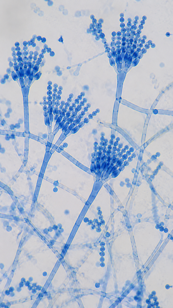
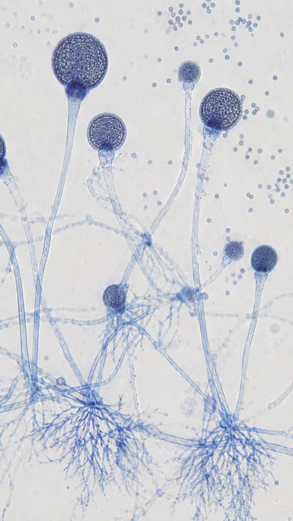
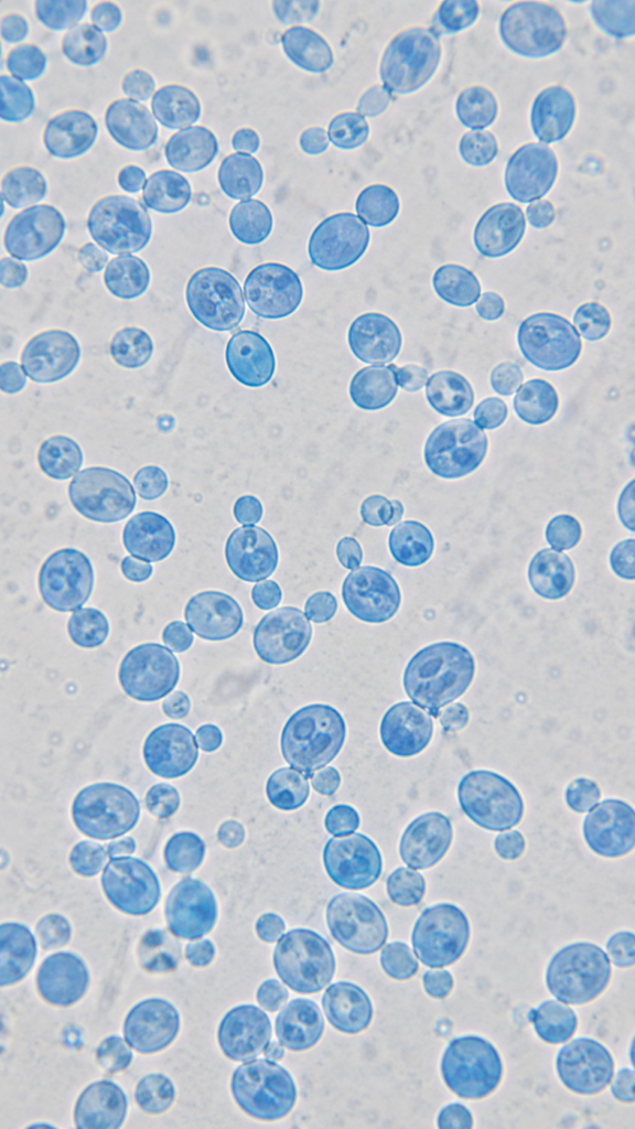
BNO-IMG-080884-01
- Magnification
- 400x
- Image dimensions
- 941 × 1672 px
- Caption
- Aspergillus Fungus Under Microscope

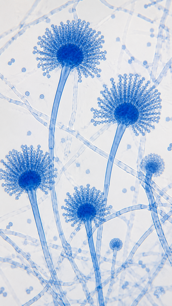


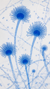
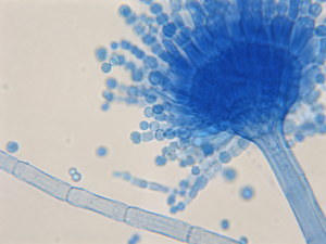
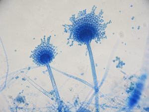

Aspergillus is a genus of filamentous molds recognizable microscopically by septate hyphae and characteristic asexual conidial structures. In this lactophenol cotton blue preparation viewed with brightfield microscopy, 100× shows the overall distribution of hyphae and sporulating conidiophores. At 400×, individual conidial heads, terminal vesicles, phialide-bearing regions, and conidia become substantially clearer. At 1000× using an oil-immersion objective, the much smaller field provides improved resolution of individual conidia, phialides where favorably positioned, and fungal septa without revealing structures beyond conventional brightfield resolution.
Septate hyphae, conidiophores, terminal vesicles, phialide-bearing regions, and conidia; exact conidial-head arrangement varies by species
Sourav Pan. (2026). Aspergillus Under Microscope – 100×, 400× and 1000× [BNO-IMG-080884]. Biology Notes Online. https://biologynotesonline.com/microscopic-image/aspergillus-fungus-under-microscope/
This microscopic image is an original work created by Sourav Pan for Biology Notes Online and published on June 21, 2026.
Scientific information associated with this image is provided for educational and student reference. Microscopy details such as specimen, stain, microscopy technique and magnification are shown where available.
The image may be cited using the citation information on this page. For copyright and reuse conditions, see Image Licensing.
If you notice an identification, labeling or scientific accuracy issue, please report it so the record can be reviewed.
 Sourav Pan