Immunodiffusion refers to the movement of antigen or antibody within the gel. Add the reactants to the wells. They diffuse to the area of lower/no concentration. A gradient of the reactants’ concentration forms as they diffuse into the gel. Immunoprecipitation takes place in a region with an equivalent concentration of both the antibody and antigen. This is represented by bands.
These bands can either be seen directly with naked eyes or stained. Autoradiography is possible if the reactants have been radioactively labeled.
Immunodiffusion
- Immunodiffusion is the term for a precipitation test on agar gel.
- This test involves the addition of reactant to the gel, and the diffusion of the antigen-antibody mixture.
- The size of the particles, temperature, gel viscosity and amount of hydration all affect the diffusion rate.
- Agar concentrations between 0.3 and 1.5% allow diffusion of most reactants.
Types of immunodiffusion reaction
There are two types of immunodiffusion tests in gels:
- Single immunodiffusion
- Double immunodiffusion
1. Single immunodiffusion
Single/simple immunodiffusion is when one reactant (antigen), diffuses, while the other (antibody), is added to the gel and fixed. There are several types of immunodiffusion assays.
- Single diffusion in one dimension
- Single diffusion in two dimensions
a. Single diffusion in one dimension
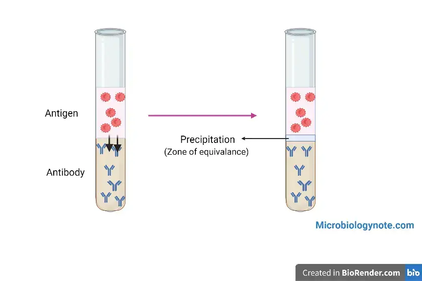
In this method, the diffusing reactant only diffuses in one direction. This includes the oudin tube method.
Oudin tube method:
This test involves laying a solution or gel containing the antigens onto a layer of antiserum-containing agar in a tube. Because of gravity/ concentration gradient, the antigen diffuses into an antibody-containing gel region. Initially, the antigen concentration becomes higher as it moves. The antigen concentration reaches equilibrium or is equal to the antibody concentration, at which point precipitation takes place. The precipitation is used to detect the presence or absence of antigen. Qualitative analysis.

b. Single diffusion in two dimensions:
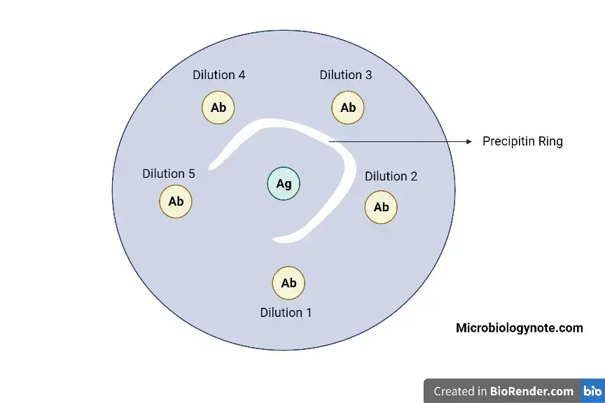
This is the single radial immunodiffusion method (SRID), also known as the Mancini method. This method incorporates the antibody against the known antigen into the gel. Wells are drilled into these gel layers, to add the antigen of your choice. The concentration gradient is formed again as the antigen diffuses radially out of the well. The precipitation takes place around the well at the point of equivalence.
SRID can be used to quantify the amount of antigen. The wells are dug into the antibody-incorporated gel, and the antigen of different concentrations are added to each of the wells. Each antigen concentration will be assigned a zone of equivalence at various distance.
A graph showing the antigen concentration against the diameter or ring area is drawn. You can find the unknown concentration by calculating the diameter of the antigen sample or the ring area, and then plotting it in the graph along with the calibration curve.
2. Double diffusion
The double diffusion technique, which involves diffusion of both reactants towards one another, is a different method from single diffusion. These are the different techniques that can be used with this technique:
a. Double diffusion in one dimension:
This is similar to single diffusion in one dimension except that there is only a layer of agar between layers of antigen or antibody. Both reactants then diffuse toward one another and the precipitation rings are formed in the middle layer. Due to the concentration gradient, each reactant diffuses in one direction: either upwards or downwards. This is why diffusion occurs in one dimension.
b. Double diffusion in two dimensions:
This is also known as the Ouchterlony method and is the most commonly used immunochemical technique. This is used to determine if two antigens have similarity or not. The Ouchterlony method can be used on a petri-dish, or even a microslide that is overlaid with agar. The antigen and antibody solutions are then added to the wells. This can be used to perform both quantitative and qualitative analysis.
Qualitative Ouchterlony technique:
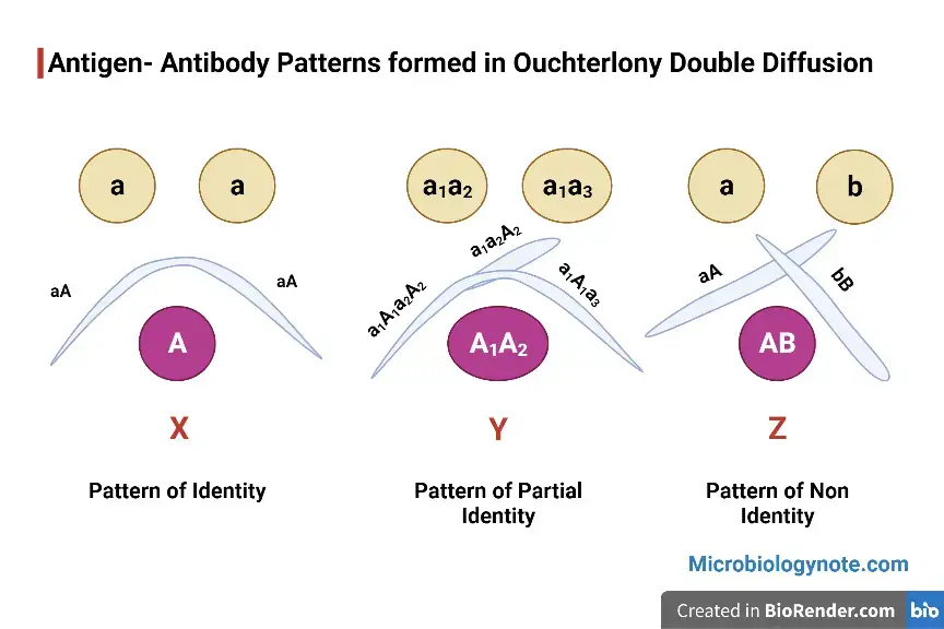
To compare antigens against identical or cross-reacting determinants, the qualitative Ouchterlony method is used. The two antigen solutions are placed in adjacent wells, and the homologous antibodies is placed in the middle well.
The precipitation line will formed because the antigens present in both wells fuse are in the same concentration, and these Ag are identical. This reaction is called a reaction of identification and the pattern is called a pattern of identity.
When two antigens of unrelated nature are added in the two wells, and a central well is filled up with antibodies for each antigen, immunoprecipitations can occur separately with the specific antibodies. These precipitation lines will cross one another following their respective zones of equivalence, and create a cross-shaped structure. This is called a pattern of non-identity.
A spur is formed when two related antigens are added to the wells with the antisera in the center well. The spur is a sign that one antigen determinant is present in each sample, while the other is only in one. The precipitation line of an additional antigen that projects toward the antigen sample containing only the common determinant is called the spur. This is known as a reaction with partial identity.

Quantitative Ouchterlony technique
Similar to SRID, this allows semi-quantitative estimation of antigen concentrations by using the relative position of the precipitation line. The precipitation line would be farther away if the antigen concentration is higher than the average concentration.
The concentration of antigen solution in the well will determine the distance of the precipitation lines. A graph showing the distance to the concentration can then be plotted. The calibration curve can be used to determine the concentration of unknown antigen samples.

Advantages
- The major advantage of immunodiffusion reaction over other methods is the ability to see the line of precipitation as a band that can be stained for preservation.
- It can also be used to identify cross reactions and non-identify antigens within a mixture.
Applications
- To determine the relative concentrations Antibodies/Antigens.
- To compare Antigens or
- To determine the purity of an Antigen preparation.
- To diagnose disease.
- Surveys of serology.
References
- https://www.biosciencenotes.com/immunodiffusion-reaction/#:~:text=Single%20diffusion%20in%20one%20dimension%3A&text=In%20this%20method%2C%20antibody%20is,line%20of%20precipitation%20is%20formed.
- https://link.springer.com/chapter/10.1007/978-1-4757-1257-5_3?noAccess=true
- https://www.sciencedirect.com/topics/immunology-and-microbiology/immunodiffusion
- https://www.clinisciences.com/en/read/serological-tests-in-mycology-1190/immunodiffusion-id-assay-2092.html
- https://thebiotechnotes.com/2019/09/19/immunoprecipitation-p2-immunodiffusion-in-gel/
- https://www.slideshare.net/suniu/immunodiffusion-principles-and-application
- https://en.wikipedia.org/wiki/Immunodiffusion
- Text Highlighting: Select any text in the post content to highlight it
- Text Annotation: Select text and add comments with annotations
- Comment Management: Edit or delete your own comments
- Highlight Management: Remove your own highlights
How to use: Simply select any text in the post content above, and you'll see annotation options. Login here or create an account to get started.