What is Clostridium?
- Clostridium is a diverse and fascinating genus of bacteria that was first described in 1880 by Prazmowski. These bacteria are characterized by their distinctive rod-like morphology. However, what makes Clostridium truly intriguing is the wide range of phenotypes they exhibit, spanning from acidophiles to psychrophiles.
- One of the notable aspects of this genus is its genetic variability. The G+C content in Clostridium species can vary significantly, ranging from about 21 to 54 percent. Despite some species appearing Gram-negative, the majority of Clostridia are Gram-positive organisms that possess a unique ability to form spores. Additionally, these bacteria thrive in anaerobic environments, where oxygen is scarce.
- While some members of the Clostridium genus are pathogenic and can cause diseases in both humans and animals, it is essential to note that a considerable number of species are non-pathogenic. In fact, some of these non-pathogenic strains have found valuable applications in various industries, showcasing their positive contributions to society.
- The medical significance of Clostridium lies in the Gram-positive species, although certain strains may exhibit Gram-variable characteristics. This variation in Gram staining underscores the diverse nature of this bacterial genus.
- Several well-known species fall under the Clostridium umbrella, including C. botulinum, which produces the deadly botulinum toxin, and C. perfringens, responsible for causing gas gangrene and foodborne illnesses. Other species like C. sporogenes, C. bifermentans, C. leptum, and C. difficile also contribute to the diversity of this intriguing bacterial group.
- In conclusion, Clostridium is a captivating genus of bacteria with rod-like morphology and a broad spectrum of characteristics. While some species pose health risks as pathogens, many others offer beneficial applications in various industries. Understanding the complexities of this diverse genus continues to be a subject of interest for researchers and scientists alike.
Morphology of Clostridium
- The morphology of Clostridium showcases a diverse array of shapes and structures among its strains, particularly those of medical significance. Most commonly, these strains appear as rod-shaped organisms, either straight rods or slightly curved, resembling cylindrical rods when observed under a microscope. However, it’s important to note that not all species or strains within the genus adhere to this typical rod-like morphology.
- While the majority of Clostridium strains are characterized by Gram-positive rods, certain colonies may present with a convex shape, and a few may even exhibit a spherical or irregular morphology. The ends of these bacterial cells also show variation, ranging from rounded to pointed ends, depending on the specific strain.
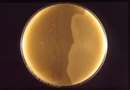
- In the case of Clostridium difficile, microscopic examination after culturing has revealed a range of shapes, including short and thick forms, as well as large Gram-positive rods with rounded ends.
- A distinguishing feature of most Clostridium species is the presence of peritrichous flagella, which allow these bacteria to move actively from one location to another, akin to swimming. Unlike other flagella structures, peritrichous flagella project from all directions of the cell, granting the bacteria the ability to move rapidly in any direction they desire.
- Additionally, some Clostridium species are capable of forming endospores, which may be observed protruding from one end of the bacterial cell. These endospores can exhibit oval or spherical shapes.
- Furthermore, it is noteworthy that the flagella of C. difficile have been found to induce a pro-inflammatory response, contributing to its pathogenicity.
- Interestingly, the process of sporulation and temperature variations have been identified as factors that can cause morphological changes in certain species, such as C. perfringens, when cultivated in lab cultures.
- In conclusion, the morphology of Clostridium is both intriguing and diverse. While the majority of medically significant strains display rod-shaped characteristics, there are variations in colony shapes and end appearances. The presence of peritrichous flagella facilitates their mobility, and the formation of endospores adds to their adaptive capabilities. The understanding of these morphological features is vital in identifying and studying these unique bacteria for medical and scientific purposes.
Gram Staining of Clostridium
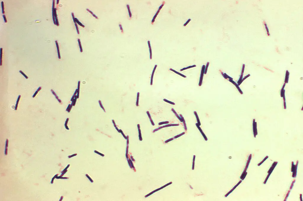
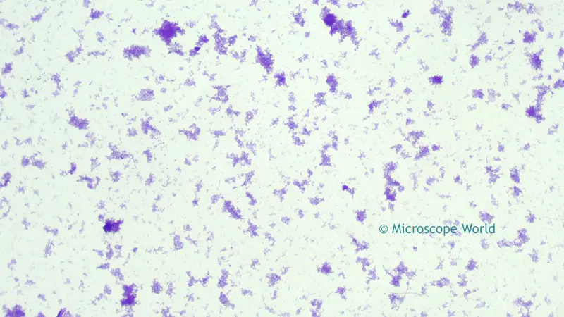
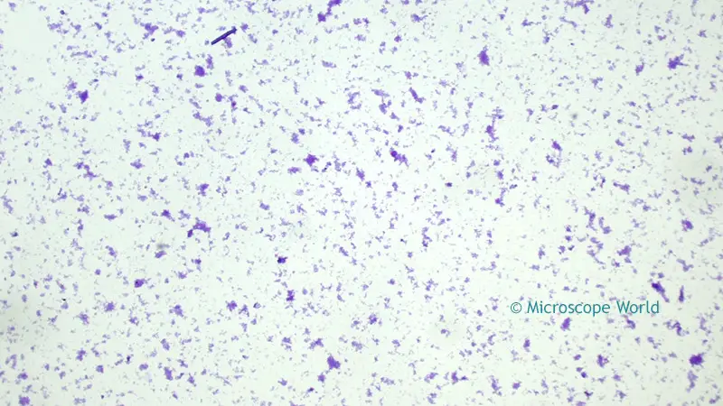
The bulk of Clostridium are Gram-positive rods. C. difficile, for example, is a Gram-positive Clostridium that will look dark blue/violet due to the retention of the main stain (crystal violet) in its thick peptidoglycan layer.
Although these bacteria appear Gram-positive, under certain conditions, they can lose this appearance. Some Clostridium species have been demonstrated to lose their Gram-positive appearance after prolonged incubation. However, a few species have been discovered to be Gram-negative and to stain pink during Gram staining.
Over 200 species of the genus Clostridium have been found to date. At least 30 of these are clinically significant. Gram staining can be used to observe and identify the species.
Requirements for Gram Staining of Clostridium
To perform Gram staining of Clostridium, several essential requirements and materials are necessary for accurate and reliable results. Gram staining is a widely used technique in microbiology to differentiate bacteria based on their cell wall composition. Here are the key requirements for conducting Gram staining of Clostridium:
- Clean Glass Slide: A clean glass slide serves as the foundation for creating a bacterial smear, allowing the sample to adhere to the surface for microscopic examination.
- Gram Stains: The Gram staining process involves multiple stains, including:
- Crystal Violet: This primary stain imparts a purple color to the bacterial cells.
- Gram’s Iodine: The iodine acts as a mordant, enhancing the retention of the crystal violet stain within the cells.
- Counterstain (Safranin): Safranin serves as the secondary stain, imparting a red color to bacterial cells after the decolorization step.
- Decolorizing Agent: Typically, ethanol or acetone is used as a decolorizing agent to differentiate between Gram-positive and Gram-negative bacteria.
- Sample: A sample of Clostridium is required, obtained either from a patient or a bacterial culture. When using a culture sample, it is crucial that it has not been incubated for an extended period, as over-incubation can affect the integrity of the bacterial cells.
- Water: Distilled or tap water is needed for various rinsing steps during the staining process.
- Staining Rack: A staining rack is used to hold and transport the glass slides during the staining procedure, ensuring efficient staining and minimizing contamination.
- Immersion Oil: Immersion oil is utilized to enhance the resolution and clarity of microscopic images. It matches the refractive index between the glass slide and the microscope lens, reducing light scattering.
- Bunsen Burner: A Bunsen burner is essential for heat-fixing the bacterial smear to the glass slide. Heat fixation kills the bacteria, immobilizes them on the slide, and helps preserve cellular structures during the staining process.
Procedure for Gram Staining of Clostridium
To perform Gram staining of Clostridium, follow this step-by-step procedure carefully for accurate and reliable results:
- Sample Preparation: Using a cotton swab, collect a small sample of Clostridium and make a smear at the central part of a clean glass slide. To create a good smear, use a circular motion while spreading the sample.
- Heat Fixation: Pass the slide, sample-side up, over the Bunsen burner flame approximately three times to heat-fix the smear. Be cautious not to overheat the slide, as excessive heat can distort the bacterial cells.
- Crystal Violet Staining: Place the slide on a staining rack and cover the smear with crystal violet. Allow the crystal violet stain to stand on the slide for about one minute to ensure proper staining of the bacterial cells.
- Rinsing with Water: Tilt the slide slightly and gently rinse off the excess crystal violet stain with water. Tipping the slide at an angle prevents washing away the stained bacteria.
- Gram’s Iodine Application: Flood the slide with Gram’s iodine and let it stand for about one minute. Gram’s iodine acts as a mordant, enhancing the binding of the crystal violet stain to the bacterial cells.
- Rinsing with Water: Tilt the slide again and rinse off the excess Gram’s iodine with water. This step helps remove any unbound stain from the slide.
- Decolorization: Tilt the slide and carefully apply the decolorizing agent, such as acetone or 95 percent ethyl alcohol, drop by drop for about 5 seconds. Take care to avoid excessive decolorization, which may lead to inaccurate results.
- Rinsing with Water: Immediately rinse the slide with water to stop the decolorization process and remove any remaining decolorizing agent.
- Safranin Counterstaining: Flood the slide with the counterstain, safranin, and allow it to stand for about 1 minute. Safranin imparts a red color to bacterial cells that have lost the crystal violet stain during the decolorization step.
- Final Rinse: Tilt the slide and rinse off the excess safranin stain with tap water.
- Blotting: Gently blot the slide with a blotting paper to remove excess water without disturbing the stained bacterial cells.
Now, your Gram-stained slide containing Clostridium samples is ready for microscopic examination. The staining process helps differentiate between Gram-positive and Gram-negative bacteria, providing valuable insights into the characteristics and structure of the Clostridium cells under the microscope.
Observation
After completing the Gram staining procedure for Clostridium samples, the next step is to observe the stained bacterial cells under a microscope. For optimal results, the slide should be viewed using immersion oil and either a compound light microscope or a phase-contrast microscope.
When observing the Gram-stained Clostridium samples under the microscope, different species may exhibit various characteristics. Here are the possible observations for two specific Clostridium species:
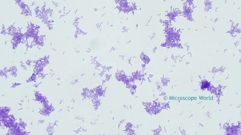
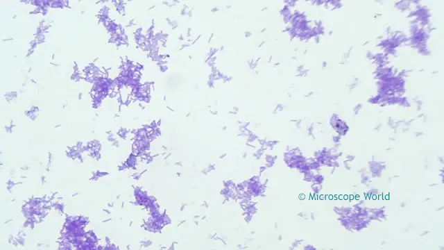
- C. tetani: When viewing C. tetani under the microscope, one will notice that the bacterial cells appear purple in color. Additionally, terminal spores may be visible, depending on the sample and sample preparation. Another potential observation is the motility of the cells, which can be observed if the sample is properly prepared. Furthermore, students will be able to identify the characteristic rod-shaped morphology of C. tetani.
- C. perfringens: Under the microscope, C. perfringens cells may present as either elongated rods or shorter in length. These bacterial cells are non-motile and do not possess terminal poles. The absence of motility is a distinctive feature of this species. Additionally, students will be able to differentiate the specific morphological characteristics of C. perfringens under microscopic examination.
It is crucial to note that the observed characteristics may vary depending on the species of Clostridium being examined. Different species within the genus Clostridium can exhibit diverse morphological features, and microscopic observation allows for their accurate identification and differentiation.
Microscopic examination of Gram-stained Clostridium samples is a fundamental technique in microbiology and plays a critical role in identifying and studying these bacteria. The observations made during this process contribute to the broader understanding of Clostridium species and their potential implications in various contexts, such as medical, industrial, or environmental settings.
FAQ
How do Clostridium bacteria appear under a microscope?
Clostridium bacteria typically appear as rod-shaped cells when viewed under a microscope.
What staining method is commonly used to observe Clostridium under the microscope?
The Gram staining method is commonly used to observe Clostridium under the microscope, allowing for differentiation between Gram-positive and Gram-negative bacteria.
Are Clostridium bacteria motile?
Some species of Clostridium are motile, while others are non-motile. Motility can be observed under the microscope if the proper conditions are met.
What distinguishes Clostridium from other bacteria under microscopic examination?
Clostridium can be distinguished by its characteristic rod shape and the presence of endospores in certain species.
Can you observe spores in all Clostridium species under the microscope?
Not all Clostridium species form visible spores under the microscope. Spore formation varies among different species.
How can you identify Clostridium difficile under the microscope?
Clostridium difficile cells may appear as large Gram-positive rods with rounded ends when observed under the microscope.
Which type of microscope is best for observing Clostridium bacteria?
A compound light microscope is commonly used for routine observation of Clostridium bacteria, but specialized microscopes like phase-contrast microscopes can also be helpful for certain studies.
What is the color of Clostridium cells after Gram staining?
Clostridium cells will retain the crystal violet stain, appearing purple or violet, when observed after Gram staining.
Can you differentiate between various Clostridium species solely based on their microscopic appearance?
While some species may have characteristic features, microscopic observation alone may not always be sufficient for definitive species identification. Additional tests may be required for accurate identification.
What is the importance of observing Clostridium under the microscope in clinical settings?
Microscopic examination of Clostridium samples is vital in clinical settings to aid in the diagnosis of infections caused by these bacteria. It helps healthcare professionals choose appropriate treatments and understand the nature of the infection.
References
- https://www.creative-diagnostics.com/blog/index.php/an-overview-of-clostridium/
- https://blog.microscopeworld.com/2015/09/tetanus-bacteria-under-microscope.html
- https://www.microscopemaster.com/clostridium.html
