The water circulation system of sponges, also known as canal system, is the defining property of the phylum Porifera. The system of canals is also known as the auriferous system. The sponge canal system aids in food uptake, respiratory gas exchange, and excretion. Many pores on the body surface of sponges allow for the admission and egress of water current; these perforations are the primary components of the canal system. Inside the body, the water circulation passes through a specific system of compartments in which food is extracted from incoming water and waste is expelled into departing water.
What is Canal system in sponges?
The canal system of sponges is a complex network of interconnected channels and pores used for water circulation and feeding.
- Sponges are filter-feeding aquatic organisms that receive their food by pumping water through their body.
- In sponges, there are three types of canals: inhalant, exhalant, and choanocyte.
- The passages via which water enters the sponge’s body are the inhalant canals. These canals are lined with specialised cells known as porocytes that control the flow of water.
- The passages through which water escapes the sponge’s body are its exhalant canals. These canals are lined with specialised cells known as oscula that control the flow of water.
- Choanocyte tubes lead to the sponge’s internal chambers, where the majority of water filtering and food capture occurs.
- Choanocytes are specialised cells that line the choanocyte canals and feature a collar of flagella that generates a current to trap food particles.
- Sponge survival is dependent on their canal system, which promotes the transport of water and nutrients throughout their bodies.
- The movement of water through the sponge’s canal system is propelled by ciliary activity and muscular contractions of the body.
- Moreover, the canal system is essential for removing waste from the sponge’s body.
- The size and structure of the canals, as well as the amount of choanocytes within the sponge’s body, determine the efficacy of its feeding system.
- The canal system of sponges is quite diverse and can vary considerably between species.
- Some sponges have a simple canal system, but others have a system with several branching canals.
- The organisation and complexity of the canal system are influenced by the feeding habits and ambient factors of the sponge.
- Based on the form and arrangement of the canals, the structure of the canal system can also be used to categorise sponges into distinct groups.
- Asconoid sponges have a simple canal system, but syconoid and leuconoid sponges have complex canal systems.
- As oxygen is absorbed from the water and carbon dioxide is released through the exhalant canals, the canal system in sponges is also engaged in gas exchange.
- The canal system is also responsible for regulating the sponge’s internal water balance and preventing the accumulation of surplus water.
- Little fish and other aquatic species that take sanctuary within the sponge’s chambers can also find protection throughout the canal system.
- Environmental problems such as pollution can disturb the canal system, which can reduce the sponge’s feeding efficiency and overall health.
- Knowing the form and function of the canal system in sponges is crucial for elucidating their ecology and evolution.
Components of canal system in sponge
- Ostia or dermal pores: A thin membrane covers the exterior grooves of the body’s surface. It has at least two or more holes, ostia or dermal pores. Around these apertures are contractile myocytes. These substances can reduce the size of skin pores, regulating the amount of incoming water. They are connected to the incurrent canals.
- Incurrent canals: These are thin spaces located radially between adjacent radial canals that are similar in size and shape. They are surrounded by pinacocytes. They open to the exterior by ostia, but their interior ends are blind.
- Prosopyles: Via prosopyles, incurrent canals communicate with radial canals. Each prosopyle is a puncture in the porocyte, a single tubular cell.
- Radial or flagellated canals: These chambers are bordered with flagellated choanocytes and are referred to as flagellated chambers or radial canals. Parallel and alternating, the radial canals and the incurrent canals are separated by the mesenchyme. On a vertical piece of the body wall, each radial canal appears to be surrounded by four incurrent canals, and each incurrent canal appears to be encircled by four radial canals. At their outside ends, radial canals end aimlessly, but at their inner ends, they open into spongocoel.
- Apopyles: The apertures of radial canals into the spongocoel are known as apopyles or gastric ostia. They are surrounded by contractile myocytes that control the apopyle diameter.
- Spongocoel: This is the huge central chamber into which the radial canals’ apopyles open. It is the longitudinal centre of the entire body.
- Osculum: The spongocoel communicates with the outside world through its terminal orifice, the osculum. Specialized contractile myocytes surround the osculum. They provide a sphincter that regulates the rate at which water leaves the body. Sponge physiology is primarily determined by the water current. The water current is created by the beating of collar cell flagella. By way of this current, all exchanges between the sponge body and external medium are maintained. This water circulation transports nutrients and oxygen into the body. Moreover, the excreta are removed from the body by this water current. The reproductive cells are conveyed by the water stream into the sponges’ bodies.
Types of canal systems in sponge
Various sponges have varying arrangements and degrees of complexity of internal channels; hence, the canal system has been categorized into four types:
- Ascon type of Canal system
- Sycon type of canal system
- Leucon type of canal system
- Rhagon Type
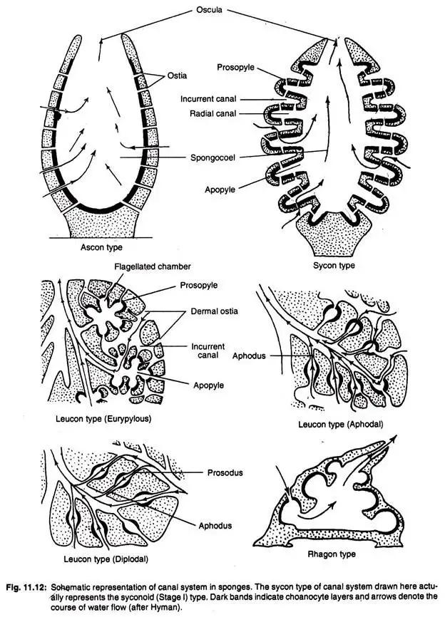
1. Ascon type of Canal system
- This canal system is the most straightforward of the three. It is found in asconoid sponges, such as Leucosolenia, as well as in various developmental stages of syconoid sponges.
- The body surface of asconoid sponges is perforated by a vast number of minute pores or ostia known as incurrent pores.
- These holes are intracellular gaps within porocytes, which are tube-shaped cells. These pores extend radially through the mesenchyme and directly into the spongocoel.
- The spongocoel is the largest and most extensive cavity in the sponge’s body. Choanocytes, which are flattened collar cells, line the spongocoel.
- Spongocoel opens to the outside through a thin circular hole known as the osculum, which is positioned at the distal end, and is bordered with massive monaxon spicules.
- Through the ostia, the surrounding seawater enters the canal system. The water flow is maintained by the beating of the collar cells’ flagella.
- Although the enormous spongocoel contains a great deal of water that cannot be squeezed out through a single osculum, the pace of water flow is slow.
- EX. – Clalhrina & Leucosoleniaand simple sponges.
Structure of Ascon type of Canal system
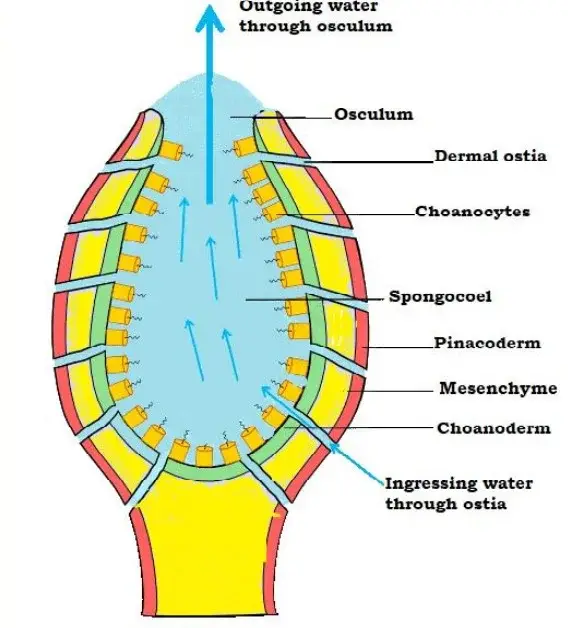
- This organism has a vase-like body with radial symmetry composed of a thin wall enclosing a huge central chamber, with the spongocoel opening at the apex through a constricted osculum.
- The wall consists of an exterior and inner epithelium separated by mesenchyme.
- The epidermis, or outside or dermal epithelium, consists of a single layer of flat cells.
- Choanocytes constitute the inner epithelium that lines the spongocoel.
- The mesenchyme is composed of skeletal spicules and numerous types of amoebocytes, all of which are embedded in a gelatinous matrix.
- Many microscopic openings called incurrent pores or ostia penetrate the asconoid sponge’s wall and stretch from the outside surface to the spongocoel.
- Each hole is intracellular, that is, it is a canal via a porocyte, a tubular cell.
- The water current propelled by the flagella of choanocytes travels through the incurrent pores into the spongocoel and through the osculum, supplying food and oxygen and transporting metabolic wastes along its path.
- Few sponges possess an asconoid canal system, including Olynthus and Leucosolenia.
Course of water current in Asconoid type canal system
Ingressing water → Ostia → Spongocoel → Osculum → outside
2. Sycon type of canal system
- Sycon canal systems are more sophisticated than ascon canal systems. This type of canal system is unique to syconoid sponges such as Scypha.
- Theoretically, the asconoid type can be created from this canal system by horizontally folding its walls. Additionally, the embryonic growth of Scypha clearly demonstrates the transformation of the asconoid pattern into the syconoid pattern.
- The body walls of syconoid sponges have two types of canals, the parallel and alternating radial canals and incurrent canals. Both of these canals stop blindly in the skin, but they are joined by minute pores.
- Incurrent pores, also known as dermal ostia, are located on the body’s outside. These incurrent pores lead to the formation of incurrent canals.
- As they are bordered by pinacocytes rather than choanocytes, the incurrent canals lack flagella. The incurrent canals connect to nearby radial canals via prosopyles, which are minute apertures.
- In contrast, radial canals are flagellated because they are lined with choanocytes. These canals are connected to the central spongocoel via ostia or apopyles.
- The spongocoel is a tiny, non-flagellated chamber lined by pinacocytes in the sycon type of canal system. It opens to the exterior by an excurrent orifice known as the osculum, which is comparable to the ascon-type canal system.
- In a few species, such as Grantia, the Sycon canal system is more complicated, with irregular and branched incurrent canals generating enormous sub-dermal voids. This is due to the formation of the sponge’s cortex, which involves the distribution of pinacoderm and mesenchyme throughout the entire outer surface.
Structure of Sycon type of canal system
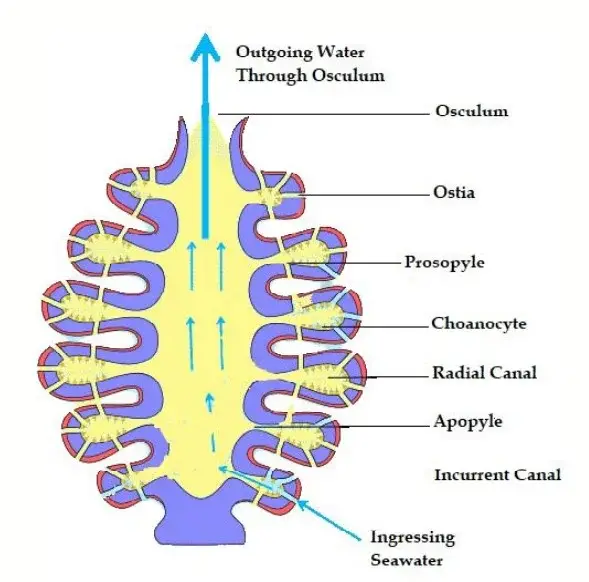
- The syconoid canal system is the first level above the asconoid canal system.
- Radial canals are generated by the outward push of the wall of an asconoid sponge at regular intervals into finger-like projections.
- Initially, these radial canals are free protrusions, and the exterior water surrounds their entire length because there are no distinct incurrent channels.
- In most syconoid sponges, however, the walls of radial canals fuse in such a way as to leave between them tubular gaps, the incurrent canals, which open to the exterior through dermal ostia or dermal pores between the blind outer ends of the radial canals.
- Since these incurrent canals represent the asconoid sponge’s original outer surface, they must be coated with epidermis. Since radial canals are the result of the expansion of the original spongocoel, they must be lined with choanocytes and are hence better known as flagellated canals.
- The inside of the syconoid sponge is hollow and forms a huge spongocoel bordered by epidermis-derived flat epithelium.
- Internal ostia are the apertures of the radial canals into the spongocoel.
- The spongocoel is connected to the outside world by a single terminal osculum.
- Many minute pores termed prospyles perforate the wall between the incurrent and radial canals.
- The syconoid structures are composed of two primary stages.
- The first kind depicted in a few heterocoelous calcareous sponges, in particular Sycon species.
- In the second stage, the epidermis and mesenchyme expand across the outer surface to form a thin or thick cortex, which frequently contains cortical spicules.
Course of water current in Syconoid type canal system
Ingressing water → dermal ostia → incurrent canal → Prosopyles → Radial canals → Apopyles → Spongocoel → Osculum → Outside
Types of Syconoid Type
The system of syconoid canals consists of three grades:
- Simple sycon type
- Complex sycon type and
- Sycon type with cortex.
(i) Simple sycon type
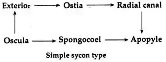
Sycetta is an example of a heterocoelous calcareous sponge with a simple canal structure. The radial canals are free projections of the wall that do not meet at any point, and the exterior surface is made up of the canals’ blind ends.
The dermal ostia are the gaps between the radial canals, as the incurrent canals have not yet formed. The radial tube is lined with flagellated cells, but the Spongocoel is lined with pinacocyte cells that are flattened.
Course of water
(ii) Complex sycon type
Scypha (= Sycon) contains a complicated canal system of the sycon type, and there is opposition. The walls of adjacent radial canals are arranged so as to leave tubular spaces, the incurrent or inhalant canals, between them. Thus, radial canals and incurrent canals are alternately organised, with the latter opening to the external via dermal ostia or incurrent pores.
The inner ends of the incurrent canals are blind and lined with pinacocytes, which are flat ectodermal cells. The intercellular dermal ostia or incurrent pores perforate a pore membrane and are surrounded by contractile myocytes. Many minute pores, known as prosopyles, perforate the wall between the incurrent and radial canals.
Through a porocyte, each prosopyle appears to have an intercellular gap or channel. The radial canal is lined by choanocytes and opens through a large opening known as the apopyle into a short, wide ex-current or exhalent canal bordered by flat epidermal cells and communicating with the spongocoel via an internal ostium. The spongocoel finally opens to the external via the osculum (Fig. 11.12).

(iii) Sycon type with cortex
Several genera of calcareous sponges possess the Syconoid (Stage III) canal system, including Grantia, Grantiopsis, Heteropia, Ute, etc. The problem is caused by the dermal membrane (composed of epidermis and a thin layer of mesenchyme) forming a variable-thickness cortex over the entire surface of the sponges.
The walls of the radial canals fuse to generate tubular gaps (incurrent canals) that communicate with the environment via dermal ostia or dermal pores. Before reaching the outer ends of the radial canals, the incurrent canals take an uneven path throughout the cortex. Sometimes enormous irregular cortical spaces or sub-dermal spaces may be created.

3. Leucon type of canal system
- This form of canal system is the consequence of further body wall folding of the sycon type of canal system.
- This canal system is a distinguishing feature of leuconoid sponges, such as Spongilla. Due to the complexity of the canal system, radial symmetry is lost in this kind, resulting in an irregular symmetry.
- The flagellated chambers are smaller than those of asconoid and syconoid organisms. These chambers have a spherical form and are lined with choanocytes.
- All remaining gaps are lined with pinacocytes. Via prosopyles, incurrent canals lead to flagellated chambers. Via apopyles, these flagellated chambers communicate with the excurrent canals.
- The excurrent canals form due to the contraction and division of the spongocoel. Absent is the large and roomy spongocoel that is present in asconoid and syconoid canal systems.
- The spongocoel is greatly diminished. Through the osculum, this excurrent canal communicates with the outer world.
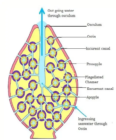
Structure of Leucon type of canal system
- The leuconoid type of canal system develops as a consequence of the continued unfolding of the choanocyte layer and the thickening of the body wall.
- The choanocyte layer of the radial canal of the syconoid stage evaginates into numerous small chambers, and these chambers may repeat the process, resulting in clusters of small rounded or oval flagellated chambers replacing the elongated chambers of the syconoid stage.
- These chambers are exclusive to choanocytes.
- Mesenchyme fills the area surrounding flagellated chambers. Typically, the spongocoel disappears, and the structure and form of the entire sponge become uneven and indeterminate.
- The sponge’s interior is infiltrated by numerous incurrent and excurrent canals that merge to generate bigger excurrent canals and spaces that lead to the oscula.
- The surface is composed of epidermal epithelium and dermal pores (ostia) and oscula.
- The dermal pores lead to irregularly branching incurrent canals within the mesenchyme.
- The incurrent canals lead into the tiny, spherical flagellated chambers through prosopyles.
- The flagellated chambers include openings called apopyles that lead to excurrent channels, which then combine to build ever-larger tubes, the largest of which leads to the oscula.
Course of water current in Leuconoid type canal system
Ingressing water → dermal ostia → incurrent canal → Prosopyles → Flagellated chambers → Apopyles → excurrent canals → Osculum → Outside
Types of Leucon type of canal system
In accordance with its evolutionary pattern, a Leucon-type canal system has three progressive grades:
1. Eurypylous type
- In the eurypylous leuconoid type of canal system, the flagellated chambers are large and thimble-shaped, with each exiting directly into the excurrent canal via apopyle and receiving water directly from the incurrent canal by prosopyle.
- This type of canal network is present in Leucilla.
- Ex: Plakina
The current of water takes the following route:-
Dermal pores or ostia → sbdermal spaces → incurrent canals → prosopyles → flagellated chambers → apopyles → excurrent canals → spongocoel → oscula → out.
2. Aphodal type
- The flagellated chambers of the aphodal leuconoid type of canal system are tiny and spherical.
- The mouth of each flagellated chamber into the excurrent canal is typically retracted into a small tube known as the aphodus.
- The relationship between the flagellated chambers and incurrent canals remains unchanged. This canal system is found in Geodia and Stelleta.
- Ex: Geodia
The route of water current is as follows:-
Dermal pores or ostia → subdermal spaces → incurrent canals → prosopyles → flagellated chamber → aphodus → excurrent cansls → spongocoel → oscula → out.
3. Diplodal type
- In certain instances, there is also a thin current tube, the prosodus, between the incurrent canal and the flagellated chambers; this situation is known as diplodal.
- This type of canal system can be found in Oscarella, Spongilla, and others
- Ex: Spongilla
The current of water takes the following route:-
Dermal pores or ostia → subdermal spaces → incurrent canals → prosodus → flagellated chambers → aphodus → excurrent canals → spongocoel → oscula → out.
4. Rhagon Type
- This form of canal system is present in demospongiae, which emerges from the direct rearranging of the inner cell mass.
- The rhagon sponge has a large base and is conical with a single osculum at the top.
- The hypophare is the basal wall that is devoid of flagellated chambers.
- The upper wall that has a series of small, oval flagellated chambers is known as spongophare.
- Spongocoel is surrounded by flagellated chambers that open into it by broad apopyles.
- Dermal pores or ostia lead to subdermal areas that extend beneath the entire body’s surface.
- From the subdermal spaces, branching incurrent canals lead to small flagellated chambers generated by the dissolution of the radial canal; the flagellated chambers are bordered by choanocytes and lead to the spongocoel.
- The spongocoel is accessed via a solitary osculum.

The course of water current of water is
Ostia → subdermal space → incurrent canals → prosopyle → flagellated chambers → apopyles → excurrent canals → spongocoel → osculum → out.
Functions of Canal System
- Nutrition or Feeding: The canal system allows sponges to filter feed by pushing water through their bodies and catching tiny food particles with their choanocytes.
- Respiration or Gas exchange: The canal system also enables sponges’ gas exchange, allowing them to collect oxygen and release carbon dioxide.
- Excretion or Waste removal: The canal system helps eliminate waste products from the sponge’s body, keeping them from accumulating and negatively impacting the sponge’s health.
- Water circulation: The canal system circulates water throughout the sponge’s body, maintaining its internal water balance and preventing the buildup of surplus water.
- Protection: Little fish and other aquatic species that seek shelter within the sponge’s chambers can find protection within the canal system.
- Adaptation: The structure and intricacy of the canal system can vary among sponge species, allowing them to adapt to various environmental conditions and feeding behaviours.
- Classification: The arrangement and structure of a sponge’s canal system can be used to categorise sponges according to their feeding and environmental preferences.
- Evolution: Understanding the canal system is essential for the study of the development of sponges and their adaptations to various aquatic conditions across time.
- Reproduction: The incoming water current transports sperm, which are caught by choanocytes and contribute to fertilisation.
Facts about Canal System in sponge
- The sponge canal system is a network of interconnecting channels and pores that aid in water circulation and feeding.
- Sponges are filter feeders that catch food particles from the water via their canal system.
- In sponges, the canal system consists of inhalant, exhalant, and choanocyte canals.
- The passages via which water enters the sponge’s body are the inhalant canals.
- The passages through which water escapes the sponge’s body are its exhalant canals.
- Choanocyte canals are the passageways that lead to the sponge’s internal chambers, where the majority of water filtration and food collection takes place.
- Choanocytes are specialised cells that line the choanocyte canals and feature a collar of flagella that generates a current to trap food particles.
- The size and structure of the canals, as well as the quantity of choanocytes present in the sponge’s body, determine the efficacy of its feeding system.
- The movement of water through the sponge’s canal system is propelled by ciliary activity and muscular contractions of the body.
- The structure and complexity of a sponge’s canal system can vary considerably between species.
- Asconoid sponges have a simple canal system, but syconoid and leuconoid sponges have complex canal systems.
- On the basis of the shape and arrangement of the canals, sponges can be classified into many types using their canal system.
- The organisation and complexity of the canal system are influenced by the feeding habits and ambient factors of the sponge.
- Little fish and other aquatic species that take sanctuary within the sponge’s chambers can also find protection throughout the canal system.
- The canal system is important in gas exchange, since carbon dioxide is released through the exhalant canals and oxygen is taken from the water.
- The canal system aids in the removal of waste materials from the sponge’s body, keeping them from accumulating and compromising its health.
- Environmental problems such as pollution can disturb the canal system, which can reduce the sponge’s feeding efficiency and overall health.
- Sponge survival is dependent on the canal system, which promotes the circulation of water and nutrients throughout their bodies.
- Knowing the canal system is essential for investigating the ecology, evolution, and adaptations of sponges to various aquatic environments over time.
- The sponge canal system is a stunning illustration of the diversity and complexity of biological systems and the astonishing adaptations that living species have evolved to survive in their environments.
MCQ on Canal System in sponge
What is the function of the canal system in sponges?
A) Reproduction
B) Gas exchange
C) Locomotion
D) Protection
Answer: B) Gas exchange
What are choanocytes?
A) Cells that produce gametes
B) Cells that line the canal system
C) Cells that protect the sponge from predators
D) Cells that form the skeleton of the sponge
Answer: B) Cells that line the canal system
What is the name of the channel through which water enters the sponge’s body?
A) Inhalant canal
B) Exhalant canal
C) Choanocyte canal
D) Collar canal
Answer: A) Inhalant canal
Which type of sponge has the simplest canal system?
A) Asconoid sponge
B) Syconoid sponge
C) Leuconoid sponge
D) All sponges have similar canal systems
Answer: A) Asconoid sponge
Which type of sponge has the most complex canal system?
A) Asconoid sponge
B) Syconoid sponge
C) Leuconoid sponge
D) All sponges have similar canal systems
Answer: C) Leuconoid sponge
What is the function of exhalant canals?
A) To absorb oxygen
B) To expel carbon dioxide
C) To capture food particles
D) To create a current for feeding
Answer: B) To expel carbon dioxide
What is the driving force behind the flow of water through the canal system?
A) Muscular contractions of the sponge’s body
B) Gravity
C) Wind
D) Electromagnetic fields
Answer: A) Muscular contractions of the sponge’s body
What environmental factor can disrupt the efficiency of the sponge’s canal system?
A) Sunlight
B) Pollution
C) High water temperatures
D) Low water salinity
Answer: B) Pollution
Which type of sponge has a canal system that resembles a series of folded tubes?
A) Asconoid sponge
B) Syconoid sponge
C) Leuconoid sponge
D) All sponges have similar canal systems
Answer: B) Syconoid sponge
Why is the study of the canal system important for understanding the ecology and evolution of sponges?
A) It provides information about the sponge’s feeding habits and environmental preferences
B) It helps to classify sponges into different categories
C) It reveals how sponges have adapted to different aquatic environments over time
D) All of the above
Answer: D) All of the above
FAQ
What is the canal system in sponges?
The canal system in sponges is a network of water-filled canals that run throughout the sponge’s body and facilitate gas exchange, feeding, and waste removal.
What is the function of the canal system in sponges?
The main function of the canal system in sponges is to facilitate gas exchange by allowing water to flow through the sponge’s body, bringing in oxygen and expelling carbon dioxide.
How does the canal system in sponges aid in feeding?
The canal system in sponges creates a current that draws water and food particles into the sponge’s body. Choanocytes, specialized cells that line the canals, capture and digest the food particles.
What is the relationship between choanocytes and the canal system in sponges?
Choanocytes line the canals of the sponge’s body and play a crucial role in capturing and digesting food particles. They also aid in gas exchange by creating a current that drives water through the canal system.
How does the canal system in sponges contribute to waste removal?
The canal system in sponges allows waste products, such as carbon dioxide and ammonia, to be expelled from the sponge’s body along with the water that flows through the canals.
What are the different types of canal systems in sponges?
There are three main types of canal systems in sponges: asconoid, syconoid, and leuconoid. Asconoid sponges have the simplest canal system, while leuconoid sponges have the most complex.
How does the structure of the canal system differ in asconoid, syconoid, and leuconoid sponges?
Asconoid sponges have a single, large inhalant canal that opens into a chamber lined with choanocytes. Syconoid sponges have a series of folded canals that increase the surface area for feeding and gas exchange. Leuconoid sponges have a highly branched and interconnected system of canals that allow for efficient feeding and waste removal.
How do sponges control the flow of water through their canal system?
Sponges use muscular contractions of their body wall to create a current that drives water through the canal system.
How does pollution affect the canal system in sponges?
Pollution can disrupt the efficiency of the canal system in sponges by clogging the canals with particles or interfering with the sponge’s ability to maintain a healthy microbial community.
How does the canal system in sponges contribute to their role in the ecosystem?
Sponges are important members of many marine ecosystems because they filter large volumes of water and can remove nutrients and pollutants from the water column.
How does the canal system in sponges differ from other types of respiratory systems?
The canal system in sponges is unique among respiratory systems because it does not involve specialized organs or tissues for gas exchange. Instead, gas exchange occurs across the surface of the choanocytes that line the canals.
How do sponges adapt their canal system to different aquatic environments?
Sponges can adapt their canal system to different aquatic environments by changing the size and shape of the canals, adjusting the flow rate of water through the canals, and altering the number and distribution of choanocytes.
Can sponges regenerate their canal system?
Sponges have a remarkable ability to regenerate their bodies, including their canal system, after injury or damage.
References
- https://prgc.ac.in/uploads/study_material/CANAL%20SYSTEM%20IN%20SPONGES.pdf289.pdf
- https://www.studyandscore.com/studymaterial-detail/phylum-porifera-canal-system-in-sponges-types-of-canal-systems-in-sponges-functions-of-water-current
- https://www.vedantu.com/question-answer/give-a-brief-account-on-the-canal-system-in-class-11-biology-cbse-5fc9c505592a523706101539
- https://bncollegebgp.ac.in/wp-content/uploads/2020/04/BSc-Part-I-Canal-system-in-Porifera.pdf
- http://zoology.uok.edu.in/Files/cae2d08f-4f62-428e-b6ea-cf46cdccbf42/Custom/102CR-1.3%20Canal%20system.pdf
- http://www.ppup.ac.in/download/econtent/pdf/CANAL%20SYSTEM%20IN%20SPONGES_B.Sc-Part%201-%20Dr.Rita%20Gupta,%20BS%20College.pdf
- https://marwaricollege.ac.in/study-material/1605458648Lecture%209.pdf
- http://www.soghracollege.com/img/Lectures_26Apr/2_Canal_system_in_Sponges-1-merged.pdf
- https://www.bmdcollegebu.in/Notes/aqar/Zoology/zool_tdc1_note1.pdf
- https://www.iaszoology.com/canal-system/
- http://rsmraiganj.in/wp-content/themes/raiganj-surendranath-mahavidyalaya/pdf/1612432144_Porifera-Canal%20system.pdf
- https://www.studocu.com/in/document/mahatma-gandhi-university/zoology/canal-system-in-sponges/41230387
- https://www.biologydiscussion.com/invertebrate-zoology/sponges/role-and-significance-of-canal-system-in-sponges/32608
- https://www.biologydiscussion.com/invertebrate-zoology/sponges/canal-systems-encountered-in-different-sponges/32624
- https://silapatharcollege.edu.in/online/attendence/classnotes/files/1628324973.pdf
- https://bpchalihacollege.org.in/online/attendence/classnotes/files/1628186019.pdf
- https://biozoomer.com/2014/04/canal-system-in-sycon-sponge.html
- http://dhingcollegeonline.co.in/attendence/classnotes/files/1603909755.pdf