Type II (Cytotoxic) Hypersensitivity is an antibody mediated hypersensitivity reaction in which IgG or IgM antibodies react with antigen present on the surface of body cell or extracellular matrix.
In this reaction, the antibody binds with the normal host cell antigen. Then the immune system identifies this cell as harmful cell. It causes injury of cell by different cytotoxic mechanism.
The cell damage may occur by complement activation, phagocytosis, or local inflammatory reaction. In complement mediated cytotoxicity, complement system make pores on the cell membrane and cell lysis takes place. In phagocytosis, the antibody coated cell is engulfed and destroyed by phagocytic cells.
Type II hypersensitivity is mainly divided into Type IIa and Type IIb reaction. Type IIa reaction causes direct destruction of cell. It produces tissue injury and cell lysis. Examples are autoimmune hemolytic anemia and immune thrombocytopenia.
Type IIb reaction does not mainly destroy the cell. It changes the normal function of the cell receptor. The antibody may stimulate or block the receptor. This type of reaction is seen in Graves’ disease and myasthenia gravis.
Mechanism of Type II Hypersensitivity Reactions
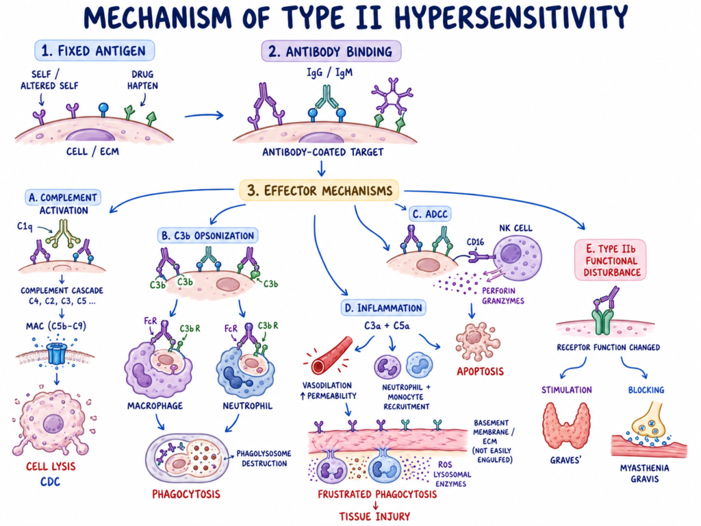
The following are the step by step mechanism of Type II hypersensitivity reaction.
Step 1- Antigen recognition
In this step, the immune system identifies some antigen as harmful antigen. These antigen may be self antigen, altered self antigen or some foreign substance attached with host cell like drug hapten.
The antigen is present on the surface of cell or on extracellular matrix. So it is a fixed antigen. It is not free circulating antigen.
Step 2- Antibody binding
After antigen recognition, IgG or IgM antibody binds with the antigen. This binding takes place directly on the host cell membrane or tissue structure.
After this antibody binding, the target cell becomes coated with antibody. This is the main starting point of the reaction.
Step 3- Activation of complement pathway
The IgG or IgM antibody attached on target cell activates classical complement pathway. In this process, C1q binds with Fc portion of antibody.
Then complement proteins are activated one after another. This forms a proteolytic cascade. At last membrane attack complex (MAC) is formed.
Step 4- Cell lysis by MAC
The MAC is made by complement proteins C5b, C6, C7, C8 and C9. It is inserted into the cell membrane.
It forms pore in the membrane of target cell. Water enters into the cell. The cell swells and finally cell lysis takes place. This is called complement dependent cytotoxicity (CDC).
Step 5- Opsonization of target cell
During complement activation, C3b is formed and it binds with the target cell membrane. C3b acts as an opsonin.
The antibody attached cell is now more easily recognized by phagocytic cells. This process is called opsonization.
Step 6- Phagocytosis of antibody coated cell
The phagocytic cells such as macrophages and neutrophils recognize the coated target cell. They use Fc receptors for IgG and complement receptors for C3b.
Then the target cell is engulfed by phagocyte. It is destroyed inside the phagolysosome by lysosomal enzymes. This causes removal of the host cell.
Step 7- Antibody dependent cellular cytotoxicity
In some condition, target cell is coated mainly by IgG antibody. Then NK cells and other cells recognize the Fc part of antibody by CD16 receptor.
After receptor binding, the effector cell releases cytotoxic granules. These granules contain perforin and granzymes.
Perforin forms channel in the target cell membrane. Granzymes enter inside the cell and activates caspase pathway. So the target cell undergoes apoptosis. This is called antibody dependent cellular cytotoxicity (ADCC).
Step 8- Complement mediated inflammation
When antibody binds with large fixed tissue like basement membrane, the tissue cannot be engulfed easily. Complement activation produces C3a and C5a.
These are called anaphylatoxins. They cause vasodilation and increase vascular permeability. C5a also attracts neutrophils and monocytes at the site.
Step 9- Tissue injury by frustrated phagocytosis
In this condition, leukocytes try to engulf the large tissue structure but they cannot do it properly. This is referred to as frustrated phagocytosis.
So the cells release reactive oxygen species (ROS) and lysosomal enzymes outside. These substances damage nearby normal host tissue. It produces inflammation and tissue destruction.
Step 10- Functional disturbance of cell receptor
In Type IIb reaction, the antibody does not destroy the cell directly. It binds with cell surface receptor and changes its normal function.
The antibody may stimulate the receptor abnormally. This is seen in Graves’ disease, where antibody stimulates thyroid receptor and excess thyroid hormone is formed.
The antibody may also block the receptor. This is seen in myasthenia gravis, where antibody blocks acetylcholine receptor and neuromuscular transmission is decreased.
So Type II hypersensitivity causes injury by cell lysis, phagocytosis, inflammation, ADCC and receptor function disturbance.
Clinical manifestation of Type II Hypersensitivity Reactions
- AIHA– RBC are destroyed by antibody. Weakness, fatigue, dizziness and pallor are seen. Shortness of breath and abdominal pain may be present. Jaundice, icterus and dark urine occur due to RBC lysis. In cold type, acrocyanosis and Raynaud phenomenon are found.
- ITP– Platelet are destroyed by antibody. Bleeding occurs in skin and mucous membrane. Petechiae and purpura are common. Hemorrhagic blister may seen in mouth. Clotting becomes poor.
- Goodpasture syndrome– Antibody acts against glomerular basement membrane (GBM). Lung and kidney are affected. Hemoptysis, shortness of breath and fatigue are seen. Hematuria, proteinuria, edema and hypertension are also present.
- Myasthenia gravis– Antibody blocks acetylcholine receptor. Skeletal muscle weakness is the main feature. Weakness is fluctuating type. Eye, bulbar and limb muscles are involved. Diplopia, dysphagia and dysarthria are seen.
- Graves disease– Antibody stimulates TSH receptor. Thyroid gland becomes overactive. Weight loss occurs with increased appetite. Tachycardia, widened pulse pressure and sweating are seen. Proptosis, lid lag, stare and periorbital edema are important eye signs.
- Pemphigus vulgaris– It is blistering disease of skin and mucous membrane. Flaccid blisters are formed. Painful erosion is seen after rupture of blister. Oral cavity is commonly affected.
- Acute rheumatic fever– It occurs after Streptococcus infection. Cross reacting antibody is formed. Fever, fatigue and migratory polyarthritis are seen. Large joints are mainly affected. Heart involvement may show tachycardia and new murmur. Sydenham chorea shows involuntary movement, muscle weakness and emotional lability.
- HDN– Maternal antibody destroys fetal RBC. Newborn shows jaundice and unconjugated hyperbilirubinemia. Anemia is present. Lethargy and tachycardia may occur. Severe condition causes kernicterus and hydrops fetalis. In hydrops, diffuse edema and fluid collection around lungs and heart are seen.
- Pernicious anemia– Antibody acts against intrinsic factor or parietal cell. Vitamin B12 absorption is decreased. Megaloblastic anemia is seen. RBC becomes large and survival of RBC is decreased. Long deficiency causes paresthesia, loss of vibration sense and ataxia.
Diagnosis of Type II Hypersensitivity Reactions
- General diagnosis– Diagnosis depends on finding the specific antibody. Tissue injury and cell destruction are also checked. Different disease has different test. So single test is not enough for all Type II hypersensitivity reaction.
- AIHA– Direct Coombs test is the main test. It detects antibody or complement attached on RBC surface. Indirect Coombs test detects free antibody in serum or plasma. Blood hemolysis panel is also done. LDH, haptoglobin, indirect bilirubin and reticulocyte count are checked. Peripheral blood smear may show spherocytes.
- Drug induced hemolytic anemia– Direct Coombs test is used. It shows antibody or complement on drug coated RBC. Indirect Coombs test may detect free antibody. Hemolysis profile is done to confirm RBC destruction.
- Paroxysmal cold hemoglobinuria– Donath-Landsteiner test is done. It is specific test for this condition. Hemolysis markers and urine hemoglobin may also be checked.
- Goodpasture syndrome– Kidney biopsy is the most accurate test. It shows linear deposition of IgG and C3 along basement membrane. ELISA is used to detect circulating anti-GBM antibody. Urinalysis, BUN, creatinine and chest X-ray are done to see kidney and lung damage.
- Myasthenia gravis– Blood test is done for anti-acetylcholine receptor (AChR) antibody. Anti-MuSK antibody may also be detected. Edrophonium test and EMG are used to see neuromuscular transmission defect.
- Graves disease– Blood test is done for thyroid stimulating immunoglobulin (TSI) or TSH receptor antibody (TRAb). Thyroid function test is also done. TSH becomes low and Free T4 becomes high in hyperthyroid condition.
- ITP– CBC is the main test. It shows low platelet count. Peripheral blood smear is done with it. Other blood cells are usually normal. Bone marrow biopsy may be done in atypical case to exclude marrow disease.
- Acute rheumatic fever– Recent Streptococcus infection is confirmed. Throat culture, rapid streptococcal antigen test, ASO titre and anti-DNase B titre are used. Echocardiogram is done to detect carditis and valvular damage.
- Pernicious anemia– Blood test detects parietal cell antibody (PCA) and intrinsic factor antibody (IFA). Vitamin B12 level is decreased. Serum gastrin may be increased. Peripheral blood smear shows large RBC due to megaloblastic anemia.
- HDN– In mother, indirect Coombs test is done during pregnancy. It detects Rh sensitization and anti-D antibody. In newborn, direct antiglobulin test (DAT) is done. CBC and bilirubin level are checked for anemia and hyperbilirubinemia.
Treatment / Management of Type II Hypersensitivity Reactions
- Inciting agent removal– Offending drug is stopped first. In transfusion reaction, incompatible blood transfusion is stopped immediately. This is the first important management.
- Glucocorticoids– Prednisone or methylprednisolone are commonly used. These drugs suppress immune reaction. Inflammation is reduced. Destruction of target cell is also decreased.
- Immunosuppressive drugs– Mycophenolate mofetil, azathioprine and cyclophosphamide are used in some cases. These drugs inhibit T-cell and B-cell proliferation. So autoantibody production becomes less.
- Plasmapheresis– It is also called plasma exchange. Patient blood is filtered to remove harmful autoantibody. It is used in acute and life threatening condition. Fast improvement may occur.
- IVIG– Intravenous immunoglobulin (IVIG) is given in high dose. It neutralizes autoantibody. It also decreases macrophage mediated destruction. It is used in severe immune reaction.
- Biological therapy– Rituximab is commonly used biological drug. It destroys B-lymphocytes. So source of autoantibody formation is reduced. It is useful in refractory cases.
- AIHA– Autoimmune hemolytic anemia is treated first with glucocorticoids. If response is poor, rituximab, immunosuppressive drugs or splenectomy may be done. Blood transfusion is given in life threatening anemia.
- ITP– Immune thrombocytopenia is treated with glucocorticoids in first line. IVIG may be used in severe bleeding. Rituximab, immunosuppressive drug or splenectomy are used when steroid does not work.
- Cold agglutinin disease– Cold exposure is avoided strictly. Patient should keep body warm. Rituximab is used. In some case chemotherapy is combined. Complement inhibitor like sutimlimab may be given.
- HDN– Hemolytic disease of newborn is prevented by Rh immune globulin (RhIg or RhoGAM) in Rh-negative mother. It prevents formation of anti-Rh antibody. Affected newborn is treated by phototherapy, blood transfusion or IVIG according to jaundice and anemia.
- Myasthenia gravis– Pyridostigmine is used first. It is acetylcholinesterase inhibitor. It increases neuromuscular transmission and muscle strength. Severe case needs plasma exchange, biological drugs or thymectomy.
- Graves disease– Methimazole or propylthiouracil are used to stop thyroid hormone synthesis. Beta blocker like atenolol is used for symptom control. Long term treatment is radioactive iodine ablation or thyroid surgery.
- Goodpasture syndrome– This condition needs aggressive treatment. Plasmapheresis, glucocorticoids and cyclophosphamide are given together. It helps to stop lung and kidney damage quickly.
- Acute rheumatic fever– Antibiotic is given to remove Group A Streptococcus infection. Penicillin is commonly used. Long term antibiotic prophylaxis is needed. It prevents recurrence and further heart valve damage.
Examples of Type II Hypersensitivity Reactions
- AIHA– Autoantibody acts against own RBC. RBC are destroyed. Hemolytic anemia is formed.
- ITP– Autoantibody acts against platelet glycoprotein. Platelet are rapidly removed from blood. Thrombocytopenia occurs.
- Autoimmune neutropenia– Antibody acts against neutrophils. Neutrophil number becomes low. Infection chance may increase.
- HDN– Maternal antibody crosses placenta and destroys fetal RBC. It is commonly due to Rh or ABO incompatibility. This is also called erythroblastosis fetalis.
- Acute transfusion reaction– Recipient antibody attacks incompatible transfused RBC. Rapid hemolysis takes place. It is serious reaction.
- Drug induced cytopenia– Some drugs act as hapten. Penicillin, cephalosporins or methyldopa may cause antibody formation. RBC or platelet are destroyed.
- Goodpasture syndrome– Antibody acts against basement membrane of kidney and lung. Tissue damage occurs. Bleeding and renal injury may seen.
- Myasthenia gravis– Antibody blocks or destroys acetylcholine receptor at neuromuscular junction. Muscle contraction becomes poor. Weakness is produced.
- Graves disease– Autoantibody binds with TSH receptor. Receptor is stimulated continuously. Thyroid gland becomes overactive.
- Pernicious anemia– Antibody acts against gastric parietal cell or intrinsic factor. Vitamin B12 absorption is decreased. Megaloblastic anemia occurs.
- Pemphigus vulgaris– Antibody acts against desmogleins in skin and mucous membrane. Cell adhesion is lost. Blister formation occurs.
- Acute rheumatic fever– Antibody formed against Group A Streptococcus cross reacts with host tissue. Heart valve may be affected. Rheumatic carditis may occur.
- Hyperacute transplant rejection– Preformed antibody attacks endothelial cell of transplanted organ. Graft injury occurs very rapidly. Graft failure may happen.
- Hashimoto thyroiditis– Autoantibody acts against thyroid gland cell. Thyroid tissue is damaged. Hypothyroid condition may develop.
- Chronic spontaneous urticaria– Antibody binds with receptor on mast cell and basophil. It causes release of inflammatory mediator. Chronic hives are formed.
References
- Labcorp. (n.d.). 010413: Intrinsic factor blocking antibodies.
- Zhuang, S., Guo, D., & Yu, D. (2025). A mini review of the pathogenesis of acute rheumatic fever and rheumatic heart disease. Frontiers in Cellular and Infection Microbiology, 15, 1447149. https://doi.org/10.3389/fcimb.2025.1447149
- PubMed Central. (n.d.). A review of hypersensitivity methods to detect immune responses to SARS-CoV-2. National Center for Biotechnology Information.
- PubMed Central. (n.d.). Acantholysis may precede elevation of circulating anti‐desmoglein 3 antibody levels in pemphigus vulgaris presenting with desquamative gingivitis. National Center for Biotechnology Information.
- Seshadri, D., Kumaran, M. S., & Kanwar, A. J. (2013). Acantholysis revisited: Back to basics. Indian Journal of Dermatology, Venereology and Leprology, 79(1), 120-126. https://doi.org/10.4103/0378-6323.104688
- UWorld. (n.d.). Acute rheumatic fever – Allergy & immunology. UWorld Medical Library.
- McLendon, K., & Sternard, B. T. (2023). Anaphylaxis. In StatPearls. StatPearls Publishing. https://www.ncbi.nlm.nih.gov/books/NBK482124/
- AMBOSS: Medical Knowledge Distilled. (n.d.). Antibody-dependent cell-mediated cytotoxicity: Type II hypersensitivity reaction [Video]. YouTube.
- Gerber, G. F. (2026). Autoimmune hemolytic anemia. Merck Manual Professional Edition.
- Dispenza, M. C. (2019). Classification of hypersensitivity reactions. Allergy and Asthma Proceedings, 40(6), 470-473. https://doi.org/10.2500/aap.2019.40.4274
- Cigna. (n.d.). Coombs test.
- Cigna. (n.d.). Coombs test.
- Johns Hopkins Rheumatology. (n.d.). Coombs’ test. Johns Hopkins Lupus Center.
- Ali, U. (n.d.). Direct Coombs test: Detecting hemolytic anemia. King of the Curve.
- Cornell University College of Veterinary Medicine. (n.d.). Direct Coombs testing. Animal Health Diagnostic Center.
- Fernandez, J. (2024). Drug hypersensitivity. MSD Manual Professional Edition.
- Oncohema Key. (2019). Drug-induced hemolytic anemia.
- AccessHemOnc. (n.d.). Drug-induced hemolytic anemia. Williams Manual of Hematology, 9e.
- UF Health. (2025). Drug-induced immune hemolytic anemia. University of Florida Health.
- PubMed. (n.d.). Drug-induced haemolytic anemia. National Center for Biotechnology Information.
- PubMed. (n.d.). Drug-induced immune hemolytic anemia. National Center for Biotechnology Information.
- Cleveland Clinic. (2024). Erythroblastosis fetalis: Causes, symptoms & treatment.
- PubMed. (n.d.). Evaluating antibody-dependent cell-mediated cytotoxicity by flow cytometry. National Center for Biotechnology Information.
- Author Unknown. (n.d.). General patholog diseases of the immune system I- Hypersensitivity reaction (allergy) II- Transplantation immunopathology [PDF].
- Wikipedia contributors. (2026). Goodpasture syndrome. In Wikipedia, The Free Encyclopedia.
- Pixorize. (n.d.). Hyperacute transplant rejection mnemonic for USMLE.
- Becker, A. F. (2022). What are hypersensitivity reactions? Healthgrades Health Library.
- Cleveland Clinic. (2025). Hypersensitivity reactions: Types, symptoms & treatment.
- Han, J., Pan, C., Tang, X., Li, Q., Zhu, Y., Zhang, Y., & Liang, A. (2022). Hypersensitivity reactions to small molecule drugs. Frontiers in Immunology, 13, 1016730. https://doi.org/10.3389/fimmu.2022.1016730
- Gray, P. E., McCarthy, H., Siggs, O. M., Saleem, M. A., O’Brien, T., Frith, K., Ziegler, J. B., Kitching, A. R., Fogo, A. B., Hudson, B. G., & Pedchenko, V. (2019). Molecular analysis of Goodpasture’s disease following hematopoietic stem cell transplant in a pediatric patient, recalls the conformeropathy of wild-type anti-GBM disease. Frontiers in Immunology, 10, 2659. https://doi.org/10.3389/fimmu.2019.02659
- Author Unknown. (n.d.). Molecular pathobiology, clinical taxonomy, and diagnostic immunohematology of type II hypersensitivity reactions.
- Mayo Clinic Laboratories. (n.d.). PCAB parietal cell antibodies, IgG, serum. Neurology Catalog.
- Kalish, R. S. (2000). Pemphigus vulgaris: The other half of the story. Journal of Clinical Investigation, 106(12), 1433–1435. https://doi.org/10.1172/JCI11828
- PubMed. (n.d.). Pemphigus, a pathomechanism of acantholysis. National Center for Biotechnology Information.
- Thermo Fisher Scientific. (n.d.). Pernicious anemia laboratory testing.
- Sabir, S., & Jan, A. (2025). Physiology, immune response. In StatPearls. StatPearls Publishing. https://www.ncbi.nlm.nih.gov/books/NBK539801/
- Allo Hope Foundation. (n.d.). Rh immune globulin (RhIG).
- MedlinePlus. (2025). Rh incompatibility – Erythroblastosis fetalis. National Library of Medicine.
- PubMed Central. (n.d.). Rheumatic fever, autoimmunity and molecular mimicry: The streptococcal connection. National Center for Biotechnology Information.
- Picmonic. (n.d.). RhoGam (Rho(D) nursing considerations.
- Walter, E., Vielmuth, F., Wanuske, M.-T., Seifert, M., Pollmann, R., Eming, R., & Waschke, J. (2019). Role of Dsg1- and Dsg3-mediated signaling in pemphigus autoantibody-induced loss of keratinocyte cohesion. Frontiers in Immunology, 10, 1128. https://doi.org/10.3389/fimmu.2019.01128
- Beier, K., Lui, F., & Pratt, D. P. (2024). Sydenham chorea. In StatPearls. StatPearls Publishing. https://www.ncbi.nlm.nih.gov/books/NBK430838/
- PubMed Central. (n.d.). Sydenham’s chorea in children with acute rheumatic fever: An echocardiographic survey of pediatric patients in northwestern Iran. National Center for Biotechnology Information.
- Mayo Clinic Laboratories. (2026). Test definition: PCAB [PDF].
- Kedrion Biopharma. (n.d.). The science behind RhoGAM.
- Kalluri, R., Gattone, V. H., Noelken, M. E., & Hudson, B. G. (1994). The alpha 3 chain of type IV collagen induces autoimmune Goodpasture syndrome. Proceedings of the National Academy of Sciences, 91(13), 6201-6205.
- PubMed Central. (n.d.). The alpha 3 chain of type IV collagen induces autoimmune Goodpasture syndrome. National Center for Biotechnology Information.
- Merck. (n.d.). The alpha 3 chain of type IV collagen induces autoimmune Goodpasture syndrome.
- WebPath. (n.d.). Transplant rejection.
- Histocompatibility & Immunogenetics. (n.d.). Transplantation immune reactions.
- Abbas, M., & Goldin, J. (2025). Type I hypersensitivity reaction. In StatPearls. StatPearls Publishing. https://www.ncbi.nlm.nih.gov/books/NBK560561/
- MD Searchlight. (2023). Type II hypersensitivity reaction.
- Saavedra Torres, J. S., & Goldin, J. (2025). Type II hypersensitivity reaction. In StatPearls. StatPearls Publishing. https://www.ncbi.nlm.nih.gov/books/NBK563264/
- Kamiya, A., & Cheprasov, A. (n.d.). Type II hypersensitivity | Types, reactions & examples – Lesson. Study.com.