Bacteria constitute a vital element in the ecology. They are essential to our health as well as the environmental health, play an essential role in the production of food, and offer bioengineers the tools to harness their abilities and produce compounds. However, they could also be harmful, causing harm and diseases. The capacity to cultivate the microbes that cause harm is an essential element in being capable of harnessing their power, recognize the most harmful causes and improve our knowledge and capabilities. This article will look at the complexities of bacterial culture and the factors that affect conditions in culture as well as common issues and a few of the many applications.
Cultivation of bacteria Definition
Microbial cultures are the process of multiplying microorganisms through letting them reproduce in a predetermined medium in controlled lab conditions. Microbial cultures can be utilized to identify the kind of organism, the amount of the sample being tested or both.
Purpose of Bacterial Cultivation
- The isolation of bacteria.
- Characteristics for bacteria i.e. culture of bacteria is the primary stage in studying the structure and determination.
- Maintenance of stock culture.
- Estimate viable counts.
- For testing for sensitivity to antibiotics.
- To create antigens for laboratory use.
- Certain genetic research and manipulations of cells require that the bacteria be cultured in the laboratory in.
- Growing on solid substrates is a effective method to separate bacteria that are mixed.
Common problems with bacterial culture
1. Contamination
Contamination of bacterial strains is a major issue especially if it is unnoticed. It could result in the need to reisolate an undiluted culture however, it could cause illness and expensive remedial efforts should it occur in a production or food environment. The contamination of culture can originate from multiple sources, starting from the initial sample during the process of cultivating or even storage. An aseptic procedure that is well-executed will help prevent contamination of bacterial culture.
2. Overgrowth of some species
Some bacteria grow easily and quickly. When trying to isolate one species from a mixture, the vigorous species could overgrow and hide the slow-growing species that are targeted for. Making use of selective media and the optimal conditions for growth for your targeted species (if identified) will help reduce this. It is recommended to try to grow the sample as quickly as possible after it’s been collected to ensure that it’s as exact as is possible.
3. Antibiotic treatment prior to sampling
In a diagnosis setting it is essential to determine if antibiotics was administered prior to taking samples. If that is the case the failure to test one specific species might not be a sign that it wasn’t the source of the illness.
4. Incorrect growth conditions
Incorrect or inadequate growth conditions can cause or hinder development of the desired strain. Make sure you double-check growth requirements , or if you use antibiotics, ensure that the right antibiotic is selected for the resistance gene that is present.
5. Non-culturable and slow-growing organisms
Some species of bacterial, even today, are not able to be cultivated in the lab.10 Other species, like mycobacteria11, are extremely slow-growing and take a long time to successfully cultivate, which is especially problematic in the diagnosis of diseases.
What is Culture Media?
A culture medium that is artificial must meet the same conditions for nutrition and environment that are found in the natural habitat of bacteria. The medium for culture contains water, which is a source of energy and carbon and nitrogen. It also contains trace elements, as well as some growth factors. In addition, the pH should be set in accordance with.
Applications
- Enhance the amount of bacteria.
- Choose a specific bacteria to block other.
- Differentiate between different types of bacteria.
What is Pure culture?
- In the lab, bacteria are isolated and then grown in pure culture to investigate the function of a particular species.
- Pure cultures are the result of a cell population that is growing in the absence of different species or types. Pure culture can be derived from one single cell or organism and in this case, these cells may be genetically clones of other.
- Pure cultures can be obtained employing a variety of different methods. Glassware, media and instruments need to be cleaned i.e. methods of aseptic sterilization are employed to obtain pure culture.
- The fundamental requirements to obtain an uncontaminated culture are solid medium, a container that is maintained in an aseptic state and a method of separating each cell.
- A single bacterium that is supplied with adequate nutrients, can expand on the medium over a small area , forming colonies, which are an accumulation of cells descendant from the first.
Classification of Bacterial Culture Media
Bacterial culture media can be classified in at least three ways. 1. Consistency, 2. Nutritional component, 3. Functional use.
Classification of Bacterial Culture Media Based on Consistency
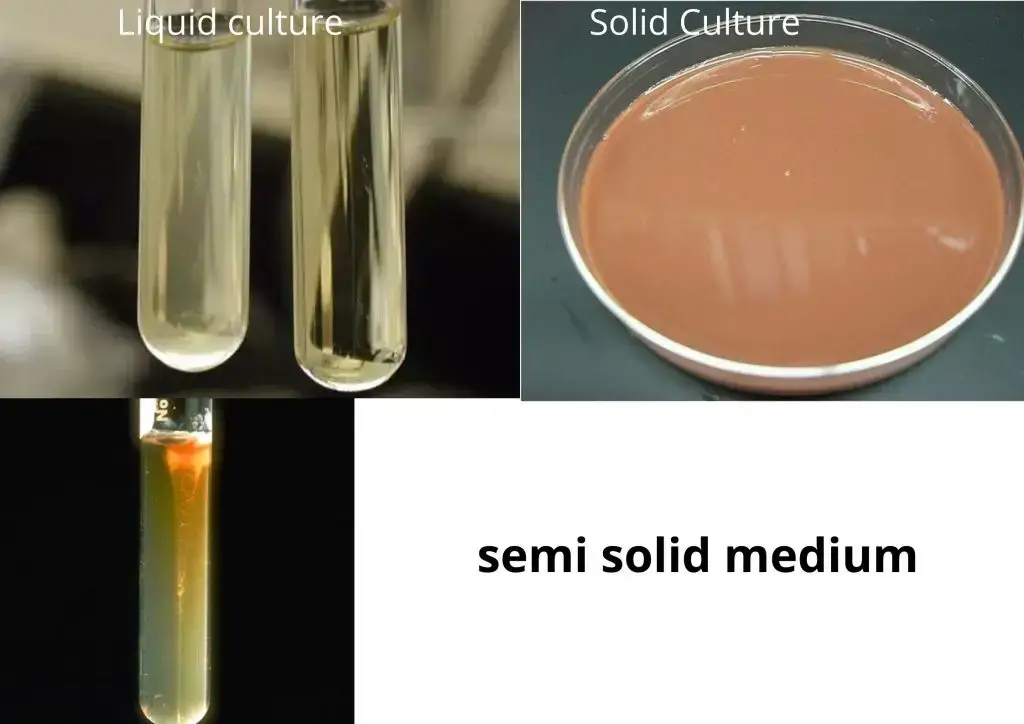
Based on Consistency Bacterial Culture Media are classified into three groups such as; 1. Liquid media. 2. Solid media. 3. Semi solid media.
- Liquid media: They can be used in bottles, test tubes or flasks. Liquid media are often referred to by the name of “broths” (e.g nutritional broth). In liquid mediums, bacteria develop uniformly, causing general turbidity. There is no agar added. It is mostly used for the preparation of inoculums.
- Solid media: Agar plate is a Petri dish that has the growth medium (typically Agar and nutrients) that is used to cultivate microorganisms. Agar contains 2% added. The most frequently utilized solidifying agent. The morphology of the colony, its pigmentation and hemolysis are all visible. Examples include Nutrient Agar as well as Blood Agar.
- Semi-solid media: Semi-solid media are quite soft and useful for showing bacterial motility and in separating motile strains from non-motile. Examples of semi-solid media (Hugh Leifson’s oxidation and Hugh). 0.5 percent agar is added.

Classification of Bacterial Culture Media Based on Nutritional Components
Based on Nutritional Components Bacterial Culture Media are classified into three groups such as; 1. Simple media. 2. Complex media. 3. Synthetic or chemically defined media.
- Simple media: Simple media like peptone, nutrients agar is able to support most non-fastidious bacteria. It’s also known the basal media. Eg: NB, NA. Nutrient Broth is composed of peptone, yeast extract , and NaCl. When 22% of agar are included in Nutrient Broth it is formed into Nutrient Agar.
- Complex media: The types of media that are not the basal media are referred to as complex media. They contain special ingredients that are used in their composition that promote the development of microorganisms. These ingredients are yeast extracts, or casein hydrolysate composed of a mix of a variety of chemicals, in a varying amount.
- Synthetic media/Chemically defined media: Synthetic media/Chemically defined media specially prepared media that are used for research, in which the composition of each ingredient is widely recognized. It is made from pure chemical compounds. For example, peptone water (1 % peptone + 0.5 percent NaCl with water).
Classification of Bacterial Culture Media Based on Functional Use or Application
Based on Functional Use or Application Bacterial Culture Media are classified into the following groups such as; 1. Enriched media. 2. Selective media. 3. Differential media. 4. Transport media. 5. Indicator media. 6. Anaerobic media.
1. Enriched media
- The addition of additional nutrients, such as serum, blood eggs, egg yolks etc. to the base medium creates richer media.
- The media used to separate pathogens in a mix culture.
- Enhance the growth of desired bacterium, and block the growth of unwanted bacteria.
- Media is mixed with inhibitory substances that kill the undesirable organism and increase the numbers of the desired bacteria.
- Examples of media that are enriched:
- Chocolate agar
- Blood agar.
- Selenite F Broth – for the isolation of Salmonella, Shigella.
- Tetrathionate Broth – inhibit coliforms .
- Alkaline Peptone Water – for Vibrio cholerae.
Chocolate Agar: Chocolate agar is a non-selective and enriched medium for growth of fastidious bacteria, like Haemophilus influenzae .
Blood Agar: Blood (BAP) is a mammalian-specific plate (usually horses or sheep) generally with 5 to 10 percent. BAP are enriched, differentiating media that are used to identify cells that are fastidious and identify hemolytic activity.
2. Selective media
- The inhibitory ingredient is added to the solid media, which causes an increase in colonies of the desired bacteria.
- The selective media and enrichment media are designed to block unintentional contaminating or commensal bacteria and assist in removing pathogens from a mix of bacteria.
- Any agar medium can be made selective through the using inhibitory substances that don’t harm the pathogen. To make a media selective includes the addition of dyes, antibiotics, chemicals and pH alteration or the mixture of all these.
- Examples of selective media:
- Thayer Martin Medium selective for Neisseria gonorrhoeae.
- EMB Agar is agar that is specific for bacteria that are gram-negative. The dye methylene blue within the medium hinders growing of the gram positive bacteria. Small amounts of this dye stop the growth of the majority of bacteria that are gram-positive.
- Campylobacter Agar (CAMPY) is used to aid in the isolation and selective treatment of Campylobacter jejuni.
3. Differential Media
- Certain media are constructed to be able to ensure that various bacteria can be distinguished by the color of their colonies. Different methods include the incorporation of metabolic substrates, dyes and so on, to make the bacteria that use them are able to appear as distinct colonies. The substances that are included in it allow it to differentiate between bacteria.
- Examples of different media: MacConkey’s Agar, CLED agar, XLD Agar and so on.
Xylose Lysine Deoxycholate Agar: Serves as a differential and selective medium for the detection of Salmonella and Shigella species.
Cysteine Lactose Electrolyte Diffecient Agar: C.L.E.D. Agar is a non-selective solid medium used for the cultivation of pathogens in urine samples. The absence in salts (electrolytes) prevents swarming by Proteus spp.
MacConkey Agar: MacConkey agar culture medium specifically designed to cultivate Gram-negative bacteria and distinguish their lactose fermentation. It is a source of Bile salts (to block the majority of Gram-positive bacteria) and the crystal violet colour (which also blocks specific Gram-positive bacteria). Lactose fermenters – pink colonies and non lactose fermenters, which are colourless colonies..
4. Transport Media
- Clinical specimens should be taken to the laboratory as soon as possible after collection in order to avoid overgrowth of organisms that are contaminating or commensals. In the case of delicate organisms, they may not be able to survive the duration of transporting the specimen without transport medium. This can be accomplished with the help of transport media.
- Transport media must satisfy the following requirements The specimens should be stored for a short period of time before that are being transferred to laboratories to be grown, ensuring the viability of all the organisms within the specimen, with no alteration in their levels Only contain salt and buffers, and lack of carbon, nitrogen as well as organic growth stimulants, so that microbial multiplication is prevented Transport media that are used for to isolate anaerobes has to have no molecular oxygen.
- Example of transport media:
- Cary Blair medium for campylobacter species.
- Alkaline peptone water medium to treat vibrio cholerae.
- Stuart’s medium – non-nutrient soft agar gel that contains an reducing agent and charcoal that is used to treat Gonnococci.
- Buffered glycerol saline to treat enteric bacteria.
5. Indicator Media
- The indicator changes its color as the bacterium is growing in them.
- Examples of indicator media:
- Wilson-Blair medium S. Typhi is a black colonies.
- McLeod’s medium (Potassium tellurite)McLeod’s medium (Potassium tellurite) Diphtheria Bacilli.
- Urease medium: Urea – CO2 + NH3. NH3 – Medium turns pink
6. Anaerobic media
- Anaerobic bacteria require special media to grow because they require low oxygen levels as well as a lower oxidation-reduction capacity and additional nutrients.
- Anaerobes’ media may need to be supplemented by nutrients such as vitamin K and hemin.
- Boiling the medium will eliminate any oxygen that is dissolved.
- An example of anaerobic media Thioglycollate media.
Culture Methods for Cultivation of bacteria
The following cultural methods are used for the Cultivation of bacteria such as;
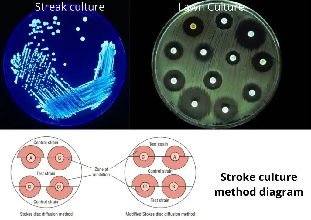
- Streak culture: Streak cultures are used to isolate bacteria from the pure form of clinical samples. The platinum wire is employed. One loop from the sample is inserted on the outside of a dried plate. It is spread over a small space near the edges. The inoculum then spreads thinly across the plate streaking it with the form of parallel lines within various segments of the dish. Incubation is completed and colonies are separated. are formed over the final sequence of streaks.
- Lawn Culture: Lawn provides a uniform growth on the surface of the bacteria. Lawn cultures are made by saturating the plate’s surface with an insoluble suspension of bacteria. Lawn Culture uses – for the typing of bacteriophage. Testing for sensitivity to antibiotics. It is also used in the preparation of bacterial antigens and vaccines.
- Stroke Culture: Stroke Stroke culture is created in tubes that contain Agar slope or slant. Stroke Culture is used to provide the pure bacteria growth for slide agglutination , as well as other tests for diagnosing.
- Stab Culture: Stab Cultures are prepared through puncturing a medium suitable for the task such as glucose agar or gelatin with a straight, long wire, charged. Stab Culture is used for demonstration of liquefaction of gelatin. The oxygen requirements of the bacterium in question. – Maintaining the culture stock.
- Pour Plate Culture: In this method, 1 ml inoculum is added to liquid Agar. Mix thoroughly and then transfer to the sterile Petri dish. Let it set. The medium’s depth. Pour Plate Culture uses to provide an estimate of the number of viable bacterial counts in the suspension. This is for the urine quantitative culture.

Colonial Morphology of Bacteria
If you look closely at the colonial growth that is visible on the surfaces of medium the characteristics of the texture of the surface, its transparency and the hue or color of the growth may be identified. The three traits listed below are easily apparent whether studying the surface of a single colony of bacteria or a more dense one, without the use of any magnifying device.
- Texture—Texture is the way your colony’s surface appears. The most common terms used to describe texture be smooth, glistening mucoid, slimy or powdery, flaky, etc.
- Transparency—colonies may be transparent (you can see through them), translucent (light passes through them), or opaque (solid-appearing).
- Color or Pigmentation—a lot of bacteria produce intracellular pigments that create colonies that have with distinct colors such as purple, pink, yellow or red. A lot of bacteria don’t produce any pigments, and appear as gray or white.

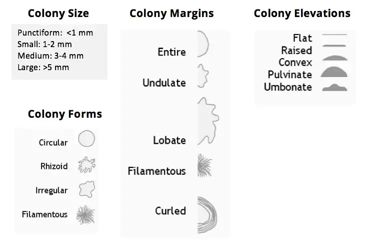
When the bacterial population expands in numbers, the colonies expand and start to adopt an appearance or shape. They can be very distinct and offer a method of distinguishing colonies that are similar in the color or texture. The three traits listed below are a good way to describe bacteria when only a one, distinct colony is able to be observed. It is possible to make use of a magnifying device for example, the colony counter or dissecting microscope, which allows an in-depth view of the colonies. Colonies must be described according by their size overall and shape as well as what a close-up view of the colony’s edges will look like (edge or the margin of the colony) and also which way the colony looks when you view on the other opposite side (elevation).
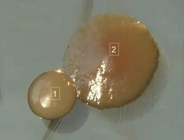
Below is the close-ups of colonies that are growing on the surface of an Agar plate. In this case the distinctions between the two bacteria are evident as each one has a distinctive colonial shape.

References
- Lagier JC, Edouard S, Pagnier I, Mediannikov O, Drancourt M, Raoult D. Current and past strategies for bacterial culture in clinical microbiology. Clin Microbiol Rev. 2015;28(1):208-236. doi:10.1128/CMR.00110-14
- https://www.technologynetworks.com/immunology/articles/an-introduction-to-culturing-bacteria-355566
- https://www2.nau.edu/~fpm/bio205/Sp-10/Chapter-03.pdf
- https://milnepublishing.geneseo.edu/suny-microbiology-lab/chapter/bacteriological-culture-methods/
- https://bio.libretexts.org/Bookshelves/Microbiology/Book%3A_Microbiology_(Boundless)/6%3A_Culturing_Microorganisms
- https://en.wikipedia.org/wiki/Microbiological_culture
- https://www.slideshare.net/AshfaqAhmad52/cultivation-of-bacteria-and-culture-methods
- Text Highlighting: Select any text in the post content to highlight it
- Text Annotation: Select text and add comments with annotations
- Comment Management: Edit or delete your own comments
- Highlight Management: Remove your own highlights
How to use: Simply select any text in the post content above, and you'll see annotation options. Login here or create an account to get started.