Major Histocompatibility Complex class I (MHC I) is a cell surface glycoprotein which is found on almost all nucleated cells and platelets. In human it is known as Human Leukocyte Antigen (HLA). The main classical forms are HLA-A, HLA-B and HLA-C.
MHC I is heterodimer in structure. It is made up of one heavy alpha chain and one small beta-2-microglobulin chain. These two chain form the groove like area for peptide binding. The groove is closed at both end and so it binds only short peptide, generally 8-11 amino acids.
The main work of MHC I is presentation of endogenous antigen. Endogenous antigen means the antigen which are formed inside the cell. These may be normal self protein or abnormal protein. Viral protein, intracellular bacterial protein and tumour protein also comes under this.
In the cytoplasm, protein are degraded by proteasome. During this process small peptide fragments are formed. These peptides pass into endoplasmic reticulum (ER). In ER, the peptide is attached with empty MHC I molecule. Then the formed peptide-MHC I complex is transported to the cell surface.
On the surface of cell, the peptide-MHC I complex is presented to CD8+ cytotoxic T lymphocytes (CTLs). The CD8+ T cell recognize the peptide. If the peptide is foreign or abnormal then the T cell is activated. It destroy the infected cell or tumour cell.
MHC I also regulate Natural Killer (NK) cell. Normal cell shows self peptide by MHC I molecule. This gives inhibitory signal to NK cell and cell is not killed. When MHC I is absent or less on cell surface, NK cell identify it as abnormal and kills the cell. This is called missing self hypothesis.
Distribution of Major Histocompatibility Complex I (MHC I)
Major Histocompatibility Complex class I (MHC I) is widely distributed in body. It is present on almost all nucleated somatic cells. So most of the body cells show MHC I on their surface.
MHC I is not found on RBCs because mature red blood cells are non-nucleated. But it is present on platelets. Platelets are non-nucleated cell fragments, but they carry MHC I molecule on their surface.
In Central Nervous System (CNS), HLA class I antigen is generally absent or very less. So the cells of brain and spinal cord show poor expression of MHC I in normal condition.
The classical MHC I molecules are distributed widely. These include HLA-A, HLA-B and HLA-C. They are present on most nucleated cells and help in showing endogenous antigen.
The non-classical MHC I molecules have restricted distribution. They are not found in all tissues like classical MHC I. Examples are HLA-G and HLA-E.
HLA-G is mainly present on fetal trophoblast cells. These cells are present at maternal-fetal interface. It is also present on thymic epithelium. Thus non-classical MHC I has tissue specific distribution.
Structure of Major Histocompatibility Complex I (MHC I)

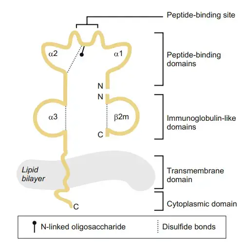
- Heterodimer– MHC I is a transmembrane heterodimer molecule. It is made up of two different polypeptide chains. One is heavy alpha (α) chain and another is small light chain, called beta-2-microglobulin (β₂m). These two chains are joined by non-covalent attachment.
- Alpha chain– The heavy α-chain is about 45 kDa protein. It is highly polymorphic. In human it is encoded by genes present on chromosome 6. These genes are HLA-A, HLA-B and HLA-C.
- Membrane part– The α-chain has a hydrophobic transmembrane part. This part passes through the cell membrane. It also has a cytoplasmic tail. The tail remains inside cytoplasm and helps in movement of the molecule inside cell.
- Outer domains– The heavy chain has three extracellular domains. These are α₁, α₂ and α₃. These domains are present outside the cell. They form the main outer structure of MHC I molecule.
- Peptide platform– α₁ and α₂ domains form the peptide binding platform. They make a groove like area. The floor is formed by eight stranded antiparallel β-sheet. The two side walls are formed by α-helices.
- α₃ domain– α₃ domain is present near the membrane. It has immunoglobulin like fold. It binds with β₂-microglobulin. It also has the binding site for CD8 co-receptor of cytotoxic T cell.
- β₂-microglobulin– β₂-microglobulin (β₂m) is the light chain of MHC I. It is about 12 kDa protein. It is invariant and non-polymorphic. In human it is encoded by gene on chromosome 15.
- Support chain– β₂-microglobulin does not enter into cell membrane. It remains attached with α₃ domain. It gives support to MHC I molecule. It is needed for proper folding and stability.
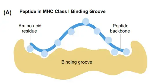
- Binding groove– The peptide binding groove of MHC I is closed at both ends. So long peptide cannot fit in it. It binds short peptide, generally 8 to 11 amino acids.
- Binding pockets– The floor of groove has special binding pockets. These are called A to F pockets. They bind with anchor residues of peptide. By this way peptide is held tightly in MHC I groove.
Mechanism of action of Major Histocompatibility Complex I (MHC I)
The following are the steps of mechanism of Major Histocompatibility Complex class I (MHC I)–
- Protein degradation– In this step, intracellular proteins are broken inside cytoplasm. These proteins may be normal self proteins, viral proteins or tumour antigens. They are degraded by proteasome. Small peptide fragments are formed.
- Peptide transport– The formed peptides are transported from cytoplasm into endoplasmic reticulum (ER). This transport takes place through TAP. TAP means Transporter associated with Antigen Processing. It is present on ER membrane.
- MHC I assembly– In ER, the heavy alpha (α) chain of MHC I starts folding. Chaperone protein like calnexin helps in this folding. Then the α-chain joins with beta-2-microglobulin (β₂m). An empty MHC I molecule is formed.
- Peptide loading complex– The empty MHC I molecule enters into Peptide Loading Complex (PLC). This complex contains TAP, tapasin, calreticulin and ERp57. Tapasin joins empty MHC I with TAP. So peptides coming inside ER can bind with MHC I groove.
- Peptide trimming– Some peptides transported by TAP are longer in size. These peptides are trimmed by ERAP enzyme. Peptides become short, generally 8 to 11 amino acids. This size is suitable for MHC I binding groove.
- Peptide editing– In this step, proper peptide is selected. Tapasin acts like peptide editor. It checks which peptide bind strongly with MHC I groove. Weak peptide is removed and strong peptide is kept. TAPBPR also helps in this editing process.
- Stable complex formation– When a good peptide binds with MHC I, the molecule becomes stable. The peptide fits inside the closed groove. Then complete peptide-MHC I complex is formed. This complex is released from PLC.
- Surface transport– The formed peptide-MHC I complex comes out from ER. It passes through Golgi apparatus. Then it reaches to plasma membrane. On the cell surface, it is displayed for immune recognition.
- CD8 T cell recognition– On cell surface, peptide-MHC I complex is recognized by CD8+ cytotoxic T lymphocyte (CTL). The T-cell receptor (TCR) binds with peptide part. CD8 co-receptor binds with MHC I molecule. If the peptide is foreign or abnormal, CTL becomes activated.
- Cell killing– Activated CD8+ T cell kills the infected cell or tumour cell. This killing occur by release of cytotoxic substances. Thus MHC I helps in removal of virus infected and malignant cells.
- NK cell inhibition– Normal cells show self peptide with MHC I. This binds with inhibitory receptor of Natural Killer (NK) cell, like KIR or CD94:NKG2A. It gives do not kill signal. So normal body cell is protected.
- Missing self– If MHC I is absent or decreased on cell surface, NK cell cannot get inhibitory signal. Then NK cell identify the cell as abnormal. It may kill that cell. This is called missing self hypothesis.
Deposition of self and non-self peptides in MHC I binding pocket
The following are the steps by which self and non-self peptides are deposited in MHC I binding pocket-
- Protein source– The proteins are present inside cytoplasm. These may be normal host protein, called self protein. These may also be viral protein, tumour protein or intracellular bacterial protein, called non-self protein.
- Proteasome action– In cytoplasm, these proteins are degraded by proteasome. The protein is cut into small peptide fragments. Both self and non-self peptides are formed by same process.
- Peptide transport– The newly formed peptides are transported into endoplasmic reticulum (ER). This transport is done by TAP. TAP means Transporter associated with Antigen Processing. It takes peptide from cytosol to ER lumen.
- MHC I folding– In ER, the MHC I alpha chain starts folding. Calnexin and ERp57 help in this folding. Then beta-2-microglobulin (β₂m) binds with alpha chain. An empty MHC I heterodimer is formed.
- PLC formation– The empty MHC I joins with Peptide Loading Complex (PLC). This complex has TAP, tapasin, calreticulin and ERp57. Tapasin keeps empty MHC I near TAP. So peptide can directly enter near the binding groove.
- Groove opening– Tapasin also keeps the peptide binding cleft in open and receptive form. The groove is ready to receive peptide. It does not select self or non-self by name. It selects peptide by proper size and strong fitting.
- Peptide trimming– Many peptides coming through TAP are longer. The MHC I groove is closed at both end. So long peptide cannot fit properly. ERAP1 trims the peptide and makes it small, generally 8 to 9 amino acids.
- Peptide loading– The trimmed peptide enters into MHC I binding groove. The peptide binds with different pockets present in the groove. Anchor residues of peptide fit into these pockets. If the fitting is strong then peptide stay in groove.
- Peptide editing– Tapasin checks the binding of peptide. Weakly bound peptide are removed. Strong peptide are kept. TAPBPR can also exchange loose peptide with better binding peptide during transport pathway.
- Stable complex– When high affinity peptide binds, the MHC I molecule become stable. The peptide is now deposited inside the closed groove. This may be self peptide or non-self peptide, depending on which protein was degraded inside the cell.
- Surface display– The stable peptide-MHC I complex leaves PLC. It passes through Golgi apparatus and reaches plasma membrane. On the cell surface it displays peptide to CD8+ cytotoxic T lymphocyte.
- Recognition– If the deposited peptide is self peptide, usually no cytotoxic response occur. If the peptide is non-self or abnormal, CD8+ T cell recognize it by TCR and the target cell may be killed.
Functions of Major Histocompatibility Complex I (MHC I)
The following are the functions of MHC I–
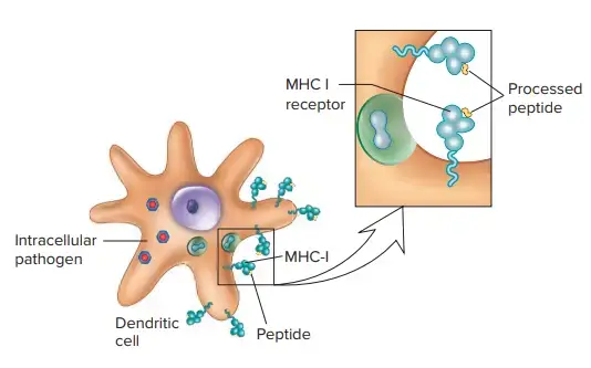
- Antigen presentation– MHC I present intracellular antigen. These antigen are formed inside the cell. It may be self protein, viral protein or tumour protein. These small peptide are shown on cell surface.
- Endogenous antigen display– MHC I mainly shows endogenous antigen. Endogenous antigen means antigen present inside cytoplasm. So the cell can show its internal protein condition to immune cells.
- CD8 cell recognition– The peptide-MHC I complex is recognized by CD8+ cytotoxic T lymphocyte (CTL). TCR of T cell bind with peptide. CD8 bind with MHC I molecule.
- Killing of infected cell– When viral peptide or abnormal peptide is present, CD8+ T cell becomes active. It kills the infected cell. It also kill tumour cell. Thus abnormal body cell are removed.
- Cell mediated immunity– MHC I helps in cell mediated immunity. It does not mainly act by antibody. It acts by activation of cytotoxic T cell and destruction of infected cell.
- NK cell inhibition– Normal cell has MHC I on surface. This MHC I bind with inhibitory receptor of Natural Killer (NK) cell. Examples are KIRs and CD94:NKG2A. So NK cell get inhibitory signal and does not kill the normal cell.
- Missing self response– Some virus infected cell and tumour cell reduce MHC I expression. Then NK cell cannot get inhibitory signal. The cell is taken as abnormal. This is called missing self response. Then NK cell kill the cell.
- NK cell education– MHC I also help in education of NK cells. During development, NK cell come in contact with self MHC I. This makes NK cell active but not harmful to normal self cell.
- Cross presentation– Some dendritic cells can present outside antigen by MHC I. This is called cross presentation. It helps in activation of naive CD8+ T cells against virus and tumour antigen.
References
- ResearchGate. (n.d.). (PDF) MHC Class I Antigen Processing Pathways – ResearchGate.
- Davis, Z. B., Cogswell, A., Scott, H., Mertsching, A., Boucau, J., Wambua, D., Le Gall, S., Planelles, V., Campbell, K. S., & Barker, E. (2016). A Conserved HIV-1-Derived Peptide Presented by HLA-E Renders Infected T-cells Highly Susceptible to Attack by NKG2A/CD94-Bearing Natural Killer Cells. PLoS Pathogens, 12(2), e1005421. https://doi.org/10.1371/journal.ppat.1005421
- Bessoles, S., Grandclément, C., Alari-Pahissa, E., Gehrig, J., Jeevan-Raj, B., & Held, W. (2014). Adaptations of Natural Killer Cells to Self-MHC Class I. Frontiers in Immunology, 5, 349. https://doi.org/10.3389/fimmu.2014.00349
- National Center for Biotechnology Information. (n.d.). Alternative Antigen Processing for MHC Class I: Multiple Roads Lead to Rome – PMC.
- ResearchGate. (n.d.). An overview of the mechanisms of antigen cross-presentation Vacuolar… – ResearchGate.
- National Center for Biotechnology Information. (n.d.). Analysis of HLA-E peptide-binding specificity and contact residues in bound peptide required for recognition by CD94/NKG2 – PubMed.
- Wikipedia contributors. (2025). CD8. In Wikipedia, The Free Encyclopedia.
- Gosset. (2026). CD8 co-receptor bound to MHC class I (CD8-MHCI).
- National Center for Biotechnology Information. (n.d.). Checking your browser – reCAPTCHA.
- National Center for Biotechnology Information. (n.d.). Cross-Presentation of Cell-Associated Antigens by MHC Class I in Dendritic Cell Subsets.
- National Center for Biotechnology Information. (n.d.). Cross-Presentation: How to Get there – or How to Get the ER – PMC.
- Wikipedia contributors. (2026). Cross-presentation. In Wikipedia, The Free Encyclopedia.
- Embgenbroich, M., & Burgdorf, S. (2018). Current Concepts of Antigen Cross-Presentation. Frontiers in Immunology, 9, 1643. https://doi.org/10.3389/fimmu.2018.01643
- GeeksforGeeks. (2025). Difference Between MHC Class 1 and MHC Class 2 Proteins.
- van den Elsen, P. J. (2011). Expression Regulation of Major Histocompatibility Complex Class I and Class II Encoding Genes. Frontiers in Immunology, 2, 48. https://doi.org/10.3389/fimmu.2011.00048
- Abcam. (n.d.). HLA-B.
- National Center for Biotechnology Information. (n.d.). HLA-E/peptide complexes differentially interact with NKG2A/CD94 and T cell receptors.
- Lutz, C. T. (2014). Human leukocyte antigen Bw4 and Bw6 epitopes recognized by antibodies and natural killer cells. Current Opinion in Organ Transplantation, 19(4), 436-441. https://doi.org/10.1097/MOT.0000000000000103
- Wikipedia contributors. (2026). Killer-cell immunoglobulin-like receptor. In Wikipedia, The Free Encyclopedia.
- Riaz, S. (2024). MHC Class I vs Class II: Key Differences & Functions [Guide]. Assay Genie.
- National Center for Biotechnology Information. (n.d.). MHC class I transactivator NLRC5 in host immunity, cancer and beyond – PMC – NIH.
- National Center for Biotechnology Information. (n.d.). Major Histocompatibility Complex (MHC) Class I and MHC Class II ….
- Unknown. (n.d.). Major Histocompatibility Complex Class I: Molecular Architecture, Biosynthetic Pathways, and Immunological Mechanisms.
- Cruz-Tapias, P., Castiblanco, J., & Anaya, J. M. (2013). Major histocompatibility complex: Antigen processing and presentation. In J. M. Anaya, Y. Shoenfeld, A. Rojas-Villarraga, et al. (Eds.), Autoimmunity: From Bench to Bedside. El Rosario University Press.
- National Center for Biotechnology Information. (n.d.). Missing or altered self: human NK cell receptors that recognize HLA ….
- Meissner, T. B., Li, A., Biswas, A., Lee, K.-H., Liu, Y.-J., Bayir, E., Iliopoulos, D., van den Elsen, P. J., & Kobayashi, K. S. (2010). NLR Family Member NLRC5 Is a Transcriptional Regulator of MHC Class I Genes. Proceedings of the National Academy of Sciences of the United States of America, 107(31), 13794-13799.
- National Center for Biotechnology Information. (n.d.). NLRC5 cooperates with the RFX transcription factor complex to induce MHC class I gene expression – PMC.
- National Center for Biotechnology Information. (n.d.). NLRC5 cooperates with the RFX transcription factor complex to induce MHC class I gene expression – PubMed.
- National Center for Biotechnology Information. (n.d.). Non-classical MHC Class I molecules regulating natural killer cell function – PMC – NIH.
- National Center for Biotechnology Information. (n.d.). Pathways of MHC I cross-presentation of exogenous antigens – PMC – NIH.
- National Center for Biotechnology Information. (n.d.). Pathways of MHC I cross-presentation of exogenous antigens – PubMed.
- Becar, M., & Kasi, A. (2022). Physiology, MHC Class I. In StatPearls. StatPearls Publishing.
- National Center for Biotechnology Information. (n.d.). Recognition of Self and Viral Ligands by NK Cell Receptors – PMC – NIH.
- Li, X.-L., Teng, M.-K., Reinherz, E. L., & Wang, J.-H. (2013). Strict Major Histocompatibility Complex Molecule Class-Specific Binding by Co-Receptors Enforces MHC-Restricted αβ TCR Recognition during T Lineage Subset Commitment. Frontiers in Immunology, 4, 383. https://doi.org/10.3389/fimmu.2013.00383
- National Center for Biotechnology Information. (n.d.). Structural Basis for Recognition of Cellular and Viral Ligands by NK Cell Receptors – PMC.
- National Center for Biotechnology Information. (n.d.). Structural Comparison Between MHC Classes I and II; in Evolution, a Class-II-Like Molecule Probably Came First – PMC.
- National Center for Biotechnology Information. (n.d.). Structural aspects of chaperone-mediated peptide loading in the ….
- Proceedings of the National Academy of Sciences. (n.d.). Structural basis for NKG2A/CD94 recognition of HLA-E | PNAS.
- National Center for Biotechnology Information. (n.d.). Structural basis for recognition of MHC and MHC-like ligands by natural killer cell receptors.
- National Center for Biotechnology Information. (n.d.). The CD8 coreceptor interaction with the alpha 3 domain of HLA class I is critical to the differentiation of human cytotoxic T-lymphocytes specific for HLA-A2 and HLA-Cw4 – PubMed.
- National Center for Biotechnology Information. (n.d.). The MHC class I antigen presentation pathway: strategies for viral immune evasion – PMC.
- National Center for Biotechnology Information. (n.d.). The Structural Basis of Ligand Recognition by Natural Killer Cell Receptors – PMC – NIH.
- Janeway, C. A. Jr., Travers, P., Walport, M., et al. (2001). The major histocompatibility complex and its functions. In Immunobiology: The Immune System in Health and Disease (5th ed.). Garland Science.
- National Center for Biotechnology Information. (n.d.). The molecular determinants of CD8 co-receptor function – PMC.
- National Center for Biotechnology Information. (n.d.). The quality control of MHC class I peptide loading – PMC.