Viroids Definition
- In 1971, a plant pathologist named Theodor Otto Diener first discovered the Viroids. He found an acellular particle when he was working in an Agriculture Research Service and named this particle as viroid, meaning “virus-like.” At present-33 species of viroid have been identified.
- Viroids are known as the smallest infectious pathogens which are made up solely of a short strand of circular, single-stranded self-replicating RNA that has no protein coating.
- Although, viroids are made of nucleic acid which does not code for any protein.
- They don’t contain any protein coat to protect their genetic information.
- Viroids are well known for causing different plant diseases. They associate with the host’s proteins by the phloem vascular channels and plasmodesmata and then move within the host’s cell (Plant).
- The symptoms of viroid disease include chlorosis, stunting, veinal discolouration, epinasty, vein clearing, localised necrotic spots, mottling of leaves and death of the host.
- The first viroid was found in potato tuber spindle disease, which causes slower sprouting and various deformities in potato plants.
- The viroid uses RNA polymerase II enzyme in its replication mechanism. In the host cell, this enzyme helps in the synthesis of messenger RNA from DNA, which instead catalyzes “rolling circle” synthesis of new RNA using the viroid’s RNA as a template.
- Sometimes they contain catalytic activity which helps them in self-cleavage and ligation of unit-size genomes from larger replication intermediates.
- There are two groups of viroids such as Avsunviroids (e.g., Avocado Sun-blotch viroid) and Pospiviroids (e.g., Potato spindle tuber viroid).
- The replication process of Avsunviroids is accomplished within chloroplasts whereas pospiviroidae replicate within nucleus and nucleolus.
Viroids Structure:

- The Electron microscope is used to observe the structure of viroid.
- They are small, circular, single-stranded RNA molecules.
- They have 50 nm long RNA.
- They are made of a short stretch (a few hundred nucleobases) of highly complementary circular ssRNA which lacks a protein coat.
- The molecular weight of their genome is about 1,07,000 and 1,27,000.
- The genome is 240 to 380 nucleotides long with dumbbell structures.
- The smallest viroid has 220 nucleobase ScRNA which is mainly found in rice yellow mottle sobemovirus (RYMV)
- The adenine: Uracil ratio is 21.7: 20.9 and the guanine: cytosine ratio is 28.9: 28.3
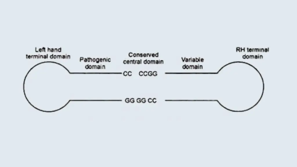
- Viroids contain five structural/functional domains such as;
- Conserved central domain.
- Pathogenic domain
- Variable domain
- Left terminal domain
- Right terminal domain.
- The Conserved central domain involved with cleavage and ligation of RNA.
- The avsunviroidae group of viroids lacks the conserved central Domain (CCR) and instead contain a ribozyme activity.
Replication of Viroids or Infection of Viroids
Viroid RNA replicates autonomously and spreads within the host by recruiting host proteins. There are three enzymes that help in RNA replication such as RNA pol. II, RNAase and RNA ligase.
Viroid RNA don’t code for any plant protein and they lack AUG initiation codon. The replication process of viroid RNA is accomplished by two different methods such as;

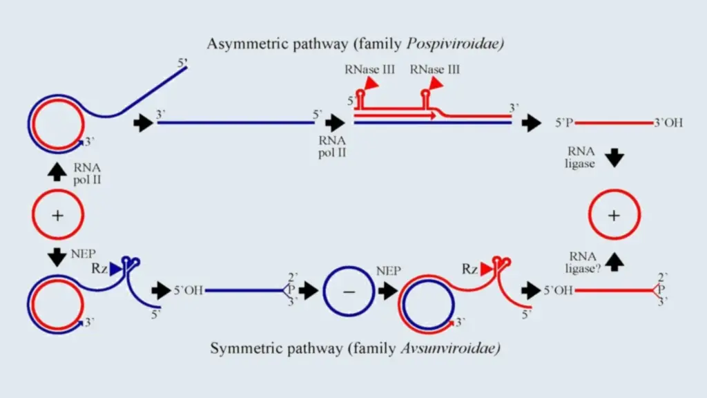
- Asymmetric Rolling circle Replication
- Pospiviroids use asymmetric Rolling circle Replication methods to replicate their RNA.
- In this method an Incoming (+)-circular RNA initially is transcribed into concatemeric linear (-)-strand RNA.
- After that it serves as the replication intermediate for the synthesis of concatemeric, linear (+)- strand RNA.
- This (+)- strand RNA subsequently is cleaved into unit length monomers that are ligated into circles.
- Symmetric Rolling Circle Replication
- Members of Avsunviroid follow this Symmetric Rolling Circle Replication method to replicate their RNA.
- In this method the circular positive sense RNA is transcribed into linear, concatemeric (-)- strand RNA
- Then Instead of serving as the direct template for the synthesis of linear concatemeric (+)- strand RNA
- After that the concatemeric (-)- strand RNA is cleaved into unit length molecules followed by circularization
- The circular (-)- RNA then serves as the template for the synthesis of linear, concatemeric (+)- strand RNA
- When subsequently is cleaved into unit-length monomers and circularized
Transmission of Viroids
- Viroids can be transmitted through seed and pollen of infected plants.
- They can be transmitted by aphids from infected plants to other plants.
- They can be transmitted from plant to plant by leaf contact.
The origin of Viroids
There are four possibility regarding the origin of Viroids;
- The viroids are considered as sub-viral agents because they occur from viruses in which the protein coat is lost.
- The low molecular weight RNAs are found in leaves infected with TMV, leaves infected with broad bean mottle virus and in Escherichia coli infected with phase Qβ. Ultimately, these might have given rise to viroids.
- The viroids may have originated from self-replicating RNAs when they become pathogenic and induce diseases. For example Potato spindle tuber viroid.
- It is estimated that viroids originate from regulatory RNAs which have become abnormal by mutation.
Viroids Characteristics
- They are known as the smallest infectious agent who mainly infects plants.
- They contain only RNA.
- They contain a nucleic acid with low molecular weight and a unique structure.
- They multiply within the host cell and as a result, it causes the death of the host.
- They have two families such as Pospiviroidae- nuclear viroids and Avsunviroidae- chloroplastic viroids.
- They move cell to cell by the plasmodesmata, and a long-distance through the phloem.
Diagnostic Methods of Viroids
- Biological indexing (bioassay) can be used to detect the presence of Viroids.
- A more precise, reliable and rapid technique is polyacrylamide gel electrophoresis (PAGE). This technique helps in differentiation of nucleic acids according to their size, based on their differential mobility in an electric field.
- Temperature gradient‐gel electrophoresis (TGGE) and analysis of heteroduplexes can be used for the detection of quasispecies form of viroids.
- There are several nucleic acid‐based techniques used for the detection of viroids such as hybridization with radioactive and chemically labelled probes, reverse transcription combined with polymerase chain reaction (RT‐PCR), and real‐time RT‐PCR (RT‐qPCR).
- Recently new methods have been developed which can enable even faster and more sensitive detection of viroid infections such as reverse transcriptase loop‐mediated isothermal amplification (RT‐LAMP), isothermal and chimeric primer‐initiated amplification of nucleic acids (ICAN), micro‐ and microarrays and next‐generation sequencing (NGS).
Viroids Disease and Examples
Some example of plant viroids are;
- Potato spindle tuber viroid (potatoes)
- Citrus exocortis (citrus plants) (sometimes called “scalybutt”) this can also infect tomato plants (sometimes called “tomato bunchy top disease”)
- Citrus gummy bark viroid
- Grapevine viroid
- Dapple peach fruit disease viroid
- Citrus cachexia viroid
- Cucumber pale fruit viroid
- Dapple plum and peach fruit disease viroid
- Cadang-cadang is a disease caused by Coconut cadang-cadang viroid (a lethal viroid of Coconut)
- Peach latent mosaic viroid (present in all peach- and nectarine-producing areas of the world)
- Eggplant latent viroid.
- Text Highlighting: Select any text in the post content to highlight it
- Text Annotation: Select text and add comments with annotations
- Comment Management: Edit or delete your own comments
- Highlight Management: Remove your own highlights
How to use: Simply select any text in the post content above, and you'll see annotation options. Login here or create an account to get started.