What is Tobacco mosaic virus (TMV)?
- Tobacco mosaic virus (TMV) is a well-known plant virus from the Tobamovirus genus, particularly affecting tobacco plants, as well as other members of the Solanaceae family, including tomatoes and potatoes. TMV infection causes characteristic symptoms such as a mosaic-like mottling and discoloration of leaves, which is how the virus got its name.
- The history of TMV’s discovery traces back to Adolf Mayer, a German chemist, who in 1886 observed that the disease could spread between tobacco plants through contact with infected sap, much like bacterial infections. However, the nature of the infectious agent was not well understood at the time. Later, in the late 19th century, Dmitri Ivanovsky, a Russian microbiologist, found that the infectious agent could pass through filters known to block bacteria, indicating that it was not a bacterial pathogen. Despite this discovery, Ivanovsky suspected that the agent was simply an exceptionally small, unculturable bacterium.
- Martinus Beijerinck expanded upon Ivanovsky’s work in 1898, conducting filtration experiments that led him to conclude that the infectious agent was neither bacterial nor cellular in nature. Beijerinck coined the term “virus” to describe this unique entity, establishing the term for a whole new category of pathogens. His work laid the foundation for the field of virology, and TMV thus became recognized as the first virus ever discovered.
- Further research into TMV revealed critical insights about viruses in general. In 1935, American biochemist Wendell Meredith Stanley successfully crystallized TMV, providing physical evidence that viruses could maintain infectivity even outside of their host. Stanley’s crystallization of TMV contributed to his receipt of the Nobel Prize in Chemistry in 1946, though some of his initial conclusions were later corrected by other researchers. In 1939, electron microscopy provided the first images of TMV, helping to refine the understanding of its structure.
- One notable discovery about TMV came in the 1950s when Heinz Fraenkel-Conrat and Robley Williams demonstrated that the virus’s RNA and protein coat could spontaneously assemble into functional viruses. Rosalind Franklin’s contributions to TMV research in the 1950s furthered the understanding of its structure, as she proposed that the virus was hollow and that its RNA was single-stranded, which was later confirmed. This was instrumental in recognizing TMV as a positive-sense, single-stranded RNA virus.
- TMV’s discovery and extensive study have provided foundational insights into viral structure, replication, and transmission, making it central to virology as a scientific discipline.

Characteristics of Tobacco Mosaic Virus (TMV)
TMV has a range of distinctive features that set it apart as a widespread and resilient plant virus, impacting several economically important crops, especially within the Solanaceae family.
- Name: Tobacco Mosaic Virus (TMV)
- Shape and Structure:
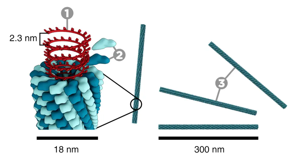
- TMV has a filamentous, rod-like shape, measuring approximately 300 nm in length and 18-20 nm in diameter.
- Its genetic material is single-stranded RNA (ssRNA) with a helical symmetry, which is tightly coiled inside the protein coat.
- The viral genome spans about 6,395 nucleotides, encoding proteins essential for replication, movement, and structure.
- Discovery:
- TMV was first identified by Martinus Beijerinck in 1898, marking it as one of the earliest viruses to be studied in detail.
- Host Range:
- TMV infects a broad spectrum of plants, with a notable preference for the Solanaceae family, including tobacco, tomato, and potato plants.
- Other susceptible hosts include peppers and certain ornamental plants, where infection results in distinctive symptoms that reduce plant vigor.
- Symptoms in Infected Plants:
- TMV produces a characteristic mosaic pattern on leaves, where light and dark green areas form mottled patches.
- Infected plants often exhibit stunted growth and leaf distortion, leading to reduced photosynthesis and lower crop yields.
- Mode of Transmission:
- TMV is primarily spread through mechanical means, not by insect vectors, which makes human handling a major factor in its transmission.
- The virus can survive on tools, clothing, and surfaces contaminated by infected plant sap, staying infectious for long periods even in dried form.
- Although insects aren’t main carriers, plant damage from chewing insects can create entry points for the virus.
- Durability and Stability:
- TMV is known for its exceptional stability, able to withstand extreme environmental conditions.
- The virus can persist in plant debris, dried sap, and contaminated tobacco products, increasing the risk of accidental transmission.
- This resilience allows it to spread easily in environments where sanitation practices aren’t rigorously applied.
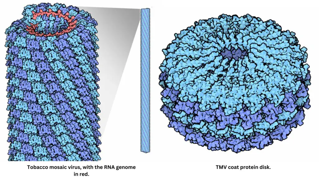
TMV’s combination of durability, wide host range, and ease of spread underscores the importance of hygiene in plant handling and management to prevent significant agricultural losses
Structure of Tobacco Mosaic Virus (TMV)
The structure of the tobacco mosaic virus (TMV) is well-suited to its role as a plant pathogen, combining a simple, rod-like form with a robust protein shell and a single-stranded RNA genome. This composition allows TMV to infect and replicate efficiently within host cells. Here’s a detailed breakdown:

- Shape and Size: TMV has a straight, rod-like shape measuring around 300 nanometers in length and 18 nanometers in diameter, with a structural design optimized for its infectious cycle.
- Capsid (Protein Coat):
- Composed of 2130 subunits called capsomeres, the protein coat, or capsid, wraps around the viral RNA in a helical arrangement.
- Each capsomere has a molecular weight of about 17,000 daltons and contains approximately 158 amino acids.
- Capsomeres form 130 turns along the length of the rod, with 16.3 proteins per helical turn.
- This protein shell accounts for roughly 94.4% of the virus’s mass, with the RNA making up the remaining 5.6%.
- RNA Genome:
- The virus’s genetic material is single-stranded RNA (ssRNA), positioned in a helical configuration within the capsid.
- TMV’s RNA strand is around 6395 nucleotides long, with some variations reporting approximately 7300 nucleotides.
- It extends slightly past one end of the rod, giving a length slightly greater than the capsid itself, about 3300 angstroms in length.
- The RNA’s molecular weight is estimated to be 25,000 daltons.
- Spatial Organization:
- Inside the rod, the RNA helix maintains a spacing of 50 angstroms from the outer surface, while a 40-angstrom core runs through the central axis.
- This precise internal arrangement maintains TMV’s stability and symmetry, facilitating interaction with host cells and enhancing its ability to withstand environmental changes.
- Chirality and Symmetry:
- TMV’s helical structure exhibits structural chirality and symmetry, making it adaptable for chemical and genetic modifications.
- This arrangement provides resilience, allowing the virus to retain infectivity under various conditions and across diverse host plants.
Genome of Tobacco Mosaic Virus (TMV)
The genome of TMV, a single-stranded RNA (ssRNA) molecule about 6.3–6.5 kilobases long, encodes the essential proteins the virus needs for replication, movement, and assembly.
- Structure of TMV RNA:
- TMV’s ssRNA has a methylated nucleotide cap at the 5’ end, identified as m7G5’pppG, which protects the viral RNA and assists in translation within the host cell.
- At the 3′ end, the RNA has a tRNA-like structure, which mimics host cellular mechanisms to stabilize the viral genome.
- Coding Regions and Open Reading Frames (ORFs):
- TMV’s genome contains four open reading frames (ORFs), each encoding essential viral proteins.
- The coding sequence begins 69 nucleotides from the 5′ end, with the first ORF encoding a replicase that includes key enzymes needed for RNA synthesis.
- This replicase is a multifunctional protein with two distinct domains: methyltransferase (MT) and RNA helicase (Hel), crucial for initiating RNA synthesis and unwinding RNA structures.
- Key Proteins Produced by TMV:
- TMV’s genome employs a leaky UAG stop codon in the first ORF, which allows the production of a second protein through ribosomal readthrough, enabling multiple protein functions from a single sequence.
- A second ORF encodes an RNA-dependent RNA polymerase, responsible for copying the viral RNA during replication.
- The third ORF encodes the movement protein (MP), allowing the virus to spread through the host plant by traveling between cells.
- The fourth ORF codes for the capsid protein (CP), which forms the protective shell around the RNA, essential for assembling new virus particles.
- Noncoding Regions:
- TMV’s RNA has noncoding regions at both the 5′ and 3′ ends, which are involved in regulating translation and genome stability.
- The 5’ end shows variability between individual virions, whereas the 3’ end remains consistent across different virions, providing stable structural integrity for the viral genome.
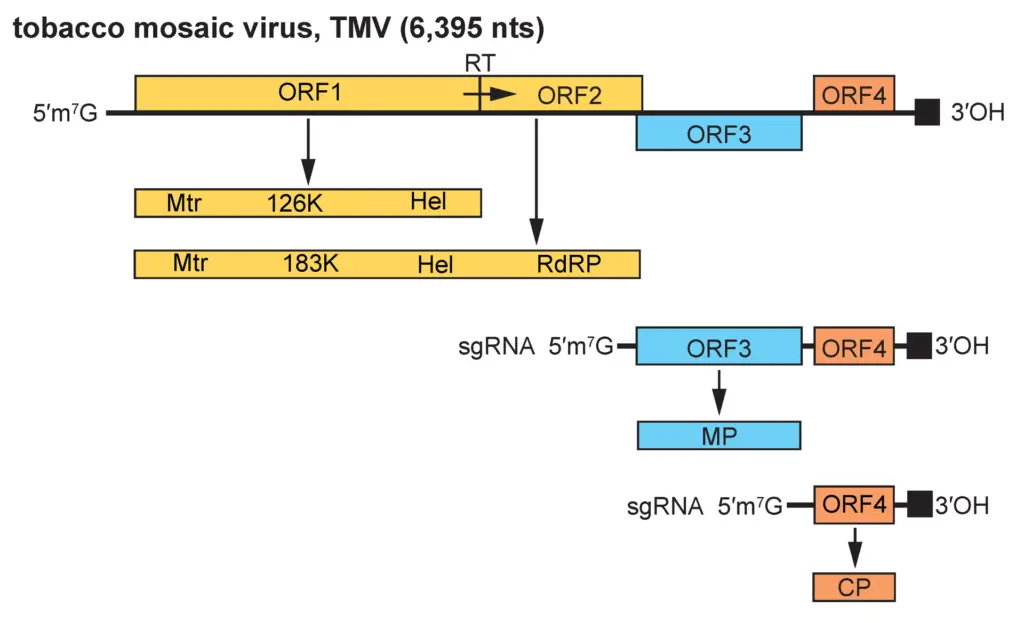
This RNA genome configuration enables TMV to hijack the plant’s cellular machinery, ensuring efficient replication and spread throughout the host.
Life Cycle of Tobacco Mosaic Virus (TMV)
TMV’s life cycle begins when the virus enters the plant’s cells, and once inside, it takes over the host’s cellular machinery to produce new viral particles. Here’s a breakdown of how TMV infects, replicates, and spreads:
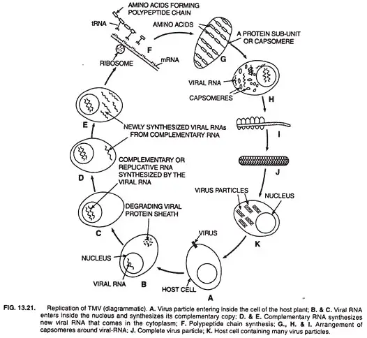
- Entry into the Host Cell:
- TMV typically enters plant cells through wounds or with the help of vectors like aphids and flies, which transfer viral particles between plants.
- Once inside, TMV moves from one cell to neighboring cells via plasmodesmata, small channels that connect plant cells.
- Uncoating of the Virus:
- Inside the cell, the virus sheds its protective protein coat, or capsid, freeing its genetic material—a single-stranded RNA molecule—into the cell’s cytoplasm.
- Replication of Viral RNA:
- The viral RNA first acts as a template to synthesize replicative RNA, which is complementary to the original viral genome.
- This replicative RNA then serves as a template to produce more copies of the viral RNA, effectively multiplying the virus’s genetic material.
- Synthesis of Viral Proteins:
- The new viral RNA functions as messenger RNA (mRNA), using the host’s ribosomes and transfer RNA (tRNA) to produce viral proteins.
- These viral proteins include capsid subunits, known as capsomeres, which will eventually form the protective protein shell around new viral RNA strands.
- Assembly of New Virus Particles:
- The viral RNA assembles with newly synthesized capsomeres to form complete virus particles, or virions, each capable of infecting other cells.
- The RNA coils inside the protein shell, ensuring each virion has both a protective coat and functional RNA genome.
- Spread to Other Cells and Plants:
- The newly formed virions do not cause lysis, or bursting, of the host cell, unlike some other viruses. Instead, they move through plasmodesmata to nearby cells, gradually spreading the infection.
- TMV can also spread to other plants through direct contact, including touch and transfer by gardening tools.
TMV’s efficient, non-lytic spread enables it to infect large areas of plant tissue, causing the characteristic mosaic mottling on leaves and compromising the health of the plant.
Transmission of Tobacco Mosaic Virus (TMV)
Tobacco Mosaic Virus (TMV) spreads primarily through mechanical means. This includes the handling of infected plants, tools, and materials. Here’s how it works:
- Mechanical Transmission:
- Human activities play a significant role in spreading the virus.
- Handling infected plants—whether through pruning, harvesting, or moving materials—can transfer the virus from one plant to another.
- Tools like pruning shears, garden gloves, or anything that comes into direct contact with an infected plant can spread the virus. If not sanitized, these tools will transfer the virus to healthy plants.
- Infected hands or even tobacco products like cigarettes and chewing tobacco can also carry TMV. When touched or used around plants, these can spread the virus.
- Contaminated Soil and Plant Residue:
- TMV can survive in plant debris left behind in the soil, making it possible for the virus to reinfect new plants in the area.
- The virus can remain viable in soil and on surfaces for extended periods, sometimes lasting years. This persistence makes it tough to eliminate completely.
- Contaminated Seeds or Seedlings:
- Infected seeds or seedlings are a common source of new infections. If new plants grow in soil or containers where TMV was previously present, they can easily become infected.
Mode of Infection of Tobacco Mosaic Virus (TMV)
TMV infects plants in a very specific way, relying primarily on mechanical transmission. Here’s how it works:
- Entry into the Plant:
- TMV typically enters plants through small wounds in the cell membrane.
- These wounds can occur from handling the plant, physical damage, or other mechanical disturbances.
- Once TMV makes its way into the plant, the virus sheds its protein coat, releasing the viral RNA into the plant cell.
- Replication in the Host Cell:
- The released RNA directs the host cell machinery to begin producing new viral particles.
- This hijacks the plant’s normal processes, shifting the cell’s functions from regular activities to viral replication.
- Movement Within the Plant:
- As the virus replicates, it spreads from one cell to another through plasmodesmata, small channels that connect plant cells.
- The virus uses a movement protein to modify and expand these channels, allowing the viral RNA to travel to adjacent cells.
- Systemic Spread:
- The virus can also travel through the plant’s phloem, which allows it to move long distances within the plant.
- This systemic spread ensures that the infection is not localized, impacting large areas of the plant.
Symptoms of Tobacco Mosaic Virus (TMV)
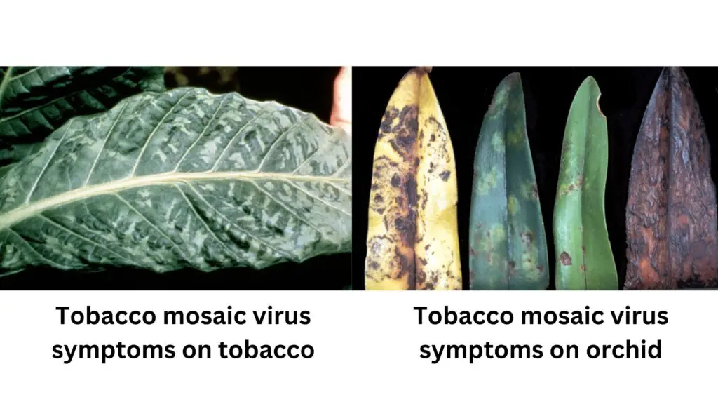
TMV infection results in a range of symptoms that disrupt the normal growth and development of plants. These symptoms can vary slightly depending on the plant species, but several key indicators remain consistent.
- Mosaic Pattern on Leaves:
- One of the most recognizable signs of TMV is the mosaic pattern on leaves.
- Infected leaves show a mix of light and dark green patches, caused by chlorosis (yellowing of the leaf tissue).
- Leaf Deformation:
- As the infection progresses, leaves may begin to curl or become distorted.
- Some leaves can develop wrinkles, altering their typical shape and structure.
- Stunted Growth:
- TMV often leads to a general stunting of plant growth.
- Infected plants are typically smaller than healthy ones, and their growth is slowed down.
- Flowering may also be delayed or reduced.
- Reduced Yield:
- The virus disrupts normal plant function, leading to a decrease in productivity.
- This can result in fewer flowers, fruits, or seeds compared to uninfected plants.
- Necrosis:
- In some cases, infected plants may experience necrosis, where portions of the tissue die off.
- This often manifests as dark spots or lesions on the leaves, signifying localized tissue death.
Treatment of Tobacco Mosaic Virus (TMV)
While there is no direct cure for Tobacco Mosaic Virus (TMV) once the plant is infected, a few strategies can be implemented to manage and prevent its spread.
- Resistant Varieties:
- One of the most effective ways to handle TMV is by growing resistant plant varieties.
- Some crops, particularly certain tobacco and tomato cultivars, have been specifically bred to resist TMV infection.
- Hygiene Practices:
- TMV spreads easily through mechanical means like contaminated tools, hands, or clothing.
- It’s critical to maintain strict hygiene in the field and greenhouse by regularly sanitizing tools and equipment.
- Disinfecting tools with bleach for at least 10 minutes can help reduce the risk of transmission.
- Removing Infected Plants:
- Once an infection is detected, it’s vital to remove and destroy the infected plants immediately.
- Isolate infected plants until they can be properly disposed of to prevent the virus from spreading to healthy crops.
- Seed Management:
- Infected seeds are a common source of TMV spread.
- Always use virus-free seeds or ensure that seeds from your own crops are properly cleaned or treated to eliminate any traces of TMV.
- Environmental Control:
- TMV can linger in plant debris and on surfaces in greenhouses for extended periods.
- Regularly clean growing environments, including removing plant debris from the soil, to minimize the chances of reinfection.
Control of Tobacco Mosaic Virus (TMV)
Controlling Tobacco Mosaic Virus (TMV) requires a solid approach that combines prevention, hygiene, and careful management practices. Since there are no chemical treatments once the virus is established, these steps are essential to minimize the spread and impact of the infection.
- Prevention:
- Inspect incoming plants carefully for TMV symptoms before introducing them to the growing area.
- Quarantine new plants until they have been tested for viruses, especially TMV.
- Vigilantly monitor plants throughout their life cycle for early signs of infection to catch any issues before they spread.
- Sanitation:
- Regularly disinfect tools, workstations, and surfaces that come in contact with plants to prevent the virus from transferring.
- Make sure that anyone entering the greenhouse follows strict hygiene practices, such as cleaning their footwear and washing their hands.
- Using footbaths with disinfectants and gloves can help prevent transmission, particularly when handling tobacco products known to carry the virus.
- Isolation and Removal:
- If plants show signs of infection, remove them immediately along with any nearby plant debris.
- Properly dispose of infected plants to prevent further contamination and spread of the virus.
- Monitoring and Testing:
- Utilize virus detection tools like Agdia TMV test strips to test plants for TMV, even if symptoms aren’t visible yet.
- Regular testing helps catch infections early, preventing the virus from spreading unchecked.
- Employee Training:
- Educate staff on TMV symptoms, proper handling techniques, and the importance of hygiene.
- Well-trained personnel are key to preventing the virus from spreading within the growing area.
Applications of Tobacco Mosaic Virus (TMV)
TMV has evolved from a plant pathogen to a powerful tool in diverse fields like biotechnology, biomedicine, and nanoelectronics. Its structure and adaptability open doors to many advanced applications.
- Genetic Engineering in Plants:
- TMV serves as an efficient viral vector, allowing scientists to introduce genes directly into plant cells.
- Through this, researchers can develop plants with enhanced resistance, increased growth rates, and better productivity.
- This gene-delivery method is used in platforms like magnICON and TRBO for effective gene expression within plant cells.
- In agricultural biotechnology, this vector technology is essential for engineering traits that boost crop resilience and yield.
- Biomaterial and Nanotechnology Applications:
- TMV’s cylindrical, rod-like structure is ideal for use in nanotechnology, thanks to its high aspect ratio and self-assembling properties.
- Scientists coat TMV particles with metals like nickel or cobalt, transforming them into effective components in nano-devices and biosensors.
- Its well-defined structure also supports its use as a template for biomaterials, making it suitable for a range of nanoscale innovations.
- Energy Storage Enhancements:
- TMV is applied in battery electrode design, where its unique structure increases the reactive surface area.
- When incorporated into battery electrodes, it can increase battery capacity by up to six times, compared to traditional electrode designs.
- By improving electrode surface area, TMV-based batteries achieve enhanced conductivity and overall energy efficiency, paving the way for more efficient energy storage solutions.
- Expression of Foreign Proteins in Fungi:
- TMV-based vectors also allow for the expression of proteins in fungal cells, an application with potential for various research fields.
- Through TMV, proteins like the green fluorescent protein (GFP) have been successfully expressed in fungi such as Colletotrichum acutatum for extended periods, offering a new pathway for fungal studies without complex transformation methods.
- This technique, known as virus-induced gene silencing (VIGS), shows that TMV can be adapted to function beyond plants, aiding in protein expression research for fungi.
- Rifkind, D., & Freeman, G. L. (2005). TOBACCO MOSAIC VIRUS. The Nobel Prize Winning Discoveries in Infectious Diseases, 81–84. doi:10.1016/b978-012369353-2/50018-7
- Sano, Y. et al. (1997). Functional Structure of Tobacco Mosaic Virus RNA. In: Carmona, P., Navarro, R., Hernanz, A. (eds) Spectroscopy of Biological Molecules: Modern Trends. Springer, Dordrecht. https://doi.org/10.1007/978-94-011-5622-6_115
- EL Sharif, Hazim. (2016). Smart materials: towards on-site detection of biomacromolecules using hydrogel-based molecularly imprinted polymers..
- https://erakina.com/plants-animal/the-viral-plant-pathogen-erakina/
- https://europepmc.org/backend/ptpmcrender.fcgi?accid=PMC1692534&blobtype=pdf
- https://www.lndcollege.co.in/syllabus/co_po/TMV-%20Structure%20and%20Replication.pdf
- https://www.apsnet.org/edcenter/disandpath/viral/pdlessons/Pages/TobaccoMosaic.aspx
- https://extension.usu.edu/pests/ipm/notes_ag/veg-TMV-ToMV
- https://www.farmersweekly.co.za/crops/field-crops/tobacco-mosaic-virus-symptoms-transmission-management/
- https://pdb101.rcsb.org/motm/109
- https://www.biologydiscussion.com/viruses/tobacco-mosaic-virus-tmv-structure-and-replication/54903
- https://www.biologydiscussion.com/plants/plant-diseases/tobacco-mosaic-virus-symptoms-and-control/58712
- https://www.brainkart.com/article/Tobacco-Mosaic-Virus-%28TMV%29-and-its-Structure_32835/
- https://byjus.com/biology/tmv-diagram/
- https://en.wikipedia.org/wiki/Tobacco_mosaic_virus
- https://ggu.ac.in/gguold/download/Class-Note13/LBC-101%20Unit%20-1%20Lect%20-6%20Botany.pdf
- https://extension.psu.edu/tobacco-mosaic-virus-tmv
- https://www.geeksforgeeks.org/tmv-diagram/
- Text Highlighting: Select any text in the post content to highlight it
- Text Annotation: Select text and add comments with annotations
- Comment Management: Edit or delete your own comments
- Highlight Management: Remove your own highlights
How to use: Simply select any text in the post content above, and you'll see annotation options. Login here or create an account to get started.