What is Muscle?
- Muscle, a fundamental component of the animal body, is a specialized tissue responsible for producing force and facilitating movement. Composed of muscle cells or fibers, these units are enveloped by protective layers of tissue. Within each muscle, numerous fibers are bundled together, all encased within a robust protective sheath. The primary energy source for muscle contraction is adenosine triphosphate (ATP), which enables the muscle to shorten and exert force on its attached structures.
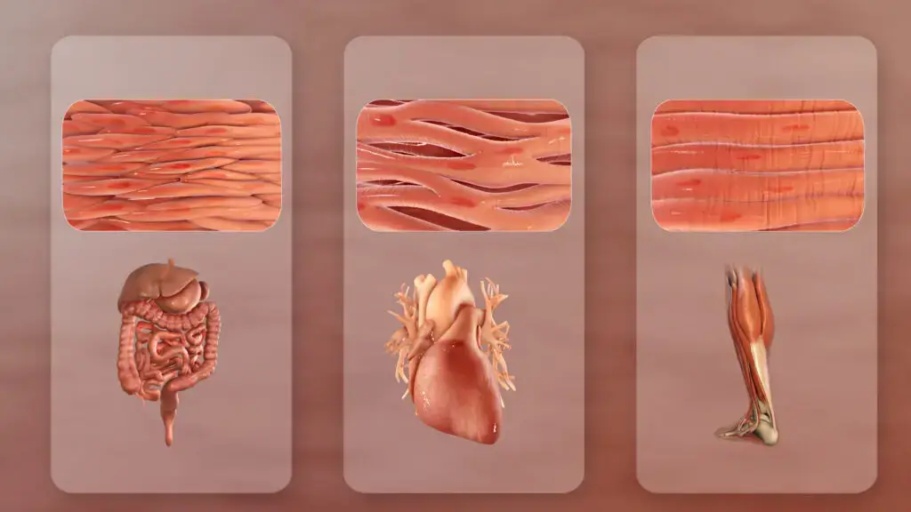
- Muscle tissue is categorized into three primary types in vertebrates: skeletal (or striated), smooth (non-striated), and cardiac. Skeletal muscle tissue, characterized by its elongated, multinucleate fibers, plays a pivotal role in bodily movements. This type of muscle operates under voluntary control, influenced by the central nervous system. In contrast, both smooth and cardiac muscles operate involuntarily. Their activation can arise from the central nervous system, peripheral plexus innervation, or endocrine stimulation. Notably, skeletal muscle can also be activated non-consciously through reflexes, although this still involves the central nervous system.
- Muscle tissue’s functionality and location in the body determine its specific characteristics. These tissues contain specialized contractile proteins, primarily actin and myosin, which interact to facilitate movement. Regulatory proteins, such as troponin and tropomyosin, are also present, playing crucial roles in muscle contraction. Furthermore, muscle tissues exhibit varied responses to neurotransmitters and hormones, including acetylcholine, noradrenaline, adrenaline, and nitric oxide, contingent on the muscle type and its precise location.
- Derived from the Latin term “musculus,” meaning “little mouse,” the word muscle alludes to the appearance or movement of certain muscles that resemble a scurrying mouse. In humans, muscles are instrumental in a myriad of functions, including locomotion, posture maintenance and alteration, blood circulation, and the movement of internal organs. For instance, the rhythmic contraction of the heart and the peristaltic movement in the digestive system are both facilitated by muscle action.
- The human body boasts over 600 muscles, accounting for approximately 40-50% of the total body weight. These muscles, intricately connected to bones, blood vessels, and internal organs, primarily consist of skeletal muscle tissue, tendons, and nerves. Every bodily movement is a consequence of muscle contraction, with muscles present in every organ, from blood vessels to the digestive tract. These muscles function by transporting substances throughout the body.
- Muscles rely heavily on the oxidation of fats and carbohydrates for energy, with the molecule adenosine triphosphate (ATP) serving as the primary energy reservoir. In summary, muscles, with their intricate structure and diverse functionality, are indispensable for the myriad movements and processes within the animal body.
Definition of Muscle
A muscle is a specialized tissue in animals that contracts to produce force and movement, composed of fibers containing proteins that enable contraction.
Structure of Muscle
- Muscles, integral components of the animal body, exhibit a sophisticated structural organization. At the core of this structure are muscle tissues, grouped together and encased within the epimysium, a robust connective tissue reminiscent of cartilage.
- Within the confines of the epimysium, bundles of nerve cells extend in elongated fibers termed fascicles. Each fascicle is safeguarded by the perimysium, a specialized protective layer that facilitates the supply of blood and nerves to the individual muscle fibers. Delving deeper into this hierarchical arrangement, every muscle fiber is enveloped by the endomysium, an additional protective sheath.
- The intricate layering and bundling within muscles enable distinct segments of a muscle to exhibit differential contractions. The protective sheaths, especially around the fascicles, ensure that adjacent bundles can glide smoothly over each other during contraction phases.
- The epimysium plays a pivotal role in connecting muscles to bones by extending to form tendons. These tendons, in turn, anchor to the periosteum, the connective tissue that encapsulates bones. This anchorage facilitates the movement of the skeletal framework upon muscle contraction.
- Furthermore, certain organs are enveloped by a unique muscle type, with the epimysium extending to other connective tissues. This arrangement exerts specific forces on these organs, orchestrating processes ranging from blood flow to the intricate mechanics of digestion.

Types of Muscle

Muscles, integral to the vertebrate anatomy, are categorized into three primary types based on their structural and functional attributes: skeletal, cardiac, and smooth muscle.
1. Skeletal Muscle:
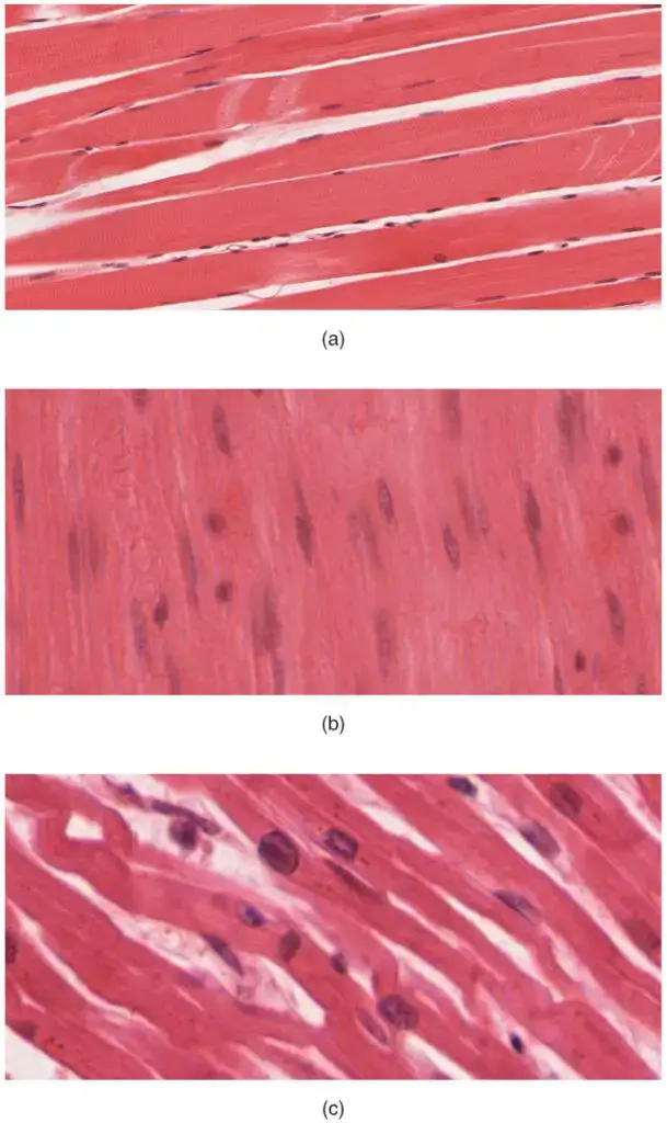
- Characteristics: Skeletal muscle, as observed under a microscope, is striated. This striation is due to the organized alignment of muscle cells, known as sarcomeres. These cells exhibit vertical bands, with alternating light and dark bands, resulting from variations in fibril composition and density. Notably, muscle-cell nuclei appear as distinct cigar-like dark patches adjacent to the myofibers.
- Structure: Comprising elongated, multinucleated muscle cells, skeletal muscles exhibit a cylindrical shape. These muscles are connected to bones via tendons, which are elastic collagen fibers. Each muscle ends in a tendon that links to the bone’s collagenous outer layer. Beneath the epimysium, muscle fibers group together to form fascicles, which are encased by another collagen protective layer.
- Classification: Skeletal muscle fibers are bifurcated into:
- Type I (slow-twitch or slow oxidative): Recognizable by its red hue, this muscle type is replete with mitochondria, myoglobin, and capillaries, enabling it to transport ample oxygen and support sustained aerobic activities.
- Type II (fast-twitch): This category further subdivides into:
- Type IIa: Aerobic in nature, it mirrors the characteristics of Type I but is faster.
- Type IIx (or IId): Characterized by reduced mitochondrial and myoglobin content, it represents the swiftest muscle type in humans, capable of rapid, forceful, but short-lived anaerobic contractions.
- Type IIb: An anaerobic, “white” muscle, it has even fewer mitochondria and myoglobin. In certain mammals like rodents, this type predominates, accounting for their flesh’s pale coloration.
- Type I (slow-twitch or slow oxidative): Recognizable by its red hue, this muscle type is replete with mitochondria, myoglobin, and capillaries, enabling it to transport ample oxygen and support sustained aerobic activities.
- Density: The density of skeletal muscle tissue in mammals approximates 1.06 kg/liter, making it about 15% denser than adipose tissue, which stands at 0.9196 kg/liter.
- Functions:
- Facilitating body movements such as writing, breathing, and extending the arm.
- Maintaining an erect body posture.
- Regulating body temperature.
- Protecting internal organs and providing support.
- Controlling entry and exit points in the body, as seen with sphincter muscles around the mouth, urinary tract, and anus.
2. Smooth Muscle:
- Characteristics: Smooth muscle is non-striated and operates involuntarily. It bifurcates into single-unit and multiunit types. In the single-unit variant, the entire muscle sheet contracts as one cohesive unit, while the multiunit type allows for nuanced control akin to skeletal muscle’s motor unit recruitment.
- Structure: Smooth muscle fibers are spindle-shaped, housing a single nucleus. They are shorter than skeletal muscles and lack specific proteins like actin and myosin. However, they produce their connective tissue.
- Functions:
- Regulating blood pressure and oxygen flow.
- Facilitating movement of fluids within organs.
- Contracting irises and raising arm hairs.
- Controlling orifice sealing and producing connective tissue proteins.
- Localization: Smooth muscle is pervasive, lining the walls of blood vessels, including arteries and veins. It’s also present in organs such as the urinary bladder, uterus, gastrointestinal tract, and respiratory system. Additionally, it’s found in the skin’s arrector pili, the eye’s ciliary muscle, and iris. While the fundamental structure and function remain consistent across organs, the stimuli inducing contractions vary to cater to specific physiological needs. Notably, the kidneys’ glomeruli house mesangial cells, resembling smooth muscle cells.
3. Cardiac Muscle:
- Characteristics:
Cardiac muscle, exclusive to the heart, particularly the myocardium, is striated and involuntary. Most cardiomyocytes, the cells constituting cardiac muscle, typically house a single nucleus, though some may contain multiple nuclei. - Structure: Cardiac muscles are specialized, designed for continuous contraction to circulate blood. They exhibit a regular fiber pattern, with cylindrical, branched fibers housing a central nucleus. The atrial muscle cells contain T-tubules rich in ion channels.
- Functions:
- Regulating heart function through rhythmic contraction and relaxation.
- Ensuring continuous blood circulation.
- Operating involuntarily, without conscious control.
- Nutrient Supply:
Distinct from other tissue types, cardiac muscle cells are reliant on a consistent supply of blood and electricity to procure essential nutrients and expel waste, a role fulfilled by the coronary arteries.

Based on Muscle Action:
- Voluntary Muscles: These muscles, primarily skeletal, are under conscious control and are responsible for body movements.
- Involuntary Muscles: Governed by the autonomic nervous system, these muscles, including cardiac and smooth muscles, function without conscious thought.
In summary, the human muscular system is intricate, with each muscle type playing a distinct role in ensuring the body’s optimal functioning. The classification into skeletal, cardiac, and smooth muscles, and further into voluntary and involuntary muscles, underscores the diversity and specialization of muscle tissues in the human body.
Comparison Between Skeletal Muscle, Smooth Muscle and Cardiac Muscle
Muscles play a pivotal role in the human body, facilitating movement, maintaining posture, and performing various other functions. The human body comprises three primary muscle types: smooth, cardiac, and skeletal muscles. Each type has distinct anatomical and physiological characteristics. Here’s a detailed comparison of these muscle types:
| Feature | Smooth Muscle | Cardiac Muscle | Skeletal Muscle |
|---|---|---|---|
| Anatomy | |||
| Neuromuscular Junction | Absent | Present | Present |
| Fibers | Fusiform and short (less than 0.4 mm) | Branched | Cylindrical and long (up to 15 cm) |
| Mitochondria | Numerous | Moderate in number | Varies from many to few, depending on the muscle type |
| Nuclei | Single | Single | Multiple |
| Sarcomeres | Absent | Present (maximum length 2.6 µm) | Present (maximum length 3.7 µm) |
| Syncytium | Absent (cells function independently) | Functionally present, though not structurally | Present |
| Sarcoplasmic Reticulum | Minimally developed | Moderately developed | Highly developed |
| ATPase | Minimal | Moderate | Abundant |
| Physiology | |||
| Self-regulation | Spontaneous slow action | Rapid spontaneous action | Requires nerve stimulus; no inherent action |
| Response to Stimulus | Unresponsive | “All-or-nothing” response | “All-or-nothing” response |
| Action Potential | Present | Present | Present |
| Workspace | Variable force/length curve | Increase in the force/length curve | Peak of the force/length curve |
Summary:
- Smooth Muscle: Found in the walls of hollow organs, these muscles are involuntary and can function without external stimuli. They have a unique fusiform shape and can generate slow, spontaneous actions.
- Cardiac Muscle: Located exclusively in the heart, these muscles are also involuntary but have a rapid spontaneous action. They possess a branched structure and play a crucial role in pumping blood throughout the body.
- Skeletal Muscle: These are the muscles attached to bones, facilitating voluntary movements. They require nerve stimuli to function and have a long, cylindrical shape.
Understanding the differences between these muscle types is crucial for comprehending the diverse roles they play in maintaining the body’s functionality and overall health.
Development of Muscle
Muscle tissue, a vital component of the human body, originates from the paraxial mesoderm during embryonic development. This mesoderm undergoes segmentation, aligning with the body’s inherent segmentation, most evident in the vertebral column. Let’s delve deeper into the intricate process of muscle formation.
1. Origin and Segmentation: The paraxial mesoderm segments into structures known as somites along the length of the embryo. Each somite is compartmentalized into three distinct sections:
- Sclerotome: Gives rise to the vertebrae.
- Dermatome: Differentiates into the skin.
- Myotome: The precursor for muscle tissue.
Within the myotome, further subdivisions occur, leading to the formation of the epimere and hypomere. These respectively give rise to the epaxial and hypaxial muscles.
2. Epaxial and Hypaxial Muscles: The epaxial muscles in humans are limited to the erector spinae and minor intervertebral muscles. These muscles receive innervation from the dorsal rami of the spinal nerves. In contrast, the hypaxial muscles, which encompass the limb muscles and others, are innervated by the ventral rami of the spinal nerves.
3. Myoblast Migration and Formation: Myoblasts, the precursor cells for muscles, play a pivotal role in muscle development. These cells either remain within the somite, contributing to muscles linked with the vertebral column, or they embark on a migration journey to other parts of the body to form the remaining muscles. This migration is not random; it is orchestrated by chemical signals that guide myoblasts to their destined locations.
Prior to the migration, connective tissue frameworks emerge, typically originating from the somatic lateral plate mesoderm. These frameworks pave the way for myoblasts. Once the myoblasts reach their designated locations, they undergo fusion, resulting in the formation of elongated skeletal muscle cells.
In conclusion, the development of muscle tissue is a meticulously coordinated process, commencing from the paraxial mesoderm and culminating in the formation of diverse muscle types throughout the body. This intricate developmental journey underscores the complexity and precision inherent in human embryology.
Function of Muscle
Muscle tissue, integral to the human body, primarily functions through the mechanism of contraction. This contraction is facilitated by the coordinated movement of actin and myosin proteins. While the three distinct types of muscle tissues—skeletal, cardiac, and smooth—exhibit varied characteristics, they all share this foundational mechanism.
1. Skeletal Muscle Functions: Skeletal muscle contraction is initiated by electrical impulses relayed by motor nerves. The neurotransmitter acetylcholine plays a pivotal role in facilitating many skeletal muscle contractions. Key functions include:
- Responding to motor nerve stimuli.
- Facilitating voluntary movements, such as walking or lifting.
- Generating force and enabling body movement.
2. Smooth Muscle Functions: Smooth muscle tissue is pervasive throughout the body, prominently in hollow organs like the stomach and bladder, tubular structures like blood vessels, and in the ducts of exocrine glands. Its functions encompass:
- Facilitating the movement of substances, such as food in the digestive tract, through peristaltic contractions.
- Regulating the diameter of blood vessels, thereby controlling blood flow and pressure.
- Sealing orifices, for instance, the pylorus in the stomach or the uterine os.
- Operating involuntarily, meaning its contractions occur without conscious thought.
- Exhibiting slower contractions compared to skeletal muscles, but these contractions are more sustained, potent, and energy-efficient.
3. Cardiac Muscle Functions: Cardiac muscle, exclusive to the heart, possesses unique attributes tailored to its critical role in circulation. Its functions include:
- Contracting autonomously, regulated by the body’s autonomic nervous system.
- Ensuring continuous, rhythmic contractions to maintain blood circulation throughout the body.
- Operating involuntarily, persistently contracting without external stimuli.
- Demonstrating resilience and endurance, given its non-stop operation throughout an organism’s lifespan.
In summation, while the foundational function of muscle tissue is contraction, the specific roles and characteristics of skeletal, smooth, and cardiac muscles vary considerably. Each type is intricately designed to fulfill its specific functions, ensuring the harmonious operation of the human body.
Quiz
Which type of muscle is responsible for the rhythmic contractions of the heart?
a) Skeletal muscle
b) Smooth muscle
c) Cardiac muscle
d) Voluntary muscle
[expand title=”Show answer” swaptitle=”Hide answer”] c) Cardiac muscle [/expand]
Which muscle type is involuntary and found in the walls of hollow organs?
a) Skeletal muscle
b) Smooth muscle
c) Cardiac muscle
d) Striated muscle
[expand title=”Show answer” swaptitle=”Hide answer”] b) Smooth muscle [/expand]
Which of the following muscles are under the control of the central nervous system and are also termed voluntary muscles?
a) Cardiac muscles
b) Smooth muscles
c) Skeletal muscles
d) Involuntary muscles
[expand title=”Show answer” swaptitle=”Hide answer”] c) Skeletal muscles [/expand]
The neurotransmitter that facilitates the contraction of skeletal muscles is:
a) Dopamine
b) Serotonin
c) Acetylcholine
d) Norepinephrine
[expand title=”Show answer” swaptitle=”Hide answer”] c) Acetylcholine [/expand]
Which muscle type is characterized by having intercalated discs?
a) Skeletal muscle
b) Smooth muscle
c) Cardiac muscle
d) Voluntary muscle
[expand title=”Show answer” swaptitle=”Hide answer”] c) Cardiac muscle [/expand]
The primary function of muscle tissue is:
a) Secretion
b) Absorption
c) Contraction
d) Insulation
[expand title=”Show answer” swaptitle=”Hide answer”] c) Contraction [/expand]
Which muscle type is spindle-shaped with a single nucleus?
a) Skeletal muscle
b) Smooth muscle
c) Cardiac muscle
d) Striated muscle
[expand title=”Show answer” swaptitle=”Hide answer”] b) Smooth muscle [/expand]
The muscles that are attached to bones and aid in body movement are:
a) Cardiac muscles
b) Smooth muscles
c) Skeletal muscles
d) Involuntary muscles
[expand title=”Show answer” swaptitle=”Hide answer”] c) Skeletal muscles [/expand]
Which of the following is NOT a function of skeletal muscles?
a) Pumping blood
b) Maintaining body posture
c) Facilitating breathing
d) Assisting in body movement
[expand title=”Show answer” swaptitle=”Hide answer”] a) Pumping blood [/expand]
All muscles are derived from which embryonic structure?
a) Ectoderm
b) Endoderm
c) Mesoderm
d) Neural crest
[expand title=”Show answer” swaptitle=”Hide answer”] c) Mesoderm [/expand]
FAQ
What are the main types of muscles in the human body?
The human body consists of three primary muscle types: skeletal, smooth, and cardiac muscles.
How do muscles work?
Muscles work by contracting and relaxing. They receive signals from the nervous system, which causes muscle fibers to shorten (contract) or lengthen (relax), producing movement.
What is the difference between voluntary and involuntary muscles?
Voluntary muscles, like skeletal muscles, are under conscious control and are used for activities such as walking or lifting. Involuntary muscles, like cardiac and smooth muscles, operate without conscious effort, performing functions like heartbeats or digestion.
Why do muscles get sore after exercise?
Muscle soreness after exercise, often referred to as delayed onset muscle soreness (DOMS), is caused by microscopic damage to muscle fibers during intense physical activity, leading to inflammation and pain.
How can I build muscle strength?
Building muscle strength typically involves resistance training, adequate nutrition, proper rest, and consistency in workouts.
What is muscle fatigue?
Muscle fatigue occurs when a muscle cannot exert force consistently and experiences a reduced ability to perform over time, often due to prolonged physical activity or overuse.
How do cardiac muscles differ from skeletal muscles?
Cardiac muscles are found only in the heart and are responsible for pumping blood. They are involuntary and have a unique structure with intercalated discs. Skeletal muscles are attached to bones and are responsible for body movement; they are voluntary.
What is the role of smooth muscles?
Smooth muscles are found in the walls of hollow organs and vessels. They help in various functions like moving food through the digestive system, constricting blood vessels, and controlling the pupil’s size in the eye.
How do muscles grow?
Muscles grow through a process called hypertrophy. When muscles experience microscopic damage from intense physical activity, the body repairs and rebuilds the muscle fibers, making them larger and stronger.
Can muscles turn into fat if I stop exercising?
No, muscles and fat are two distinct tissues. If you stop exercising, muscles may shrink or atrophy, and you might gain fat due to decreased physical activity, but muscles do not turn into fat.
References
- Nelson, D. L., & Cox, M. M. (2008). Principles of Biochemistry. New York: W.H. Freeman and Company.
- Lodish, H., Berk, A., Kaiser, C. A., Krieger, M., Scott, M. P., Bretscher, A., . . . Matsudaira, P. (2008). Molecular Cell Biology 6th. ed. New York: W.H. Freeman and Company.