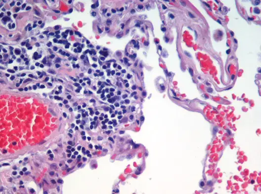
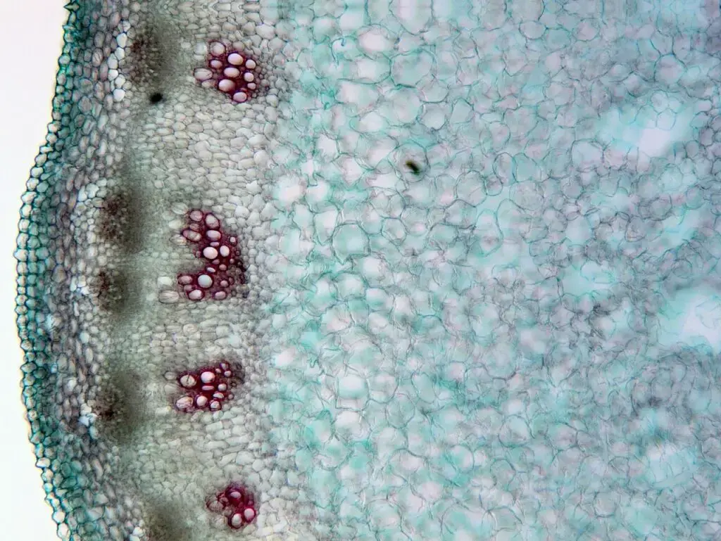
What is Histology?
- Histology, often referred to as microscopic anatomy or microanatomy, delves into the intricate realm of the microscopic structures of biological tissues. This scientific discipline stands as the microscopic counterpart to gross anatomy, which focuses on larger, macroscopic structures discernible to the naked eye. While one might be tempted to segregate microscopic anatomy into distinct subfields such as organology (study of organs), cytology (study of cells), and histology (study of tissues), contemporary scientific parlance amalgamates all these under the umbrella of histology.
- Every cellular and tissue type possesses distinct characteristics, intricately linked to the myriad functions executed by an organism.
- Advanced imaging modalities, ranging from conventional light microscopy to more specialized systems like electron microscopy, are employed to elucidate the minuscule structures housed within specially curated tissue samples.
- Such detailed histological analyses not only facilitate the identification of previously unknown tissues but also offer insights into the potential functions of specific tissues or cells. Moreover, they can be pivotal in detecting pathological changes within cellular structures, thereby aiding in disease identification.
- In the medical domain, histopathology emerges as a specialized subset of histology, concentrating on the microscopic delineation and study of diseased tissues. Furthermore, in the realm of paleontology, the term “paleohistology” is coined to describe the study of tissue structures in fossilized organisms.
- In essence, histology serves as a critical bridge, connecting the macroscopic world of anatomy with the microscopic intricacies of cellular and tissue structures, thereby enriching our understanding of biological systems and their functions.

Definition of Histology
Histology is the scientific study of the microscopic structure of biological tissues.
Sample preparation Methods in Histology
Histological analysis necessitates meticulous sample preparation to ensure accurate microscopic observation. The preparation techniques vary based on the specimen type and the intended observation method. Herein, we delineate the primary steps involved in histological sample preparation.
- Fixation: The foremost step, fixation, employs chemical fixatives to preserve cellular and tissue structures. These fixatives not only maintain tissue integrity but also facilitate the slicing of thin tissue sections for microscopy. The predominant fixative for light microscopy is 10% neutral buffered formalin (NBF). Electron microscopy, on the other hand, often utilizes glutaraldehyde or other agents like osmium tetroxide. These aldehyde fixatives primarily function by cross-linking proteins, ensuring structural preservation but potentially compromising protein functionality.
- Selection and Trimming: This involves choosing relevant tissue sections and subsequently trimming them to expose pertinent surfaces for sectioning. The trimmed samples are then sized appropriately for embedding.
- Embedding: To facilitate sectioning, tissues are embedded in a supportive medium. Prior to embedding, tissues undergo dehydration, where water is replaced either directly by the embedding medium or via an intermediary fluid. Paraffin wax is the predominant embedding material for light microscopy. However, for specific requirements, alternatives like epoxy, acrylic, and other resins might be employed. For frozen sectioning, water-based embedding mediums are used.
- Sectioning: In this phase, thin tissue sections are cut using a microtome for light microscopy or an ultramicrotome for electron microscopy.
- Staining: Given the inherent lack of contrast in biological tissues, staining is imperative. It not only provides contrast but also accentuates specific tissue features. Hematoxylin and eosin (H&E) are commonly used for general tissue structure visualization. However, myriad specialized staining techniques exist for highlighting specific cellular components or substances.
- Historadiography: This technique involves X-raying a slide, often stained histochemically. Autoradiography, a subset, visualizes locations to which a radioactive substance has been transported within the body.
- Immunohistochemistry: This recent advancement employs antibodies to visualize specific proteins, carbohydrates, and lipids in tissues. When combined with fluorescent molecules, it’s termed immunofluorescence. This technique has significantly enhanced cellular categorization under microscopy.
- Electron Microscopy: For this, heavy metals are typically employed to stain tissue sections, with uranyl acetate and lead citrate being common choices.
- Specialized Techniques:
- Cryosectioning: A rapid method to freeze, cut, and mount tissue sections. It’s particularly useful for enzyme localization studies and for rapid tumor margin identification during surgeries.
- Ultramicrotomy: This technique prepares ultra-thin sections for transmission electron microscope (TEM) analysis, typically using epoxy or other resins.
- Artifacts: These are unwanted structural features in tissues that can obscure normal histological examination. They can arise from various sources, including fixative-induced pigments, tissue shrinkage, or color changes.
In summation, histological sample preparation is a multifaceted process, demanding precision at each step to ensure that the microscopic anatomy of tissues is accurately represented and analyzed.

Basic Procedures in Histology
Histology, the study of the microscopic structure of tissues, is a cornerstone in the realm of scientific research. While the scope of histology is vast, researchers typically specialize in the histological aspects of their specific organisms of interest. For instance, while a botanist may not be well-versed in human renal histology, they would be adept at understanding plant tissue histology. The methodologies and techniques of histology have found applications across diverse scientific domains due to their pivotal role in tissue preparation and visualization.
- Light Microscopy Observations: The journey of histology began with rudimentary observations using light microscopy. Over time, this evolved into a plethora of techniques and procedures tailored for specific cellular observations.
- Staining Procedures: One of the foundational techniques in histology is staining. A basic staining procedure involves the application of specialized dyes to cells mounted on a slide. These dyes are designed to selectively bind to specific cellular components, such as DNA. Post staining, the unbound dye is rinsed off, leaving behind a stained component that enhances visualization under the microscope. It was through such staining techniques that cellular processes like mitosis were first elucidated.
- Sectioning: To delve deeper into the cellular architecture, sectioning is employed. This technique necessitates the encapsulation of a cell or tissue in a solid matrix, facilitating the slicing of thin sections. While rudimentary sectioning can be achieved with sharp blades on simple specimens like onion cells, intricate cellular structures demand more sophisticated approaches. Techniques have been developed wherein the cellular cytoplasm is substituted with substances like plastic epoxy. Once solidified, precise sections can be obtained without compromising the cellular integrity. Alternatively, rapid freezing followed by fracturing can also unveil the cell’s internal milieu without causing damage.
- Freeze Fracture Technique: Paired with advanced imaging modalities like electron microscopy, the freeze fracture technique has revolutionized histological observations. This method has unveiled intricate cellular details, such as the elaborate protein configurations within cell membranes.
- Applications of Histology: The versatility of histological techniques has made them indispensable across various scientific sectors. In agriculture, plant histology aids in early detection of nutrient deficiencies and monitors water consumption. The medical realm leverages histology for disease diagnosis and therapeutic interventions. Furthermore, biologists employ histological techniques to understand organismal interactions and devise strategies against pests.
In essence, histology, with its myriad of techniques, has been instrumental in answering foundational questions across diverse scientific disciplines. Whether it’s understanding cellular dynamics, diagnosing diseases, or exploring organismal interactions, histology remains at the forefront of scientific inquiry.
Tissue Preparation
Tissue preparation is a meticulous process, pivotal for ensuring the accurate microscopic examination of biological samples. This process encompasses several stages, each tailored to preserve, enhance, and reveal the intricate details of tissue samples. In contemporary histology laboratories, automation has streamlined many of these steps. Here’s a detailed breakdown of the stages involved:
- Fixation: This is the foundational step in tissue preparation. Fixation employs chemicals to preserve the tissue’s structural integrity and prevent its degradation. By irreversibly cross-linking proteins, fixation ensures the tissue retains its natural form. While various specialized fixatives exist, Neutral Buffered Formalin is frequently employed. The significance of fixation extends beyond mere preservation; it hardens the tissue, facilitating subsequent sectioning. Other fixatives, like Paraffin-formalin, are preferred for immunostaining, albeit necessitating immediate preparation. Bouin’s solution, renowned for preserving embryonic and brain tissues, is less favorable for kidney tissues and can alter mitochondrial structures.
- Dehydration: Post fixation, the tissue undergoes dehydration, primarily achieved using ethanol. This step expels water from the tissue, further solidifying it for microscopic examination. Subsequent to ethanol application, xylene is introduced to eliminate the ethanol residues.
- Embedding: At this juncture, the tissue sample is encased in a medium, typically paraffin wax or a plastic resin. This encapsulation accentuates the visualization of cellular structures. However, caution is imperative during embedding, especially if immunostaining is the end goal. Paraffin wax can hinder antibody penetration, potentially skewing results.
- Sectioning: With the specimen embedded, it’s mounted onto a microtome for sectioning. Precision is key, with the ideal section thickness being 4-5 micrometers. This ensures optimal staining and subsequent microscopic examination.
- Antigen Retrieval: This step is pivotal for unmasking antigens that might have been obscured during fixation and embedding. Cross-linking of proteins during earlier stages can conceal antigenic sites, potentially diminishing the efficacy of immunohistochemical reactions. Antigen retrieval employs heat and proteolytic techniques to dismantle these cross-links, unveiling previously concealed epitopes and antigens. While this step carries the inherent risk of denaturing both the fixative and the antigens, adept antigen retrieval can significantly amplify immunostaining intensity.
In summation, tissue preparation in histology is a series of orchestrated steps, each designed to optimize the microscopic visualization of biological samples. Mastery over these stages ensures that the intricate details of tissues are accurately represented and analyzed under the microscope.
Careers in Histology
Histology, the study of the microscopic structure of tissues, offers a range of career opportunities for those interested in the cellular and molecular aspects of biology and medicine. Professionals in this field play a crucial role in disease diagnosis, research, and understanding the fundamental processes of life at the cellular level. Here are some of the prominent careers in histology:
- Histotechnologist/Histotechnician: These professionals prepare tissue samples for examination by pathologists. Their responsibilities include fixing, staining, and sectioning tissues to produce high-quality slides that can be analyzed under a microscope.
- Pathologist: A medical doctor specializing in the diagnosis of diseases by examining tissues, cells, and bodily fluids. They often collaborate with histotechnologists to interpret the microscopic images of tissue samples.
- Cytopathologist: A subspecialty of pathology, cytopathologists focus on the diagnosis of diseases at the cellular level, often using fine needle aspirations or other methods to obtain cells.
- Histology Supervisor/Manager: Individuals in this role oversee the operations of histology laboratories, ensuring quality control, managing staff, and ensuring compliance with regulations.
- Research Scientist: Many scientists in academia and industry use histological techniques to study diseases, develop new treatments, or delve into basic biological processes.
- Sales Representative for Laboratory Equipment: Those with a background in histology can work for companies that manufacture and sell histological equipment and supplies, leveraging their expertise to inform potential customers about the products.
- Histology Educator: With a deep understanding of histological techniques and processes, some professionals choose to teach in academic institutions, training the next generation of histotechnologists and researchers.
- Quality Control Specialist: In this role, professionals ensure that histology labs maintain high standards, produce accurate results, and comply with industry regulations.
- Molecular Biologist: Many molecular biologists employ histological techniques to study gene expression and cellular processes at the microscopic level.
- Forensic Histologist: In forensic labs, histologists help in determining causes of death or contributing factors by examining tissue samples.
- Veterinary Histologist: Similar to human medicine, veterinary medicine also requires histological analysis to diagnose diseases in animals.
- Technical Specialist: These professionals provide technical support in histology labs, ensuring that equipment operates correctly and assisting with troubleshooting.
- Histology Consultant: With vast experience, some histologists offer consultancy services to labs, helping them set up, improve efficiency, or maintain quality standards.
- Immunohistochemist: Specializing in immunohistochemistry, these professionals study the distribution and localization of biomolecules in tissue sections using labeled antibodies.
The field of histology offers diverse career opportunities, from hands-on laboratory work to research, teaching, and management roles. As medical and biological research continues to evolve, the demand for skilled histology professionals remains robust, ensuring a promising career trajectory for those in the field.
Histochemistry and Cytochemistry
Histochemistry and cytochemistry are specialized fields within histology that focus on the chemical composition of cells and tissues. Through various staining techniques, these disciplines allow for the visualization and differentiation of cellular and extracellular components. Here’s an overview of some of the prominent staining techniques:
- Hematoxylin and Eosin (H&E): A foundational staining method, H&E employs two dyes. Hematoxylin, a basic dye, targets acidic structures, rendering them blue or purple. Such structures are termed Basophilic and include components like DNA and RNA. Eosin, on the other hand, is an acidic dye staining basic structures pink or red, with cytoplasm being a prime example.
- Gram Stain: Specifically designed for bacterial differentiation, this sequential staining technique discerns bacterial species based on their cell wall composition. The process involves primary staining with crystal violet, followed by secondary staining, decolorization, and counterstaining. The outcome reveals gram-positive bacteria in purple and gram-negative bacteria in pink.
- Giemsa Stain: Predominantly used in hematology, this stain excels in highlighting bone marrow, plasma cells, and mast cells. Additionally, it’s instrumental in detecting blood parasites and visualizing chromosomal abnormalities.
- Periodic Acid Schiff (PAS) Reaction: This technique is tailored to visualize structures rich in carbohydrates. Structures like the intestinal brush border and renal tubular cells are stained red or magenta, offering insights into their composition and functionality.
- Masson’s Trichrome: A multicolored staining method, it’s renowned for its ability to stain collagen fibers blue, aiding in the identification of various fibrotic conditions.
- Congo Red: This dye is pivotal for detecting amyloid fibers, with its characteristic red and orange hues. Under polarized light, amyloid-rich tissues exhibit a distinctive “apple” green birefringence.
- Prussian Blue: Employed to detect iron stores, this stain reveals bright blue pigments, assisting in diagnosing conditions like hemochromatosis or hemosiderosis.
- Mucicarmine: Specializing in staining mucin, this technique is invaluable in identifying carcinomas and inflammatory conditions marked by excessive mucin production.
- Sudan Black & Oil Red O: Both these dyes are adept at staining lipid-rich structures, providing insights into conditions like atherosclerosis and lipid accumulation in various tissues.
- Silver Stain: A broad category, silver stains are employed predominantly in neurology to study accumulation-based diseases. The staining intensity and color vary based on the size and amount of accumulated substances.
- Nissl Stain: Employing Cresyl Violet, this stain is tailored for studying neuronal structures, particularly in the brain and spinal cord.
- Papanicolaou Stain: Commonly known as the Pap smear, this cytological staining technique is pivotal for detecting cervical cancer. It employs a combination of dyes to stain various cellular components, aiding in the identification of precancerous and cancerous processes.
In essence, histochemistry and cytochemistry provide a window into the intricate world of cells and tissues. Through a myriad of staining techniques, they illuminate the chemical composition and structural nuances, enabling accurate diagnosis and fostering a deeper understanding of biological processes.
Importance of Histology
Histology, the study of the microscopic anatomy of tissues and cells, plays an indispensable role in the realm of biological sciences and medicine. Its importance can be understood through the following facets:
- Foundation of Pathology: Histology provides the groundwork for pathology, the study of disease. By examining tissue samples, pathologists can diagnose diseases, determine their severity, and guide treatment decisions. For instance, cancer biopsies are analyzed histologically to determine the type, grade, and stage of the tumor.
- Understanding Physiology: Histology offers insights into the normal structure of tissues and organs, which is essential for understanding their function. For instance, the histological study of the alveoli in the lungs provides insights into the mechanism of gas exchange.
- Developmental Biology: Histological techniques allow researchers to track the development of tissues and organs during embryogenesis, shedding light on congenital anomalies and the processes of differentiation and morphogenesis.
- Pharmacological Research: In drug development, histology is used to study the effects of potential new drugs on tissues, helping to determine their therapeutic efficacy and potential side effects.
- Comparative Anatomy: Histology aids in comparing the microscopic structures of tissues and organs across different species, providing insights into evolutionary biology and the functional adaptations of different organisms.
- Forensic Investigations: Histological examinations can be pivotal in forensic science, helping determine the cause of death or the nature of injuries.
- Innovative Techniques: Advanced histological techniques, such as immunohistochemistry, allow for the localization of specific proteins or other molecules in tissues, furthering our understanding of cellular functions and disease mechanisms.
- Educational Tool: Histology is a fundamental component of medical and biological education, helping students understand the intricate relationships between structure and function at the microscopic level.
- Basis for Advanced Imaging: Understanding the basic histological structure of tissues is crucial for interpreting results from advanced imaging techniques like MRI, CT scans, and PET scans.
- Research and Discovery: Histology plays a pivotal role in biomedical research, aiding in the discovery of new knowledge about health and disease. It helps validate findings from molecular and genetic studies by placing them in the context of whole tissues and organs.
In essence, histology serves as a bridge between the macroscopic world that we can observe with the naked eye and the microscopic world that remains hidden. By revealing the intricate details of tissues and cells, histology provides invaluable insights into health, disease, and the very essence of life.
Quiz
What is the primary purpose of fixation in histology?
a) To stain the tissue
b) To magnify the tissue
c) To preserve and maintain the structure of tissues and cells
d) To make the tissue transparent
Which stain is commonly used to visualize DNA in cell nuclei?
a) Eosin
b) Hematoxylin
c) Congo Red
d) Sudan Black
The study of tissues at a microscopic level is termed:
a) Cytology
b) Pathology
c) Histology
d) Physiology
Which of the following is NOT a basic step in tissue preparation for histological study?
a) Embedding
b) Sectioning
c) Fixation
d) Amplification
Which stain is specifically used for detecting fungal infections?
a) Giemsa
b) Mucicarmine
c) PAS (Periodic Acid Schiff)
d) Prussian Blue
Which technique is used to differentiate bacterial species based on the composition of their cell walls?
a) Nissl Stain
b) Gram Stain
c) Congo Red
d) Silver Stain
Which structure would be stained by Sudan Black or Oil Red O?
a) DNA
b) Lipids
c) Proteins
d) Carbohydrates
In which condition is the Prussian Blue stain primarily used?
a) Detection of lipids
b) Detection of iron stores
c) Detection of carbohydrates
d) Detection of proteins
What is the primary target of the Eosin stain?
a) Cell nuclei
b) Lipids
c) Cytoplasm
d) Cell wall
Which of the following is NOT a characteristic of histological studies?
a) Examination of tissues at a macroscopic level
b) Use of stains to enhance tissue contrast
c) Examination of tissue sections under a microscope
d) Analysis of cellular structure and organization
FAQ
What is histology?
Histology is the study of the microscopic structure of tissues.
Why is histology important in medical science?
Histology provides insight into the structure and function of organs, allowing for the diagnosis and understanding of diseases at the cellular level.
What is the difference between histology and cytology?
While histology studies the microscopic structure of tissues, cytology focuses on individual cells.
How are tissues prepared for histological examination?
Tissues undergo a series of steps including fixation, dehydration, embedding, sectioning, and staining before examination under a microscope.
What is the purpose of staining in histology?
Staining enhances the contrast of the tissue, allowing for the differentiation of various cellular structures and components.
What is the difference between a histopathologist and a histotechnologist?
A histopathologist is a medical doctor who interprets and diagnoses the changes caused by disease in tissues, while a histotechnologist prepares tissue samples for examination.
Why is fixation crucial in histology?
Fixation preserves the structural integrity and biochemical composition of tissues, preventing decomposition and autolysis.
What is immunohistochemistry?
Immunohistochemistry is a technique that uses antibodies to detect specific proteins, antigens, or molecules within tissue sections.
How does electron microscopy differ from light microscopy in histology?
Electron microscopy uses a beam of electrons to visualize structures at a much higher resolution than light microscopy, allowing for the observation of sub-cellular details.
What is the significance of the Gram stain in histology?
The Gram stain differentiates bacterial species based on the composition of their cell walls, aiding in the diagnosis and treatment of infections.
References
- Brusca, R. C., & Brusca, G. J. (2003). Invertebrates. Sunderland, MA: Sinauer Associates, Inc.
- McMahon, M. J., Kofranek, A. M., & Rubatzky, V. E. (2011). Plant Science: Growth, Development, and Utilization of Cultivated Plants (5th ed.). Boston: Prentince Hall.
- National Society for Histology. (2018, 1 22). Schools & Programs of Histotechnology. Retrieved from NSH.org: https://nsh.org/content/schools
- Gurina TS, Simms L. Histology, Staining. [Updated 2023 May 1]. In: StatPearls [Internet]. Treasure Island (FL): StatPearls Publishing; 2023 Jan-. Available from: https://www.ncbi.nlm.nih.gov/books/NBK557663/
- Text Highlighting: Select any text in the post content to highlight it
- Text Annotation: Select text and add comments with annotations
- Comment Management: Edit or delete your own comments
- Highlight Management: Remove your own highlights
How to use: Simply select any text in the post content above, and you'll see annotation options. Login here or create an account to get started.