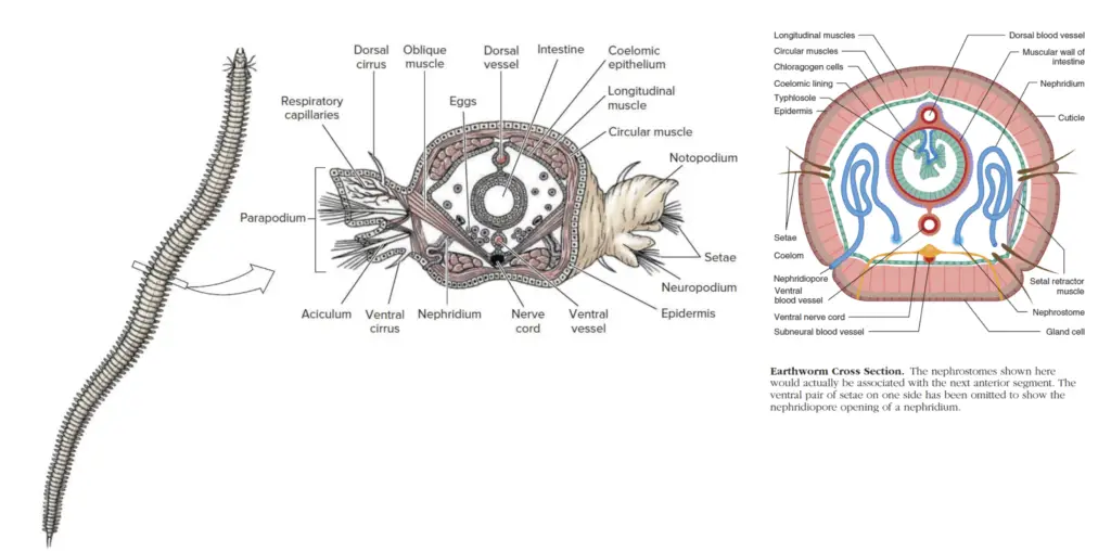
Excretion in Annelida
- Excretion in annelids is a crucial process for maintaining homeostasis and removing nitrogenous waste produced during metabolism. The excretory system in these organisms primarily functions through specialized structures called nephridia. These minute, coiled tubes are segmentally arranged along the length of the body, with each segment possessing its own pair of nephridia.
- Nephridia are internally lined with cilia and open into the coelom through an opening known as the nephrostome. The nephrostome acts as a funnel, drawing in the coelomic fluid, which contains metabolic waste. The cells lining the nephridia perform ultrafiltration, a process where waste products are separated from the haemocoelomic fluid. This filtration process helps eliminate excess nitrogenous compounds, such as ammonia or urea, depending on the habitat of the species.
- The nephridia not only remove nitrogenous waste but also regulate the osmotic pressure of the organism by managing the excess water within the body. After the filtration, the excretory fluid passes through a central canal, where further processing may occur, and is eventually collected in a terminal vesicle. From there, the waste is expelled to the external environment through an opening called the nephridiopore.
- The type of nitrogenous waste excreted by annelids varies based on their environment. Aquatic species typically excrete ammonia, a compound that is diluted in the surrounding water. In contrast, terrestrial species produce urea, a more energy-intensive compound that is less toxic and can be retained longer in the body, reducing water loss. Therefore, the excretory system of annelids is adapted to their environmental conditions, allowing them to efficiently remove waste and regulate bodily fluids.
What is nephridia?
Nephridia are specialized excretory structures found in many invertebrates, including annelids, that play a crucial role in eliminating metabolic wastes and maintaining osmotic balance. These tubular organs function similarly to kidneys in higher animals, facilitating the removal of waste products and regulating internal fluid composition. In annelids, such as earthworms, nephridia are the primary organs responsible for these functions. Below is a detailed explanation of nephridia and their various forms.
- Definition of Nephridia:
- Nephridia are excretory tubules that open to the exterior through a pore known as the nephridiopore. The inner end of the tubule is either closed and associated with terminal cells (as in protonephridia) or opens into the coelom through a ciliated funnel called the nephrostome (as in metanephridia).
- Types of Nephridia:
- Protonephridia:
- In this type, the nephridium opens internally to the coelom but lacks a nephrostome. Instead, it is connected to terminal cells or solenocytes that aid in the movement and filtration of waste through the tubule.
- Metanephridia:
- This more advanced type has a nephrostome, a ciliated funnel that opens into the coelom. The nephrostome facilitates the collection of waste from the body cavity and channels it into the excretory tubule.
- Protonephridia:
- Nephridia in Earthworms:
- Earthworms, like many annelids, are both ammontelic (excreting ammonia) and ureotelic (excreting urea), depending on their environmental conditions. Nephridia play a central role in these processes by filtering nitrogenous wastes and helping to maintain water balance.
- Structure and Function:
- Earthworm nephridia are slender, coiled tubules that serve dual purposes—excretion of waste and osmoregulation (control of water balance within the body).
- Types of Nephridia in Earthworms:
- Nephridia are found in all segments of the earthworm’s body except the first two segments. There are three primary types of nephridia based on their location:
- Septal Nephridia:
- Located in the septa between segments, these nephridia are involved in filtering waste from the coelomic fluid. They are enteronephric, meaning they release nitrogenous waste into the alimentary canal, from where it is expelled through the gut.
- Pharyngeal Nephridia:
- These are found near the pharyngeal region and are responsible for excreting nitrogenous waste products directly into the digestive tract.
- Integumentary Nephridia:
- Located in the skin (integument), these are exonephric nephridia that directly discharge waste products to the exterior of the body. They play a crucial role in the removal of nitrogenous wastes like ammonia from the earthworm.
- Septal Nephridia:
- Nephridia are found in all segments of the earthworm’s body except the first two segments. There are three primary types of nephridia based on their location:

Structure of nephridia
The structure of nephridia in annelids, such as earthworms, represents a highly specialized system for excretion and osmoregulation. These tubular organs consist of multiple components working together to filter waste products and regulate the organism’s internal fluid balance. Nephridia vary in their complexity and location within different segments of the body, but their basic structural elements are consistent across the system. The following is a detailed description of the key structural components and the types of nephridia found in annelids.
- General Structure of Nephridia:
- A typical nephridium consists of several integral parts:
- Nephrostome: This is a funnel-like, ciliated opening that hangs into the coelom. It functions as the site of waste collection, drawing excretory matter from the coelomic fluid and surrounding blood.
- Nephridial Duct: The nephrostome leads into a tubular structure known as the nephridial duct, which can vary in length and configuration, depending on the type of nephridium. It is ciliated internally to facilitate the movement of waste material through the tube.
- Nephridiopore: The nephridial duct ultimately opens to the exterior through an opening called the nephridiopore, allowing the waste to exit the body.
- A typical nephridium consists of several integral parts:
- Septal Nephridia:
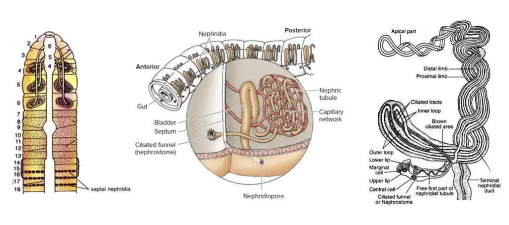
- Septal nephridia are a well-organized type of nephridium, also referred to as holonephridia, and are found attached to the septa between segments, beginning behind the fifteenth segment in earthworms.
- Each septum typically bears 40 to 50 septal nephridia, resulting in 80 to 100 septal nephridia per segment.
- The structure of septal nephridia can be broken down into four primary components:
- Nephrostome: A fat, funnel-like structure that is ciliated and responsible for collecting excretory matter from both coelomic fluid and blood.
- Neck: A short, narrow, ciliated tubule that connects the nephrostome to the body of the nephridium, allowing for waste transport.
- Body: The body of the nephridium is divided into two lobes. One lobe is small and straight, while the other is long and twisted. The twisted lobe consists of a proximal and a distal limb, each of which is coiled around each other 9 to 13 times.
- Terminal Duct: The terminal duct is short, narrow, and ciliated. It serves as the final section of the nephridium, where waste is transported before being expelled through the nephridiopore.
- Function of Septal Nephridia:
- The terminal ducts of the septal nephridia from each segment connect to loop-like excretory canals. These canals form a pair in each segment and empty into a pair of supra-intestinal excretory ducts, which run along the dorsal surface from segment 15 to the posterior end.
- The supra-intestinal ducts ultimately discharge nitrogenous waste into the intestine, which then eliminates the waste through the digestive system. Therefore, septal nephridia are classified as enteronephric, as they release waste into the alimentary canal.
- Pharyngeal Nephridia:
- These nephridia are located in segments 4, 5, and 6 and are as large as the septal nephridia.
- Unlike septal nephridia, pharyngeal nephridia lack a nephrostome and a neck. The terminal ducts of all pharyngeal nephridia unite to form a common duct. This common duct has three pairs, two of which open into the buccal chamber (mouth), while the third opens into the pharynx. These nephridia are responsible for excretion in the pharyngeal region.
- Integumentary Nephridia:
- Integumentary nephridia are scattered across the inner surface of the body wall (integument) in each segment, except for the first two segments.
- The number of integumentary nephridia varies based on the region. In most segments, there are 200 to 250 nephridia, while in the clitellar region (segments 14 to 16), their number increases dramatically to 2000 to 2500, forming what is known as the “forest of nephridia.”
- Integumentary nephridia are the smallest of the three types, and unlike septal and pharyngeal nephridia, they lack a neck and nephrostome. Their terminal ducts are not ciliated, and they open directly to the exterior through nephridiopores. As a result, they are classified as exonephric or ectonephric, meaning they release waste directly outside the body.
Functions of nephridia
The following points elaborate on the essential roles of nephridia, based on their biological structure and function:
- Excretion of Liquid Nitrogenous Waste:
- One of the primary functions of nephridia is to eliminate liquid nitrogenous waste products from the body to the exterior. These waste products typically include substances like ammonia, urea, or other nitrogenous compounds, which are by-products of metabolic processes.
- The nephridia filter these waste products from the coelomic fluid, processing them before expelling them out of the body through openings such as nephridiopores.
- Elimination of Basic and Non-Volatile Acid Radicals:
- In addition to nitrogenous waste, nephridia help remove basic and non-volatile acid radicals from the body. These are important to remove because they can alter the pH balance of the internal environment, potentially leading to toxicity or metabolic disruptions.
- The removal of these substances ensures that the organism maintains a stable internal environment, crucial for cellular function and overall organism health.
- Maintenance of Water Balance:
- Nephridia play a vital role in regulating the water balance within the body. This function is especially important in organisms living in environments where water availability varies or where osmotic stress can be an issue.
- By selectively reabsorbing water and excreting waste, nephridia help ensure that the organism maintains an appropriate internal fluid balance, preventing dehydration or overhydration.
- Regulation of Osmotic Relations:
- Nephridia help regulate the osmotic relationship between the blood and tissue fluids, which is essential for the proper functioning of cells and tissues.
- By maintaining this osmotic balance, nephridia prevent excessive water loss or gain, ensuring that the cells are not exposed to osmotic stress, which could impair cellular processes.
- Function as Gonoducts (Coelomoducts) in Some Cases:
- In some species, nephridia have a dual role and act as gonoducts, or coelomoducts. In this capacity, they are involved in the transportation of reproductive units, such as gametes, from the gonads to the exterior.
- This dual function highlights the versatility of nephridia and their involvement in both excretory and reproductive processes, contributing to the overall reproductive strategy of the organism.
Physiology of Excretion
The physiology of excretion in nephridia is a highly specialized process that allows earthworms to maintain homeostasis, particularly through the removal of nitrogenous waste and the regulation of water balance. This process involves the selective excretion and reabsorption of metabolic by-products, thus maintaining the internal environment of the worm. The following points provide a detailed overview of the physiological mechanisms involved in the excretion process in nephridia.
- Nitrogenous Waste Production:
- Like many animals, earthworms produce nitrogenous waste as a by-product of protein catabolism. In earthworms, this waste primarily consists of free ammonia and urea.
- Free ammonia is the most abundant nitrogenous waste under well-fed conditions, constituting about 72% of the excreted nitrogenous compounds. In contrast, urea makes up around 5%, with other nitrogenous compounds present in smaller quantities.
- During periods of starvation, the nitrogenous waste profile shifts significantly, with ammonia dropping to 8.6% and urea increasing to 84.5%.
- Overall, the nitrogen excreted in the well-fed earthworm is primarily in the form of ammonia (72%), with urea and other nitrogenous compounds making up the remainder.
- Excretory Products:
- Earthworms excrete nitrogenous waste primarily in the form of urine, which contains urea, water, traces of ammonia, and creatinine.
- While amino acids are not typically excreted, traces of creatinine may be present in the urine.
- The presence of ammonia and urea indicates that earthworms exhibit both ammonotelic and ureotelic modes of excretion, depending on their nutritional state. In well-fed worms, they are considered ammonotelic (mainly excreting ammonia), while starved worms are ureotelic (mainly excreting urea).
- Role of Nephridia in Excretion:
- The nephridia, specialized excretory organs, play a key role in filtering and excreting nitrogenous waste products. These structures are abundantly supplied with blood vessels, allowing them to extract excess water and nitrogenous wastes from the blood.
- Septal nephridia are responsible for eliminating excretory materials from the coelomic fluid. These nephridia help in maintaining the balance of waste products in the body.
- Integumentary nephridia, being exonephric, discharge excretory materials directly to the outer surface of the body via nephridiopores. This allows the earthworm to expel waste to the environment.
- Pharyngeal and septal nephridia, which are endonephric, direct their waste products into the gut lumen, where they are ultimately eliminated through feces.
- Osmoregulation:
- In addition to their excretory functions, nephridia also serve an important osmoregulatory role in earthworms. They help in maintaining water balance by reabsorbing water from the excreted products, which is especially important during dry periods.
- During the summer and winter, when water conservation is vital, nephridia excrete hypertonic urine in relation to the blood. This means the urine contains less water and more waste products, conserving bodily fluids.
- Conversely, during the rainy season, when water is more abundant, nephridia release more dilute urine due to the reduced need for water conservation. This process allows the earthworm to maintain optimal hydration levels.
- Conservation of Water:
- The enteronephric nature of pharyngeal and septal nephridia provides an additional mechanism for conserving water. By discharging excretory products into the gut, these nephridia facilitate the reabsorption of water from the waste material, thereby minimizing water loss.
- This mechanism is particularly important for the earthworm’s survival in varying environmental conditions, as it enables the organism to adapt to changes in water availability, ensuring proper hydration during different seasons.
Excretion in Earthworm
Excretion in earthworms is primarily mediated by the nephridia, specialized excretory organs that play a crucial role in the elimination of waste and the regulation of water balance. The earthworm possesses three distinct types of nephridia: septal, integumentary, and pharyngeal, each performing specific functions in different segments of the worm’s body. Below is a detailed description of the structure and functioning of these nephridia, highlighting their vital role in the earthworm’s physiology.
- Excretory Organs: Nephridia
- The primary excretory organs of the earthworm are nephridia, which are distributed throughout the body, though their presence is absent in the first two segments.
- Three types of nephridia occur in the body: septal nephridia, integumentary nephridia, and pharyngeal nephridia.
- Septal Nephridia:
- These nephridia are attached to the septa, the internal partitions separating segments of the earthworm. They are found starting from the 15th segment backward, and each segment contains 80 to 100 of these nephridia.
- Structure of Septal Nephridium:
- A typical septal nephridium consists of a main body made up of a straight lobe and a spirally twisted loop. The nephrostome, a funnel-like structure, connects to the main body via a short neck.
- The nephrostome is ciliated, with a large upper lip and a small lower lip that help in drawing coelomic fluid into the nephridium. The lips are lined with several rows of ciliated marginal cells.
- The ciliated neck leads to the main body, which includes both a straight lobe and a loop that is twisted 9 to 13 times. The proximal limb of the twisted loop receives fluid from the nephrostome, while the distal limb leads into the terminal duct.
- The terminal ducts of the septal nephridia empty into the septal excretory canals, which run parallel to the commissural vessels. These canals connect to the supra-intestinal excretory ducts running along the dorsal side from the 15th segment to the posterior end, ultimately opening into the intestine.
- Excretion Pathway:
- The septal nephridia are classified as “enteronephric” because they open into the alimentary canal. This system allows the earthworm to conserve water by reabsorbing it from the excretory product in the intestine.
- Integumentary Nephridia:
- These nephridia are smaller than septal nephridia and are found scattered on the inner surface of the body wall in all segments except the first two.
- There are 200-250 integumentary nephridia in each segment, though the numbers increase significantly in the 14th, 15th, and 16th segments.
- Structure:
- Integumentary nephridia resemble septal nephridia but lack the nephrostome and are simpler in structure. They open directly to the exterior via nephridiopores located on the outer surface of the body wall.
- Excretion Pathway:
- These nephridia are considered “exonephric” as they release waste directly to the exterior, without involvement in the alimentary canal.
- Pharyngeal Nephridia:
- Found in the 4th, 5th, and 6th segments, pharyngeal nephridia are large and occur in the form of three pairs of tufts on either side of the pharynx and esophagus. They are structurally similar to septal nephridia but lack nephrostomes.
- Function:
- The terminal ducts of the pharyngeal nephridia join to form a slender duct that subsequently combines to form a thick-walled duct that opens into the alimentary canal. This structure aids in digestion, and for this reason, pharyngeal nephridia are sometimes referred to as ‘peptic nephridia’.
- Excretion Pathway:
- Like the septal nephridia, the pharyngeal nephridia are also enteronephric, as they open into the alimentary canal, playing a role in water conservation.
- Water Conservation and Osmoregulation:
- The enteronephric system, consisting of septal and pharyngeal nephridia, assists in water conservation. Water present in the excretory products is reabsorbed in the intestine, reducing water loss, particularly advantageous in dry conditions.
- Chloragogen Cells and Nitrogenous Waste:
- Chloragogen cells are specialized cells that play a role in the formation of urea and in the collection of nitrogenous waste from the bloodstream. These cells deaminate amino acids and absorb ammonia, which is eventually converted into urea.
- Once these cells are laden with excretory products, they release the waste into the coelomic fluid, where it is then eliminated through dorsal pores or nephridiopores.
Excretion in Earthworm
Excretion in earthworms involves a highly specialized and efficient system for the removal of metabolic waste products. This system primarily consists of nephridia, which are distributed across different segments of the earthworm’s body. The nephridia perform crucial roles in both excretion and osmoregulation. The earthworm exhibits three distinct types of nephridia, each with unique structures and functions, contributing to the organism’s overall waste management system.

- Excretory Organs: Nephridia:
- The primary excretory organs in earthworms are the nephridia, which are involved in filtering waste from the body and maintaining osmotic balance.
- There are three types of nephridia in earthworms:
- Septal Nephridia
- Integumentary Nephridia
- Pharyngeal Nephridia
- Septal Nephridia:
- Septal nephridia are attached to the two faces of the septa, which divide the segments of the earthworm’s body. They begin from the 15th segment and continue to the posterior end.
- Each septum holds 40-50 nephridia on both its anterior and posterior faces. This results in around 80-100 nephridia in each segment beyond the 14th segment.
- Structure:
- A typical septal nephridium consists of a main body, which is formed by a straight lobe and a spirally twisted loop.
- The nephrostome, a funnel-like structure, is ciliated and is connected to the body of the nephridium via a short neck. The nephrostome draws coelomic fluid and waste products into the nephridium.
- The straight lobe extends into the distal limb of the twisted loop, which consists of a proximal and distal limb. The limbs are coiled around each other, enhancing the efficiency of filtration.
- The terminal duct of the septal nephridium opens to the exterior through a nephridiopore. The terminal ducts of multiple nephridia converge into septal excretory canals.
- These excretory canals are connected to supra-intestinal excretory ducts that run along the mid-dorsal line, eventually opening into the intestine through small ducts.
- The septal nephridia, therefore, are enteronephric, meaning their excretory products are emptied into the alimentary canal for further processing or reabsorption.
- Integumentary Nephridia:
- Integumentary nephridia are smaller than septal nephridia and are located on the inner surface of the earthworm’s body wall (integument) in every segment, except for the first two segments.
- They number between 200 and 250 in most segments, though the clitellar segments (14th to 16th) contain significantly more nephridia.
- These nephridia resemble septal nephridia in structure but lack the nephrostome. They open directly to the outside of the body through nephridiopores.
- As exonephric structures, they expel waste products directly to the exterior, without passing through the alimentary canal. They play a role in eliminating nitrogenous waste directly from the body.
- Pharyngeal Nephridia:
- Pharyngeal nephridia are found in the 4th, 5th, and 6th segments, arranged in three pairs on either side of the pharynx and esophagus.
- These nephridia are of similar size to the septal nephridia but do not have nephrostomes. They function differently from other types of nephridia by aiding in digestion, which earns them the alternative name “peptic nephridia.”
- The terminal ducts of the pharyngeal nephridia join to form a single duct in each segment, which opens into the alimentary canal. Thus, they are also classified as enteronephric.
- The pharyngeal nephridia help facilitate the digestion process by participating in the removal of digestive waste products.
- Excretory Pathways and Functions:
- Enteronephric System: The septal and pharyngeal nephridia discharge their waste products into the alimentary canal, making them part of the enteronephric system. This system allows the earthworm to reabsorb water and essential nutrients from the excretory fluids as they pass through the digestive system.
- Exonephric System: The integumentary nephridia, in contrast, are exonephric, meaning they expel waste directly to the outside environment. This differentiation allows the earthworm to regulate its water balance more efficiently, especially under varying environmental conditions.
- Chloragogen Cells and Urea Formation:
- Chloragogen cells play an essential role in the formation of urea within the earthworm. These cells are responsible for deaminating amino acids and absorbing ammonia from the coelomic fluid and blood.
- When chloragogen cells become saturated with waste products, they pinch off from the coelomic fluid and release these materials into the nephridia for excretion.
- Nitrogenous Waste:
- The earthworm excretes a mixture of nitrogenous compounds, including urea (40%), ammonia (20%), and amino acids (40%), but it does not excrete uric acid or urates. This composition is indicative of its ammonotelic and ureotelic nature.
- The earthworm’s ability to conserve water is critical, especially in dry conditions, as the enteronephric system reabsorbs water from the excretory fluid, ensuring that excess water loss is minimized.
Excretion in Leech
Excretion in leeches is carried out through a specialized excretory system consisting of nephridia, which are responsible for filtering waste products from the body and maintaining osmotic balance. The structure and function of these nephridia are tailored to the unique biology of leeches, which differ from other invertebrates, such as earthworms, in several key ways.

- Excretory Organs: Nephridia:
- The excretory system in leeches is primarily composed of nephridia.
- There are seventeen pairs of nephridia in total, with one pair located in each of the 6th and 22nd segments.
- Notably, the first five segments and the last four segments of the body lack nephridia, making their distribution specific and segmented.
- These nephridia can be classified into two categories:
- Pre-testicular Nephridia: The first six pairs of nephridia, located in the segments anterior to the testis.
- Testicular Nephridia: The remaining eleven pairs, which are positioned within the testicular segments.
- Structure of Testicular Nephridium:
- A typical testicular nephridium consists of several distinct parts, each contributing to the overall function of the nephridium.
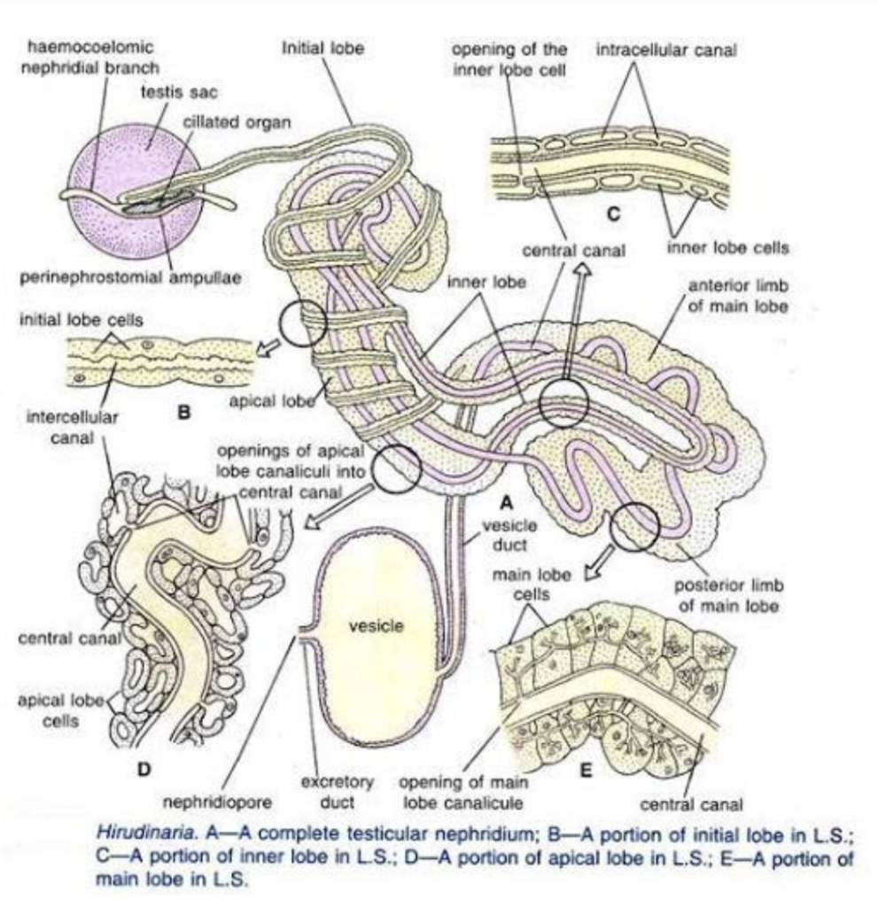
- Main Lobe:
- The main lobe is a horse-shoe-shaped structure located ventro-laterally in the body.
- It consists of two unequal limbs, with the anterior limb being larger than the posterior limb. Together, they form the bend of the horse-shoe shape.
- Apical Lobe:
- The posterior limb of the main lobe extends forward to form a stout apical lobe.
- This lobe is positioned beneath the gut, with its anterior end bent upon itself, contributing to the complex structure of the nephridium.
- Initial Lobe:
- The initial lobe is long and slender, wrapping around the apical lobe.
- It runs toward the testis sac at the anterior extremity and terminates near the perinephrostomial ampulla, while its posterior extremity connects to the main lobe near the vesicle duct.
- Ciliated Organ:
- The ciliated organ is located within the peri-nephrostomial ampulla, and it is composed of a central spongy reservoir surrounded by ciliated funnels.
- Each funnel has a broad distal end and a narrow neck, fitting into pores in the reservoir wall. These funnels are densely ciliated and composed of several layers of cells.
- Despite its proximity to the nephridia, the ciliated organ does not play a direct role in excretion in adult leeches. Instead, it is connected to the haemocoelomic system and is involved in the manufacture of haemocoelomic corpuscles.
- Inner Lobe:
- The inner lobe is situated within the concavity of the main lobe and extends forward along the outer border of the apical lobe for about half its length.
- Main Lobe:
- A typical testicular nephridium consists of several distinct parts, each contributing to the overall function of the nephridium.
- Central Canal in Nephridium:
- The nephridium contains a long, zigzagging central canal that follows a complex course throughout the structure.
- The canal begins as an intracellular lumen at the anterior end of the apical lobe and extends through the apical lobe, passing into the inner lobe, and then into the posterior limb of the main lobe.
- After making several loops within the main lobe, the canal eventually emerges from the posterior lip of the anterior limb of the main lobe, continuing into the vesicle as the vesicle duct.
- The canal’s path within the nephridium is quite intricate, making one complete loop and another incomplete loop through the structure, contributing to the efficiency of filtration.
- Role of Nephridium and Ciliated Organ:
- In leeches, the haemocoelomic fluid, which includes both blood and coelomic fluid, is not separated into distinct entities as seen in earthworms. Instead, these fluids mix within the leech’s body.
- Although the ciliated organ is considered an excretory structure, it does not participate in the actual process of excretion. Instead, it is associated with the haemocoelomic system, where it is involved in the production of haemocoelomic corpuscles.
- The main body of the nephridium, excluding the ciliated organ, is richly supplied with lateral channels, which play an important role in the excretory and osmoregulatory functions of the nephridium.
- The nephridia, therefore, serve primarily to regulate the balance of water and waste removal from the body. They filter out metabolic waste from the leech’s coelomic fluid and help in maintaining internal homeostasis.
- Botryoidal Tissues:
- In addition to the nephridia, the botryoidal tissues, which are specialized clusters of cells found in the leech’s body, are sometimes considered to have excretory functions.
- These tissues may contribute to waste processing and excretion, though their precise role in comparison to the nephridia remains an area of interest and study.
- https://www.bpchalihacollege.org.in/online/attendence/classnotes/files/1625504042.pdf
- https://www.slideshare.net/slideshow/excretion-in-annelidapdf/258186234
- http://www.rajasinghcollegesiwan.com/Zoology/B.Sc%20First%20Year%20Zoology%20(Hons)%20Excretory%20System%20in%20Annelida%201.pdf
- https://tmv.ac.in/ematerial/zoology/sm/UG%20SEM-2%20C%203%20T%20UNIT%202%20ANNELIDA.pdf
- https://content.patnawomenscollege.in/zoology/EXCRETION%20IN%20ANNELIDA.pdf
- https://www.westgoalparacollege.ac.in/upload/e_content/1685617089.pdf
- Text Highlighting: Select any text in the post content to highlight it
- Text Annotation: Select text and add comments with annotations
- Comment Management: Edit or delete your own comments
- Highlight Management: Remove your own highlights
How to use: Simply select any text in the post content above, and you'll see annotation options. Login here or create an account to get started.