What is Euglena?
- Euglena, these tiny enigmatic organisms, captivate the curious minds of scientists and students alike. Belonging to the genus protist, they defy classification as plants, animals, or fungi. Instead, Euglena showcases a unique blend of characteristics from both plants and animals. Like plants, they possess the ability to synthesize their own food, but at the same time, they display animal-like traits by actively moving and consuming food.
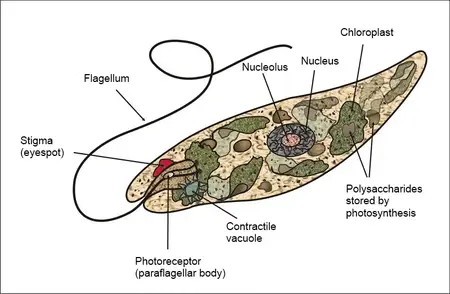
- Under the lens of a light microscope, Euglena appear as elongated unicellular beings, swiftly gliding through their aqueous environment. A peculiar feature that distinguishes them is their tear-drop shape, with a rounded head housing the whip-like tail, also known as a flagellum. Though one flagellum is typically visible, they possess two, with one usually hidden within a reservoir. The rapidly twirling flagellum propels Euglena gracefully across the water’s surface, a mesmerizing spectacle to behold.
- Unlike most plant cells, Euglena lack a cell wall. Instead, their organelles and cytoplasm are enclosed by a plasma membrane, endowing them with greater flexibility and ease of movement. Electron microscopes have revealed an ornamented pellicle beneath the plasma membrane. This thin protein layer not only provides protection and shape to the cell but also enhances its ability to compress and maneuver through tight spaces.
- Upon closer inspection, a reddish spot at the anterior end catches the eye. This crucial organelle contains carotenoid granules, enabling Euglena to sense and move towards sunlight. Known as the eyespot, it also filters the light wavelength reaching the paraflagellar body, a light-detecting structure at the base of the flagellum. In response, Euglena showcases positive phototaxis, moving towards the light source to maximize photosynthesis.
- Moreover, scattered throughout the organism’s body, dark greenish spots reveal the presence of chloroplasts containing chlorophyll. This green pigment plays a vital role in photosynthesis, converting sunlight into energy. Some Euglena species possess both chlorophyll A and B, with the latter enhancing light absorption. Through photosynthesis, they synthesize glucose and release oxygen, making them autotrophic, capable of producing their own food when exposed to sunlight.
- However, despite their autotrophic abilities, Euglena are also heterotrophic, meaning they consume food. The process of food consumption, known as phagocytosis, involves engulfing food particles within a vacuole for digestion. Enzymes released within the vacuole break down the ingested food, providing essential nutrients for the organism. Additionally, Euglena possess a contractile vacuole that collects and expels excess fluids, safeguarding the cell from rupturing due to excessive water intake.
- In conclusion, Euglena represents a fascinating amalgamation of plant and animal-like features. Their tear-drop shape, flagella-driven mobility, and ability to harness sunlight for photosynthesis showcase their plant-like attributes. Meanwhile, their eyespot-driven phototaxis, heterotrophic feeding, and contractile vacuole reveal their animal-like traits. Such versatility and adaptability make Euglena a true wonder of the microscopic world, worthy of our admiration and continued study.

Requirements for Microscopy of Euglena Under The Microscope
- Microscope Glass Slides: The first requirement is a set of microscope glass slides. These small, flat, and transparent pieces of glass serve as the platform on which the Euglena sample will be mounted for observation. Carefully placing a droplet of the sample on the center of the slide ensures that the Euglena organisms are well-distributed and positioned for optimal viewing.
- Microscope Cover Slips: Cover slips, also known as cover glasses, are crucial companions to microscope glass slides. These thin and transparent squares of glass delicately cover the specimen on the slide, holding it in place and preventing any distortion or damage during the observation process. Cover slips provide a clear and stable surface for the microscope to focus on, allowing for a detailed examination of Euglena.
- A Compound Microscope: The heart of any microscopy endeavor is the compound microscope. This sophisticated optical instrument magnifies the microscopic world, enabling us to explore the intricacies of Euglena’s structure and behavior. When selecting a compound microscope, it’s important to consider its magnification capabilities and resolution power to ensure a clear and vivid view of these minute organisms.
- A Dropper: A dropper, also known as a Pasteur pipette, is an indispensable tool for handling and transferring liquid samples. In the case of Euglena microscopy, the dropper comes in handy when placing a small droplet of the sample onto the microscope slide. This ensures that the Euglena are well-distributed and evenly spread out, allowing for a comprehensive examination without overcrowding.
With these simple yet indispensable requirements, the microscopic realm of Euglena can be unlocked. By skillfully preparing a well-mounted slide with a carefully placed cover slip, the compound microscope becomes a portal to the hidden wonders of the single-celled organisms. The dropper serves as a delicate guide, ensuring that the Euglena are presented in their full splendor, ready to reveal the captivating blend of plant and animal-like characteristics they embody. As one peeks through the eyepiece, a world of discovery unfolds, shedding light on the unique and enigmatic life of Euglena.
Sample Preparation
- Collecting Euglena from Pond Water: If the intention is to directly observe Euglena from pond water, one can start by obtaining a sample of pond water from any suitable water body, preferably one with an abundance of aquatic vegetation. Ponds, lakes, or slow-moving streams are ideal places to find Euglena. Using a clean container, carefully collect a sample of pond water without disturbing the sediment at the bottom.
- Microscope Slide Preparation: Once the pond water sample is collected, it’s time to prepare the microscope slide. Place a clean microscope glass slide on a flat surface. Using a dropper or a pipette, carefully transfer a small droplet of the pond water onto the center of the slide. Be cautious not to overfill the slide, as this can lead to overcrowding and hinder clear observations.
- Cover Slip Placement: Next, gently place a microscope cover slip over the droplet of pond water on the slide. The cover slip helps create a thin and even layer of the sample, preventing distortion and providing a clear surface for observation. Ensure there are no air bubbles trapped between the slide and the cover slip, as these can obstruct the view and distort the sample.
- Cultivating Euglena Using Pond Weed: Alternatively, to increase the number of Euglena for a more comprehensive study, a simple culture experiment can be carried out using pond weed (preferably Ceratophyllum). Take a few healthy strands of pond weed and place them in a clean jar or petri dish. Cover the pond weed with water, providing a suitable aquatic environment for Euglena to thrive.
- Creating the Ideal Environment: To promote Euglena growth, place the jar or petri dish in a dark room for a few days. The absence of light triggers the growth of Euglena, leading to the formation of a brown scum on the water’s surface. This scum indicates an increase in Euglena numbers and readiness for observation.
Procedure
- Obtain the Sample: Using a dropper, carefully collect a small amount of pond water directly from a pond or, if available, gather the brown scum that formed on the water’s surface through the culture experiment. Euglena can be found in both these sources, and each offers a unique insight into their behavior and characteristics.
- Prepare the Microscope Slide: Place a clean and dry microscope glass slide on a flat and stable surface. With the dropper, deposit a small droplet of the collected sample at the center of the slide. Take care not to oversaturate the slide, as this might hinder proper observation by causing overcrowding.
- Position the Cover Slip: Gently and carefully lower a microscope cover slip onto the droplet of the sample. Ensure that it makes contact with the sample at one edge and then slowly lower it to avoid trapping air bubbles. The cover slip acts as a thin and transparent layer, providing a clear surface for microscopic examination.
- Microscope Setup: Once the slide is prepared, it’s time to set up the compound microscope for observation. Place the slide on the microscope stage and secure it in place using the stage clips. Adjust the position of the slide so that the area of interest, where Euglena are expected to be, is beneath the objective lens.
- Illumination and Magnification: Adjust the microscope’s light source to provide adequate illumination for the sample. Start with the lowest magnification objective lens (usually 4x or 10x) and focus the microscope on the sample. Gradually increase the magnification to higher levels (40x, 100x) for more detailed examination of Euglena.
- Observing Euglena: Peek through the eyepiece of the microscope and marvel at the microscopic world that unfolds before your eyes. Observe the distinctive tear-drop shape of Euglena and their rapid movement using their whip-like flagellum. Take note of the reddish eyespot at the anterior end, aiding in their positive phototaxis towards sunlight.
- Record Your Findings: During the observation, document your findings, noting any interesting behaviors, patterns, or characteristics displayed by the Euglena. Drawing sketches or taking photographs through the microscope can help in creating a visual record of the observation.
- Clean Up: Once you have completed your observation, carefully remove the slide from the microscope stage and place it in a designated area for cleaning. Wipe the microscope lenses and stage to ensure they are free from any residue or contaminants.
Observation
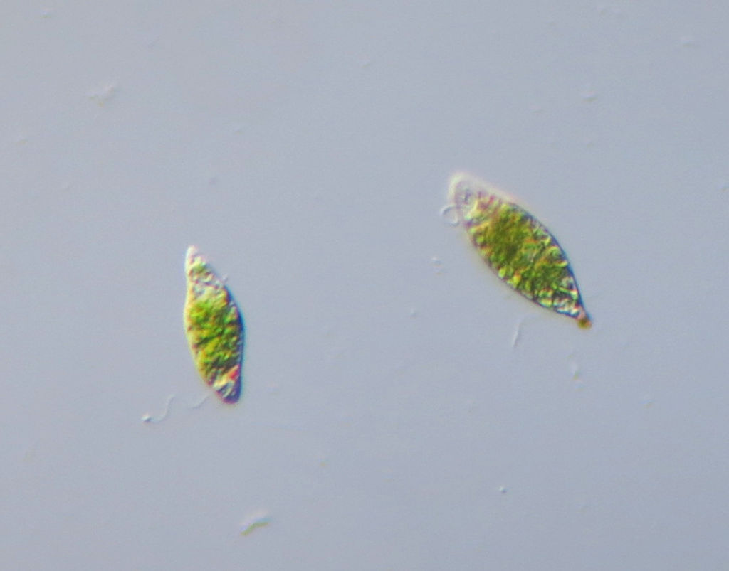
As students peer through the microscope, the fascinating microscopic world of pond water and pond weed unfolds before their eyes. Among the common inhabitants, both amoeba and Euglena grace the slide, each revealing their unique characteristics. While amoeba may also be present, distinguishing them from Euglena becomes an intriguing challenge. However, with a keen eye and careful observation, Euglena can be identified as elongated organisms with a distinctive whip-like tail.
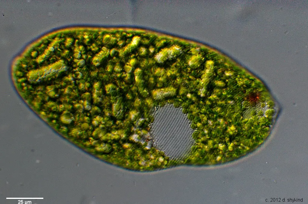
At 40X magnification, Euglena appear as minute particles, exhibiting slight movements in the water. As the magnification increases to 100x and 400x, a delightful transformation takes place. Euglena come into focus, displaying a captivating green or light green hue with dark spots scattered within their elongated bodies. The whip-like tail at one end becomes more evident, swaying gently as if guiding their graceful movements.
Among the features that become apparent at higher magnifications is the presence of colored granules, aptly named the eyespot. This vital organelle allows Euglena to sense and move towards sources of light, playing a crucial role in their phototactic behavior. The eyespot stands as a testament to the remarkable abilities of these single-celled wonders to adapt and survive in their aquatic environment.
As students delve deeper into the observation, they may notice the subtlety of the whip-like tail located at the front end of the organism. While it may appear colorless or transparent, careful scrutiny allows its presence to be discerned. This remarkable structure serves as a propeller, propelling Euglena gracefully through the water, exhibiting their unique blend of plant and animal-like characteristics.
As the microscopy session unfolds, students may find themselves captivated by the enigmatic beauty of Euglena. Their elongated forms and whip-like tails, accompanied by the presence of the eyespot and colored granules, showcase the complexity of these seemingly simple organisms. The microscopic world offers a glimpse into the astonishing diversity of life, reminding us of the wonders that lie beyond what the naked eye can behold.
With every observation, students venture deeper into the mysteries of Euglena, discovering the remarkable adaptations that allow them to thrive in their watery realm. This microscopic journey sparks a sense of wonder and curiosity, leaving a lasting impression on young minds, and instilling a passion for exploring the hidden intricacies of the natural world.


FAQ
What is Euglena, and why is it interesting to observe under the microscope?
Euglena is a single-celled organism belonging to the genus protist. It is intriguing to observe under the microscope because it exhibits characteristics of both plants and animals, making it a unique and fascinating microorganism to study.
How do I collect Euglena for observation under the microscope?
Euglena can be collected from pond water or through a simple culture experiment using pond weed. The brown scum formed during the culture process is rich in Euglena and can be used for observation.
What tools do I need to observe Euglena under the microscope?
To observe Euglena, you will need a compound microscope, microscope glass slides, microscope cover slips, and a dropper for sample preparation.
How can I differentiate Euglena from amoeba under the microscope?
Euglena can be distinguished from amoeba by its elongated shape and the presence of a whip-like tail at one end. Additionally, Euglena usually appear green or light green in color with dark spots and a distinct eyespot.
At what magnification can I observe Euglena in detail?
Euglena can be observed in more detail at higher magnifications, typically at 100x and 400x. At these magnifications, the green color, dark spots, eyespot, and whip-like tail become more prominent.
What is the role of the eyespot in Euglena?
The eyespot in Euglena helps the organism sense and move towards sources of light. It plays a crucial role in phototactic behavior, guiding Euglena towards areas with optimal light conditions for photosynthesis.
Can I observe Euglena’s movement under the microscope?
Yes, Euglena’s movement can be observed under the microscope. The whip-like tail propels Euglena through the water in a graceful manner, showcasing its unique motility.
How do Euglena obtain their food?
Euglena are both autotrophic and heterotrophic. They can manufacture their own food through photosynthesis when exposed to sunlight. However, they can also consume food through phagocytosis, where they engulf and digest food particles.
What are the greenish spots observed inside Euglena under the microscope?
The greenish spots inside Euglena are chloroplasts containing chlorophyll, which is responsible for the organism’s green color. Chloroplasts are essential for photosynthesis, where Euglena convert sunlight into energy.
Why is studying Euglena under the microscope important?
Studying Euglena under the microscope provides valuable insights into the diversity and adaptability of microscopic life. It helps us understand the complex relationships between organisms and their environments, shedding light on the intricate processes that sustain life on Earth.
- Text Highlighting: Select any text in the post content to highlight it
- Text Annotation: Select text and add comments with annotations
- Comment Management: Edit or delete your own comments
- Highlight Management: Remove your own highlights
How to use: Simply select any text in the post content above, and you'll see annotation options. Login here or create an account to get started.