What is a Dissecting microscope or a Stereo microscope?
A dissecting microscope—also known as a stereo microscope—is an optical microscope study apparatus that enables the observation of an object at low to moderate magnification (typically 5x to 250x) through reflective light versus transmitted light. Thus, it’s a microscope made for viewing little details that could be overlooked on these surfaces, making it the perfect equipment for dissection. Progress toward a dissecting microscope came in 1677.
In 1677, Cherudin d’Orleans created a basic dissecting microscope with eyepieces set apart to accommodate the distance and their objective lenses. A general outline, but it would serve as the foundation for all dissecting microscopes to come. Then, almost two centuries later, great progress happened in 1852. 1852 was the year Charles Wheatstone proposed the idea of binocular vision. He explained how the brain recognizes and processes two distant images according to how each eye perceives one image. Wheatstone’s findings were innovative and published in 1852 as ‘On Some Remarkable, and Hitherto Unobserved, Phenomena of Binocular Vision.’
But the idea of stereoscopic vision permeated even further. In 1860, John Leonard Riddell extended Wheatstone’s findings and published in the Journal of Microscopical Science his article, ‘On Binocular Microscope’. He tinkered with the capacity to view in a binocular fashion and how it might extend the field of depth for micro images, a critical component of stereo microscopes used in the industry today.
Much later still, American biologist Horatio S. Greenough defined it further. He worked with the Carl Zeiss Company to remap the arrangement of the stereo microscope. Using two like optical paths, he established an elaborate journey to view an object in a three-dimensional reality. Currently, this formula is the foundation for a dissecting microscope. Its intent is to dissect.
From low-power magnification for broader views to high-power magnification to assess surface detail, such microscopes exist in every field. Ultimately, this microscope boasts a clear advantage over the others in the ability to view samples in three dimensions, whereas regular microscopes only offer two-dimensional fields of view. The applications range from cellular biology to materials science to even nanotechnology, more than one would expect.
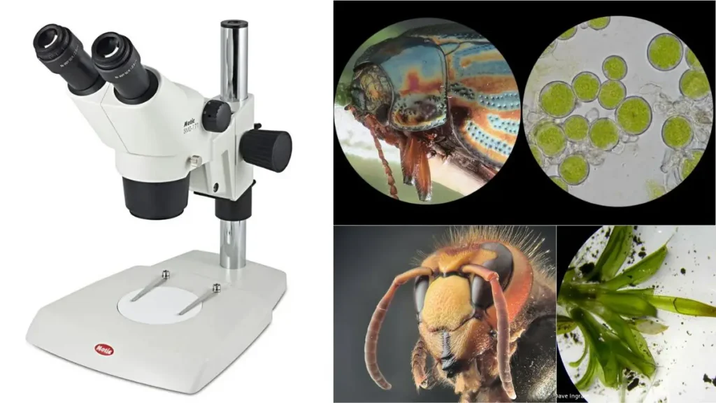
Principle of Dissecting microscope
The dissecting microscope is a unique optical device used for particular means of examination for larger, more intricate biological specimens. For instance, it transmits light through different pathways, as a light source/projector is situated above the stage and below the stage. These facilitate different angles of illumination that may be ideal for viewing specific types of samples. Nevertheless, two ocular lenses at the eyepiece merge it all into a binocular stereoscopic view.
The biggest benefit of the dissecting microscope is the magnified image. The image is projected onto the computer screen digitally and often appears in real-time 3D on the school computer. This is true for certain experiments—examining insects—for the image can be larger than the actual sample, essentially rendering it macro photography. In addition, because the dissecting microscope examines outside morphology, it’s easier to evaluate and see into more complicated samples in 3D.
When it comes to magnification, the stereoscope contains two types: it contains fixed and zoom. Fixed means two objective lenses to achieve a specific level of magnification. Zoom refers to a selection of magnification levels from which one can choose as it can be adjusted up or down in a controlled manner. Furthermore, depending upon need, supplemental objectives can enhance the overall magnification beyond that of the provided one.
Maybe the most groundbreaking element of this microscope is that it runs off a Galilean optical system, which is a compromise between normal, unmovable magnification levels or zooming. The optical system consists of fixed focus lenses to produce different levels of magnification—the two lenses produce four magnifications, or three sets of lenses produce six magnifications. Thus, by altering the ocular lenses, one can achieve the necessary and proper magnifications for viewing delicate internal cellular structures or for dissection of easily damaged tissues. Ultimately, viewing both inside and outside of the louse is easier through the compound microscope with its more sophisticated optical arrangement and ensuing magnification.
Dissecting Microscope Parts (Parts of Stereo microscope)
| No. | Part | Description |
|---|---|---|
| 1 | Light Source | Dissecting microscopes have a built-in or external light source, typically an LED or lamp, to illuminate the specimen. It may be located above, below, or on the side of the stage, and often involves a diaphragm and condenser to control and focus light onto the specimen. The quality and distribution of light are crucial for image clarity. |
| 2 | Stereo Head | Contains the eyepieces and objective lenses, providing a three-dimensional view of the specimen. It’s adjustable for height and angle, and may include features like a tilting or rotating head for different viewing perspectives. The stereo head affects the magnification and field of view. |
| 3 | Eyepieces | Also known as ocular lenses, they magnify the image produced by the objective lens. They have a specified eye relief to accommodate users with eyeglasses and contain multiple lenses for desired magnification and field of view. They may include features like diopter adjustment and interpupillary distance adjustment. |
| 4 | Diopter Setting | Allows fine-tuning of eyepiece focus to match individual eyesight. Each eyepiece has its diopter adjustment for achieving a sharp, clear view. |
| 5 | Objective Lens | Primary lenses that magnify the specimen and are crucial for image formation. In dissecting microscopes, they are housed within a cylindrical cone and are not individually visible. Magnification can be altered by rotating this cone, and the total magnification is the product of the objective and ocular lens magnifications. Additional lenses like Barlow lenses can modify overall magnification. |
| 6 | Adjustment Knobs | Include focus and zoom knobs to adjust magnification and focus the image. They may be located on various parts of the microscope such as the base, head, body, or stand. |
| 7 | Specimen Stage | A flat platform where the specimen is placed, often equipped with a mechanical stage for movement and focusing. The stage is crucial for positioning the specimen within the field of view and may include a fine focus mechanism. |
| 8 | Stage Clips | Devices to hold the specimen in place on the stage, ensuring it remains stationary during examination. They are adaptable to various specimen sizes and shapes. |
| 9 | Optical System | Provides fixed-focus lensing for the microscope. |
| 10 | Digital Camera | Included in most dissecting microscopes for capturing and recording images of the specimens. |
| 11 | Base | The lower support structure that also functions as the stage in dissecting microscopes. It may include stage clips for securing specimens and is designed for stability and support. |
| 12 | Stand | Connects the base to the microscope’s head and serves as the structural support. It may include the power cord, allow vertical movement for focusing, and serve as a handle for transportation. |
| 13 | Mirror | Used to reflect light onto the specimen, especially useful when the light source is below the stage. Various types of mirrors (flat, concave, convex) are used depending on the desired lighting effect. |
| 14 | On/Off Switch | Controls the microscope’s illumination and is typically located on the side of the base. Light intensity should be reduced before turning off the microscope. |
| 15 | Light Intensity Control | Adjusts the brightness of the microscope’s light source. May include separate controls for overhead and stage lighting, allowing customization of illumination levels. |
| Others | Internal Optical Components | Includes prism, relay lens, and reticle among others, housed within the microscope’s body/head. These components contribute to image orientation, extension, and measurement capabilities. |
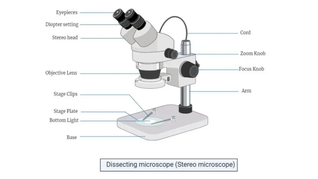
- Light Source– Illuminates the specimen, often LED or lamp, placed above, below, or beside the stage.
- Stereo Head– Houses eyepieces and objective lenses, providing a 3D view. Adjustable for height and angle.
- Eyepieces – Magnify the image and accommodate users with glasses. Some have focus adjustments.
- Diopter Setting– Fine-tunes focus for each eyepiece to match individual eyesight.
- Objective Lens– Magnifies the specimen, with the magnification adjustable by rotating the lens.
- Adjustment Knobs – Used to adjust magnification and focus. Located on various parts of the microscope.
- Specimen Stage -Platform where the specimen is placed, often with movement for easier focus.
- Stage Clips– Hold the specimen in place on the stage.
- Optical System – Provides fixed-focus lenses for magnification.
- Digital Camera– Captures and records images of specimens.
- Base– Supports the microscope and includes stage clips for securing specimens.
- Stand– Connects the base to the head and allows vertical movement for focusing.
- Mirror– Reflects light onto the specimen, useful when the light source is below.
- On/Off Switch– Controls the illumination, usually located on the base.
- Light Intensity Control– Adjusts the brightness of the light source.
- Internal Optical Components– Includes parts like the prism and relay lens for image clarity and measurements.
Dissecting microscope Magnification
A dissecting microscope or stereo microscope achieves total magnification by adding together the magnification of the eyepiece and the objective lens. For example: A dissecting microscope with a 10x eyepiece and 4x objective lens produces the following total magnification:
Dissecting microscope Magnification = 10 + 4 = 14x Magnification.
Two standard types of magnification systems exist in stereo microscopes. One is a fixed magnification system. This means that the primary source of magnification comes from dedicated objective lenses that possess a relatively fixed magnification power per lens. The other type is a zoom or pancratic magnification. This system possesses a relative range of magnification that can be determined at any given time. However, with the zoom system, there is an additional degree of magnification that can be reached with accessory objectives, which adds to the cumulative magnification by a fixed amount.
In both cases, interchangeable objective eyepieces allow for different levels of total magnification. Thus, the “Galilean optical system,” which resides between fixed and zoom magnification, is the best system credited to Galileo. It features a convex lens that gives a fixed-focusing ability, which creates a fixed magnification, but the same optical pieces at the same distance between lenses will—if reversed—create another fixed magnification.
Thus, where two options exist, a turret of one lens system creates four that the human eye cannot detect. In addition, if three options can exist in one plane, six fixed magnifications are beyond sight. Thus, applications of such Galilean optical systems exist just as beneficial as a vastly more expensive zoom system, the only disadvantage being that no one ever knows what the fixed magnification is, as it is not read through an analog scale. (In addition, it’s more durable, which makes it more appropriate for off-the-beaten-path use.)
Auxiliary lens
Auxiliary lens used to alter the total magnification power of Dissecting microscope. The magnification power of an Auxiliary lens will be multiplied with the total magnification power of a stereomicroscope.
For example; if a stereo microscope with 10x eyepieces, the zoom knob is set to 5x and you also have a 0.3x auxiliary lens on the microscope.
The total magnification would be determined with the following formula 10 x 5 x 0.3 = 15x magnification.
Types of Dissecting Microscope or Stereo Microscope
- Stereo Zoom Dissecting Microscope
Zoom range of 6.7x-45x. Can be connected to a digital camera. Dual LED lights and 360° rotation. Magnification adjustable with extra lenses. - Digital Tablet Dissection Microscope
Touch screen LCD tablet with magnification 6.7x-45x. 5.0-megapixel camera for capturing images and videos. Separate top and bottom LED lights. - Stereo Zoom Boom Stand Microscope
Large base and stage for big samples. Zoom range of 6x-45x. Can be adjusted with extra lenses or eyepieces. - Stereo Zoom Dissecting Microscope
Compact with 10x-30x magnification. Rotating head for better view. Uses 10-watt halogen and 5-watt fluorescent lights. - Dual Power Dissecting Microscope
Magnifications of 10x and 30x. 360° rotation. High-intensity LED ring light. Flexible stand for larger specimens. - Single Power Stereo Dissection Microscope
Magnification range of 10x-40x. 45° inclined eyepieces. Diopter adjustments from 50mm to 70mm. - Single Magnification Handheld Pocket Microscope
- Compact and portable with two magnification options. No light required. High-quality optical glass.
Operating Procedure of Dissecting microscope
- Purpose of Dissecting Microscope
- A dissecting microscope is used to examine larger specimens like fossils, rocks, insects, and plant pieces.
- It can also magnify specimens placed on slides.
- Types of Stages
- Two stage types: one for observation and another for dissection.
- Black or white opaque stages are used for non-transparent specimens.
- Glass stages allow light to shine from below, useful for specimens on slides.
- Installing the Appropriate Stage
- Select the correct stage before turning on the microscope.
- Loosen the stage plate lock screw to change the stage.
- Insert a blue filter if switching to a glass stage.
- Lock the stage in place by tightening the screw.
- Turn on the Microscope
- Press the on/off switch on the base to turn on the microscope.
- Activate either the incident or transmitted light, depending on the specimen.
- Adjusting the Head/Body
- Use the coarse focus knob to slowly lower the microscope’s head and body.
- This step is crucial for establishing the starting position.
- Adjusting Eye Distance
- Look through the eyepieces and adjust the distance between them.
- Move the eyepieces in or out, like adjusting binoculars, until a single circle appears.
- This ensures proper interpupillary distance.
- Mount the Specimen
- Place the specimen on the stage and secure it with stage clips if necessary.
- Use the best lighting for each specimen: incident light for opaque specimens and both incident and transmitted light for others.
- Position the specimen in the focal plane before focusing.
- Focusing
- Turn the focus knob to gradually bring the image into sharper focus.
- If starting with the head/body at its lowest position, slowly raise it until the image is clear.
- Zooming In
- After focusing, use the zoom knob to focus on a specific area of the specimen.
- For detailed study, like looking at a hydra’s tentacles, zoom in.
- If the image becomes blurry, refocus with the focus knob.
- Adjusting Light Intensity
- Use intensity controls to adjust the amount of light and contrast.
- This helps to see details clearly.
- Finishing Up
- At the end of the session, return the zoom to its lowest setting.
- Lower the light intensity, turn off the microscope, and cover it with a protective bag.
Applications of Dissecting microscope (Stereo microscope)
- Study surface features of solid samples.
- Used for dissection of specimens.
- Aid in microsurgical procedures.
- Inspect watches and circuit boards during manufacturing.
- Analyze fractures in materials.
- Help in forensic engineering investigations.
Advantages of Dissecting Microscope (Stereo Microscope)
- Provides 3D images, making details easier to see.
- Has a large working distance for easier manipulation of objects.
- Offers a wide field of view, great for larger objects.
- Easy to use, with minimal focus adjustments.
- Portable and easy to carry around.
- Versatile, suitable for many fields like biology, electronics, and forensics.
Disadvantage of Dissecting Microscope (Stereo Microscope)
- Low magnification limits viewing of very small objects.
- Limited depth of field makes focusing difficult.
- Image distortion can occur at higher magnifications.
- More complex to use, especially for beginners.
- Can be expensive compared to other microscopes.
Compound vs Dissecting microscope
There are several key differences between compound microscopes and dissecting microscopes:
- Magnification: Compound microscopes can magnify objects much more than dissecting microscopes. While dissecting microscopes typically have magnifications ranging from 10x to 50x, compound microscopes can magnify objects up to 1000x or more.
- Depth of field: Dissecting microscopes have a greater depth of field, meaning that objects at different distances from the lens are in focus at the same time. This makes it easier to view larger, three-dimensional objects. In contrast, compound microscopes have a narrow depth of field, meaning that only objects at a specific distance from the lens are in focus at any given time.
- Illumination: Dissecting microscopes use transmitted light, meaning that light passes through the specimen and is then focused by the lenses. Compound microscopes can use either transmitted or reflected light, depending on the type of specimen being observed.
- Resolution: Compound microscopes have a higher resolution, meaning that they can distinguish between small details more easily than dissecting microscopes. This makes them ideal for studying small structures such as cells and tissues.
- Uses: Dissecting microscopes are typically used to examine larger specimens, such as plants, insects, and other small organisms. Compound microscopes are used to study smaller structures such as cells and tissues, and are often used in scientific research and medical settings.
Dissecting microscope price
The price of a dissecting microscope can vary widely depending on the features and quality of the instrument. Basic models may cost a few hundred dollars, while more advanced models with additional features can cost several thousand dollars. Some factors that may affect the price of a dissecting microscope include the type and quality of the lenses, the type of illumination system, the magnification range, and the overall durability and build quality of the microscope. It is generally advisable to choose a microscope that is appropriate for the intended use and budget, rather than selecting the cheapest option available.
Dissecting Microscope Worksheet
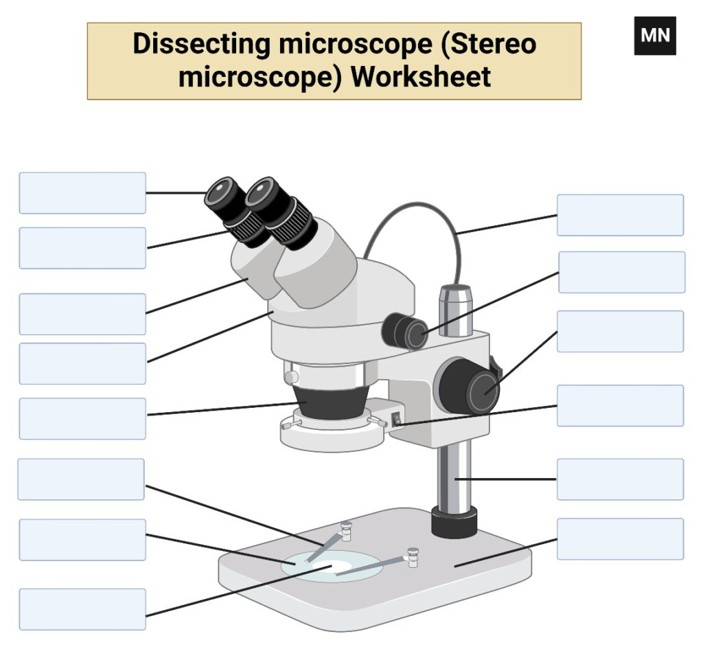
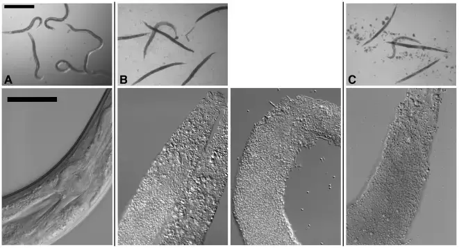
Dissecting microscope images

Reference
- https://www.slideshare.net/waleedtareen2/stereo-microscope-or-dissecting-miscrscope-54430261
- https://en.wikipedia.org/wiki/Stereo_microscope
- https://www.microscopeinternational.com/what-is-a-stereo-microscope/
- https://www.news-medical.net/life-sciences/What-are-Stereo-Microscopes-Used-For.aspx
- https://www.microscope.com/education-center/five-things-you-should-know/about-stereo-microscopes
- https://www.microscope-detective.com/stereo-microscope.html