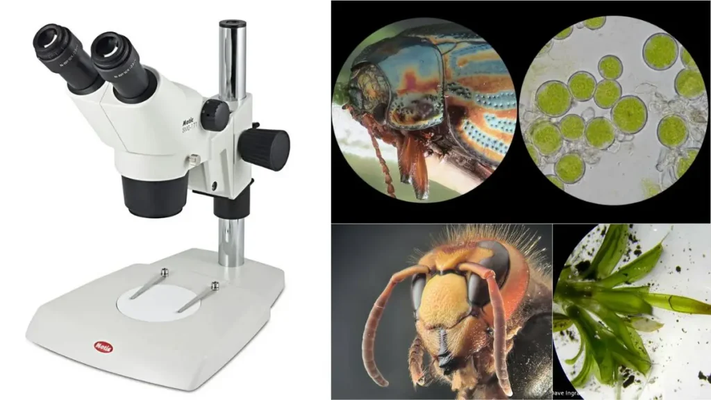
Dissecting Microscope (Stereo Microscope) Definition, Uses, Parts, Principle.

Stereo microscopes, in contrast to compound light microscopes, often employ reflected illumination, or light reflected off an object’s surface, rather than transmitted (diascopic) illumination. When a specimen is too thick or opaque for compound microscopy, reflected light from the object enables for investigation. Unlike a compound microscope, transmitted illumination in most stereo microscopes is not focussed by a condenser, but rather is provided by a lamp or mirror placed beneath a transparent stage placed beneath the object. Dark field microscopy makes use of stereoscopes with specialised illuminators to produce either reflected or transmitted light for observation.

Features such as a large working distance and depth of field are particularly valuable in a microscope of this sort. To put it another way, resolution (the distance at which two neighbouring points may be identified as independent) decreases depth of field and working distance. Although stereo microscopes typically provide significantly lower magnification, they are capable of providing magnifications of up to 100x, which is equivalent to a 10x objective and 10x eyepiece in a standard compound microscope. The resolution of a standard compound optical microscope is around ten times higher than this.

Using fiber-optic lighting, as will be detailed more below, the large working distance at low magnification is ideal for inspecting large solid objects like fracture surfaces. It’s also simple to alter such samples to zero in on the relevant details.

What is a Dissecting microscope (Stereo microscope)?

Definition of dissecting microscope

A dissecting microscope, also known as a stereo microscope, is a type of microscope that uses two separate optical paths to create a three-dimensional image of an object. It is often used for examining small objects or for tasks that require a high level of detail, such as dissection or soldering.

Dissecting microscopes typically have low magnification, typically ranging from 4x to 50x, and a relatively large working distance, which allows the user to manipulate the object being viewed with tools such as forceps or tweezers. They are often used in fields such as biology, entomology, geology, and engineering.

The stereo microscope consists of two eyepieces, each with its own objective lens, which are mounted on a stand that can be adjusted for height and angle. The object being viewed is placed on a stage, which can be moved up and down or side to side to focus the image. Some dissecting microscopes also have a light source, such as a lamp or LED, to illuminate the object being viewed.

The first recorded use of a dissecting microscope dates back to the 17th century, when the Dutch scientist Antonie van Leeuwenhoek used a simple single-lens microscope to observe and describe microorganisms. However, the stereo microscope as we know it today was not developed until the 19th century.

In the early 19th century, several scientists and inventors began experimenting with ways to create a microscope that could provide a more realistic, three-dimensional image of an object. One of the first to succeed was the English scientist William Brewster, who developed a stereo microscope in 1827. Brewster’s design used two lenses mounted on a single stand, and it was the first microscope to use both eyepieces to create a three-dimensional image.

In the decades that followed, several other inventors and companies developed their own versions of the stereo microscope. By the late 19th and early 20th centuries, stereo microscopes had become a common tool in scientific and industrial laboratories around the world. Today, they are used in a wide variety of fields, including biology, engineering, and manufacturing.

Differences from normal optical microscopes

There are several key differences between a normal optical microscope and a dissecting microscope (also known as a stereo microscope). Some of the most important differences include:

  1. Magnification: Normal optical microscopes typically have much higher magnification than dissecting microscopes, with magnification ranging from 100x to 1000x or more. In contrast, dissecting microscopes typically have magnification ranging from 4x to 50x.
  2. Objective lenses: Normal optical microscopes have one or more objective lenses that are used to focus the image, while dissecting microscopes have two objective lenses, one for each eyepiece.
  3. Working distance: Dissecting microscopes typically have a much larger working distance than normal optical microscopes, which allows the user to manipulate the object being viewed more easily with tools such as forceps or tweezers.
  4. Depth of field: The depth of field, or the range of distances over which the image is in focus, is typically much larger in a dissecting microscope than in a normal optical microscope. This allows the user to see more of the object at once, rather than having to focus on a single plane.
  5. Image quality: Dissecting microscopes generally produce a less detailed and less accurate image than normal optical microscopes, due to the lower magnification and the use of two separate optical paths. However, they are still useful for examining small objects or for tasks that require a high level of detail, such as dissection or soldering.
Dissecting Microscope (Stereo Microscope) Definition, Uses, Parts, Principle.

Principle of Dissecting microscope

The objectives and eyepiece of a dissecting microscope use two different sorts of light routes, and these paths are crucial to the microscope’s operation. Each ray of sunshine offers a new perspective. Dissections can be performed with the top light, and images can be viewed with the bottom light.

This illumination is made possible by the design of a binocular stereoscope, which consists of two separate eyepieces, each of which displays a distinct kind of light pathway and each of which provides a viewing comfort region. A digital microscope allows for real-time, three-dimensional visualisation of specimens on a computer screen. Macro photography allows for a magnified view of tiny objects like insects, which would otherwise be too small to see clearly in a standard photograph.

In the case of complicated samples, the image is captured and the topography (surface) is examined in three dimensions. There are two types of magnification used in the dissecting microscope: fixed (primary) magnification, where the distance between the two objective lenses determines the magnification level, and zoom (pancratic) magnification, which provides a continuous magnification at varying ranges by employing auxiliary objectives whose purpose is to increase the total magnification.

Changing the eyepiece lenses allows for a range of magnifications, from zoom to fixed. The Galilean optical system, located between fixed and zoom magnification, consists of fixed-focus lenses that impart fixed magnification for various sets of magnification, such as two sets of magnification offering four-magnification, three sets offering six-magnification, etc.

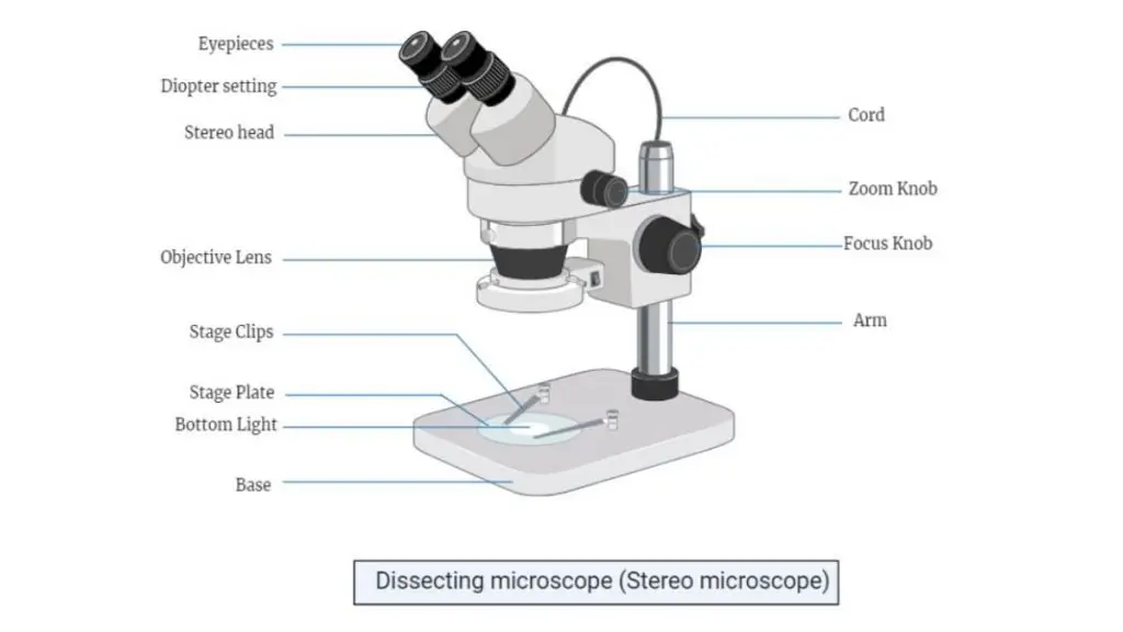
Dissecting Microscope Parts (Parts of Stereo microscope)

No.PartDescription
1Light SourceDissecting microscopes have a built-in or external light source, typically an LED or lamp, to illuminate the specimen. It may be located above, below, or on the side of the stage, and often involves a diaphragm and condenser to control and focus light onto the specimen. The quality and distribution of light are crucial for image clarity.
2Stereo HeadContains the eyepieces and objective lenses, providing a three-dimensional view of the specimen. It’s adjustable for height and angle, and may include features like a tilting or rotating head for different viewing perspectives. The stereo head affects the magnification and field of view.
3EyepiecesAlso known as ocular lenses, they magnify the image produced by the objective lens. They have a specified eye relief to accommodate users with eyeglasses and contain multiple lenses for desired magnification and field of view. They may include features like diopter adjustment and interpupillary distance adjustment.
4Diopter SettingAllows fine-tuning of eyepiece focus to match individual eyesight. Each eyepiece has its diopter adjustment for achieving a sharp, clear view.
5Objective LensPrimary lenses that magnify the specimen and are crucial for image formation. In dissecting microscopes, they are housed within a cylindrical cone and are not individually visible. Magnification can be altered by rotating this cone, and the total magnification is the product of the objective and ocular lens magnifications. Additional lenses like Barlow lenses can modify overall magnification.
6Adjustment KnobsInclude focus and zoom knobs to adjust magnification and focus the image. They may be located on various parts of the microscope such as the base, head, body, or stand.
7Specimen StageA flat platform where the specimen is placed, often equipped with a mechanical stage for movement and focusing. The stage is crucial for positioning the specimen within the field of view and may include a fine focus mechanism.
8Stage ClipsDevices to hold the specimen in place on the stage, ensuring it remains stationary during examination. They are adaptable to various specimen sizes and shapes.
9Optical SystemProvides fixed-focus lensing for the microscope.
10Digital CameraIncluded in most dissecting microscopes for capturing and recording images of the specimens.
11BaseThe lower support structure that also functions as the stage in dissecting microscopes. It may include stage clips for securing specimens and is designed for stability and support.
12StandConnects the base to the microscope’s head and serves as the structural support. It may include the power cord, allow vertical movement for focusing, and serve as a handle for transportation.
13MirrorUsed to reflect light onto the specimen, especially useful when the light source is below the stage. Various types of mirrors (flat, concave, convex) are used depending on the desired lighting effect.
14On/Off SwitchControls the microscope’s illumination and is typically located on the side of the base. Light intensity should be reduced before turning off the microscope.
15Light Intensity ControlAdjusts the brightness of the microscope’s light source. May include separate controls for overhead and stage lighting, allowing customization of illumination levels.
OthersInternal Optical ComponentsIncludes prism, relay lens, and reticle among others, housed within the microscope’s body/head. These components contribute to image orientation, extension, and measurement capabilities.
Dissecting Microscope Parts (Parts of Stereo microscope)
dissecting microscope labeled

1. Light source

  • Most dissecting microscopes have a light source, such as a lamp or LED, to illuminate the object being viewed. The light source is typically located above the stage, below the stage, or on the side of the microscope.
  • In dissecting microscopes that have a light source located above the stage, the light typically passes through a diaphragm and a condenser before reaching the object being viewed. The diaphragm is a device that controls the amount of light that reaches the object, while the condenser is a lens system that focuses the light onto the object.
  • Some dissecting microscopes have a built-in light source, such as a lamp or LED, while others require an external light source, such as a flashlight or a lamp. Some dissecting microscopes also have a reflector or a mirror to redirect the light onto the object being viewed.
  • It’s important to note that the quality and intensity of the light source can have a significant impact on the quality of the image produced by the microscope. A bright, evenly distributed light source will produce a clearer, more detailed image than a dim or uneven light source.

2. Stereo head

  • The stereo head of a dissecting microscope is the part of the microscope that contains the eyepieces and the objective lenses. It is called a “stereo” head because it allows the user to see a three-dimensional image of the object being viewed.
  • The stereo head is typically mounted on a stand that can be adjusted for height and angle, allowing the user to view the object from different perspectives. Some dissecting microscopes also have a tilting or rotating stereo head, which allows the user to change the angle of the eyepieces relative to the objective lenses.
  • The stereo head is an important part of the dissecting microscope, as it determines the magnification and the field of view of the microscope. Most dissecting microscopes allow the user to change the eyepieces or to use auxiliary lenses to adjust the magnification. The eyepieces and the objective lenses also determine the field of view, or the area of the object that can be seen at once.
  • In addition to the eyepieces and the objective lenses, the stereo head may also contain other components, such as a diaphragm or a condenser, which control the amount of light that reaches the object being viewed.

3. Eyepieces

  • The eyepieces, sometimes called ocular lenses, are the part of the microscope that the user looks into to examine the image of the object or specimen.
  • There are ocular lenses in a compound microscope, too, but due to their different diameters, you can’t swap them out with those from a compound microscope.
  • The eyepiece is essentially a magnifier that boosts the size of the picture produced by the objective. Because of this, they aid in magnifying the image so that finer details may be examined.
  • Eye relief refers to how far away from the ocular lens the user’s eye can go while still receiving a complete image. The range for this distance is 10–15 mm. This is vital, as it keeps eyelashes from touching the eyepiece lens and distorting the vision.
  • However, by simply changing the eyepiece eyeshields/eye-guards, this distance can be adjusted to somewhere between 25 and 30 mm for people who use eyeglasses.
  • Some eyepieces (such the aberration-free 10 eyepiece with diopter adjustment) contain multiple lenses—the eye lens, the lens doublet, the lens triplet, and the field lens doublet—to achieve the desired magnification and field of view.
  • The eye lens and the field lens may be all that’s installed in cheaper eyepieces.
  • Among other things, an eyepiece’s specifications should detail the magnification it offers. As an example, the typical magnification of an eyepiece is 10x (10). This is why the 10 designation will typically be found on the eyepiece.
  • Some eyepieces may also include a “WF” marking, which indicates that in addition to magnification, the eyepiece offers a broad field of vision. Simply said, this means that the user will be able to see more of the object or specimen than they would be able to with other eyepieces.
  • Eighteen and twenty millimetres are two of the most typical field numbers. While neither improves magnification nor resolution, the broader field of vision does allow for greater observation.
  • When looking through two eyepiece lenses, if one of your eyes is weaker than the other, you can adjust the diopter adjustment (found on the eyepiece’s base) to make up for the discrepancy.
  • The eyepieces’ ability to have the interpupillary distance (the distance between them) altered means that they can be used by people of varying eye sizes and shapes.
  • Adjusting this distance (by simply pulling apart or pressing the interpupillary adjuster close together) will help merge your left and right fields into a single image in the eyepieces.
  • After this is done, other settings (such as brightness, magnification, focus, etc.) can be tweaked for improved visibility.

4. Diopter setting

  • The diopter setting of a dissecting microscope, also known as a stereo microscope, is the adjustment that allows the user to fine-tune the focus of the eyepieces to match their individual eyesight. Each eyepiece of a stereo microscope has a diopter adjustment, which is usually a small knob or lever located near the eyepiece.
  • To adjust the diopter setting, the user looks through the eyepiece and focuses on an object at a fixed distance. The diopter adjustment is then used to fine-tune the focus of the eyepiece until the object appears sharp and clear. This process is typically repeated for each eyepiece to ensure that both eyes are seeing a clear image.
  • The diopter setting is an important part of the stereo microscope, as it allows the user to get the best possible image quality and comfort while using the microscope. It’s important to note that the diopter setting should be adjusted each time the eyepieces are changed, as different eyepieces may have different focal lengths and require different adjustments.

5. Objective Lens

  • Objectives are the primary lenses that magnify the object, gather light, and produce the picture visible in the ocular lenses, making them a crucial part of a dissecting microscope. The objective lenses, or “eyepieces,” of high-quality microscopes are often composed of glass, which is far superior to plastic.
  • Whereas the objectives of a compound microscope are mounted to the nosepiece and can be seen and identified individually (based on colour bands and their associated labels), the objectives of a dissecting microscope are hidden within a cylindrical cone and are not visible to the naked eye.
  • The magnification can be changed by rotating the cylindrical cone housing the objectives.
  • The microscope’s eyepiece and objective lenses work together to create a three-dimensional image. Two objective lenses can be used to increase the magnification of a stereo microscope from 1x to 4x (1x, 2x, 3x, or 4x).
  • Multiplying the objective lens’s magnification by the ocular lens’s magnifying power yields the total magnification.
  • Dissecting microscopes typically use objective lenses, but can sometimes accommodate additional lenses, such as Barlow lenses. Barlow lenses are auxiliary lenses that can be added or subtracted from the main lens system to change the overall magnification.
  • A 2x Barlow lens will enhance magnification by a factor of 2.5x, whereas a 0.5x Barlow lens will reduce magnification by a factor of 1.5x. Simply screwing it into the threaded lens port of a dissecting microscope makes it possible to use this device (in the cylindrical cone that contains the objectives).
  • Here, the working distance must be adapted upwards (with a Barlow lens) or downwards (with a close-up lens). The working distance must be increased when a 0.5x Barlow lens is attached since lower magnification necessitates a larger viewing area.
  • Using the 2.0x Barlow lens increases magnification, but reduces the working distance, necessitating a lower microscope head.

6. Adjustment Knobs

  • Some dissecting microscopes include knobs on the base, while others have them on the head, body, or stand/arm.
  • Among the many uses for these dials are:
    • Focus knob – The microscope’s focus knob (sometimes called the coarse knob) is used to adjust the magnification and is often found either on the microscope’s stand/arm (more frequent in microscopes with a stiff arm) or on the microscope’s body (more common in microscopes with a hollow cylindrical rod). Adjusting the height of the microscope’s head (and, by extension, the focal plane of the objectives) is as simple as rotating this knob. Also, the image can be brought into focus by gradually turning this knob.
    • Zoom knob – The microscope’s zoom control is a knob that sits below the eyepieces on both the left and right sides of the microscope’s head. It is possible to focus in on a specific section of the field of view by turning the zoom knob. Resolution may degrade when using the zoom control. In order to bring the image into focus, the user may need to readjust the focus knob.

7. Specimen Stage

  • The specimen stage of a dissecting microscope, also known as a stereo microscope, is a flat platform on which the object being viewed is placed. It is located below the eyepieces and the objective lenses and is usually made of glass or metal.
  • The specimen stage is typically mounted on a mechanical stage, which allows the user to move the stage up and down or side to side to focus the image. Some dissecting microscopes also have a fine focus mechanism, such as a knob or lever, which allows the user to make small adjustments to the focus without having to move the stage.
  • The specimen stage is an important part of the dissecting microscope, as it allows the user to position the object being viewed in the field of view of the microscope. It is typically large enough to accommodate a variety of objects, including slides, coins, stamps, and other small items.
  • In addition to the mechanical stage, the specimen stage may also contain other components, such as a diaphragm or a condenser, which control the amount of light that reaches the object being viewed. Some dissecting microscopes also have a light source, such as a lamp or LED, which is used to illuminate the object being viewed.

8. Stage clips: 

  • Stage clips, also known as stage clips or stage jaws, are small metal or plastic devices that are used to hold the object being viewed in place on the specimen stage of a dissecting microscope, also known as a stereo microscope. They are typically located on the sides or corners of the stage and are used to secure the object in place so that it does not move or shift while being viewed.
  • Stage clips are often used when examining small, fragile, or irregularly shaped objects that are difficult to hold in place without damaging them. They are also useful for examining objects that are too large to fit on the stage, as they can be used to hold the object in place while the stage is moved to focus the image.
  • Stage clips come in a variety of shapes and sizes to accommodate different types of objects. Some have a flat surface that can be used to hold slides, while others have curved jaws that can hold irregularly shaped objects. Some stage clips are also adjustable, which allows the user to fine-tune the position of the object being viewed.
  • In addition to stage clips, some dissecting microscopes also have a mechanical stage, which allows the user to move the stage up and down or side to side to focus the image. The mechanical stage is typically controlled by a knob or lever and is used in conjunction with the stage clips to hold the object in place.

9. The Optical system:

  • The Optical system offers fixed-focus lensing

10. Digital camera:

  • Most of the dissecting microscopes contain a digital camera, which records or captures the images of the specimens.

11. Base

  • The base of the microscope is the underside upon which the rest of the instrument rests. Its heft and width make it ideal for supporting a microscope on a table or workbench.
  • Some of these microscopes may have a more substantial foundation than others. They both do the same functions, though.

Functions of Base

  • Stage – In contrast to a compound microscope, in which the stage and the base are distinct components, the base of a dissecting/stereo microscope also serves as the stage. This is the portion of the microscope where specimens or objects are placed for viewing. Depending on the microscope, the base may additionally include a pair of clips for securing the specimen or slide. Here, the stage clips are positioned on the base/stage, directly beneath the objectives. Notably, the base/stage of a dissecting microscope cannot be raised or lowered during viewing, unlike the stage of a compound microscope.
  • Support – As previously stated, the base is the lower, heaviest component of the microscope that provides support for the entire instrument. This is the portion of the microscope to which the stand is fastened in order to secure the head. The weight and diameter of the base offer the necessary stability to keep the microscope balanced while in use. It prevents the microscope from toppling, falling, or turning over.

12. Stand

  • The stand/arm connects the microscope’s base to the microscope’s head, providing structural support for the microscope’s optics.
  • The stand can be either a solid, cuboid arm or a hollow, cylinder rod, depending on the type of microscope being used (elongated cuboid).
  • If you want your specimen to be lit from above, you can plug in the power source at the very top of the stand.
  • The arm track, which permits vertical movement of the microscope head while viewing, may also be a part of the stand/arm in addition to the power cord (focusing). This is most often found at the forearm of a stiff object.

Functions of Stand

  • Supporting the head – As previously stated, the stand is the “backbone” of the microscope, connecting the base to the head. While a result, it maintains the position of the head even as it moves up and down during viewing.
  • Focusing – The coarse focus knob on a rigid microscope stand or arm is situated on the stand. By rotating this knob, the user can raise or lower the head to focus on an object. On a hollow cylinder rod stand, the knob is not situated directly on the stand. Regardless, rotating the knob allows the focusing head to be shifted up or down.
  • Holds the power cord – The stand/arm of various dissecting microscopes holds the power cord. Typically, the cord is situated at the top of the stand.
  • Lifting/carrying – To carry, raise, or move a microscope, it is frequently advised that the user (students, technicians, etc.) hold the instrument by the stand/arm and the base. This is due to the stand’s durability and ample space for supporting the microscope when moving or lifting it.

13. Mirror

  • A mirror can be used with a dissecting microscope, also known as a stereo microscope, to reflect light onto the object being viewed. This can be useful when the light source is located below the stage, as it allows the user to view the object from above.
  • There are several types of mirrors that can be used with a dissecting microscope, including flat mirrors, concave mirrors, and convex mirrors. Flat mirrors reflect light in a straight line, while concave mirrors focus the light onto a single point. Convex mirrors spread the light out over a wider area.
  • To use a mirror with a dissecting microscope, the mirror is typically mounted above the eyepieces and angled downward to reflect the light onto the object being viewed. Some dissecting microscopes have a built-in mirror, while others require an external mirror to be attached to the microscope.
  • Using a mirror with a dissecting microscope can be useful in a variety of situations, such as when the light source is not strong enough to adequately illuminate the object being viewed, or when the object is too large or irregularly shaped to be illuminated by the light source. It can also be useful for examining objects that are transparent or have a reflective surface, as it allows the user to view them from different angles.

14. The on/off switch

  • On some microscopes, the on/off switch is situated on the side of the base (usually the right side). This is the component of the microscope that controls the illumination. Prior to turning off the microscope, it is always necessary to reduce the light intensity.

15. Light intensity regulator/control or the light adjustment knobs

  • Some dissecting microscopes just feature an overhead light, while others have both an overhead and a stage light.
  • Single-illumination (from above) microscopes typically have a single light intensity setting on one side of the stage. By adjusting this knob, the user can alter the light’s radiance and therefore the room’s overall ambience, whether it brighter, darker, more or less contrasty, etc.
  • If your microscope has both a stage light and an overhead light, you’ll likely find two knobs for adjusting the brightness (one on the other side of the stand/arm from the power button) on the base.
  • A knob controls the brightness of the lights above the stage, while the other controls the brightness of the lights on the stage itself. The two knobs function similarly to the light intensity regulator in that they allow the viewer to modify the level of illumination.
  • A rheostat is a device used to adjust the brightness of an electric light source.

Other parts of a dissecting microscope

Several crucial parts of a dissecting microscope’s body/head are housed inside the tube itself.

Among these are:

  • Prism – With a prism, you may alter the visual perspective by bending the light.
  • Relay lens – Relay lenses invert images and lengthen the reach of a camera’s imaging capabilities.
  • Reticle – You can take precise measures with a reticle, which is a tiny grid-patterned glass measuring device.

Dissecting microscope Magnification

The total magnification powers of a Dissecting microscope or stereo microscope is referred as the combination of both magnification power of eyepiece and objective lens.

For example; If a Dissecting microscope comes with 10x eyepiece and 4x objective lens, then the total magnification power will be;

Dissecting microscope Magnification = 10 X 4 = 40x Magnification.

Stereo microscopes typically use one of two different magnification systems. The primary magnification in a fixed magnification type is achieved by a pair of objective lenses, each of which has a predetermined magnification power. The other type of magnification is known as zoom or pancratic magnification, and it allows for a continuous range of magnification. An additional step up in magnification can be attained with a zoom system by way of supplementary objectives that raise overall magnification by a predetermined amount. In addition, the total magnification can be adjusted by switching eyepieces in both fixed and zoom systems.

The “Galilean optical system” is a system that falls between fixed magnification and zoom magnification, and it is commonly attributed to Galileo. In this system, fixed-focus convex lenses are used to provide a fixed magnification, but with the crucial difference that the same optical components in the same spacing will, if physically inverted, result in a different, though still fixed, magnification. As a result, two sets of lenses can be used in a single turret to offer four magnification levels, while three sets of lenses can provide six magnification levels in the same space. In use, such Galilean optics systems have shown to be just as helpful as a much more expensive zoom system, with the added benefit of knowing the magnification in use as a fixed figure without the need to read analogue scales. (The systems’ robustness is also a significant benefit in outlying areas.)

Auxiliary lens

Auxiliary lens used to alter the total magnification power of Dissecting microscope. The magnification power of an Auxiliary lens will be multiplied with the total magnification power of a stereomicroscope.

For example; if a stereo microscope with 10x eyepieces, the zoom knob is set to 5x and you also have a 0.3x auxiliary lens on the microscope.

The total magnification would be determined with the following formula 10 x 5 x 0.3 = 15x magnification.

Types of Dissecting Microscope or Stereo Microscope

1. Stereo Zoom Dissecting Microscope:

  • It is a trinocular or binocular Stereo Microscope.
  • It has a zooming range of 6.7x-45x.
  • It also contains digital camera which captures photos of the viewing images.
  • It contains a rotatable (360 degree) dual-LED illuminator.
  • The total magnification power can be changed with the addition of an auxiliary objective.

2. Stereo zoom boom stand microscopes:

  • It comes with a larger stage and base.
  • It contains optional dual-pipe lighting.
  • It has a magnification power of 6x-45x, which can be changed with the additions of an auxiliary lens.

3. Digital Tablet Dissection Microscope:

  • It is a high-end Stereo Microscopes.
  • It contains a touch screen LCD tablet and camera.
  • The camera comes with a magnification power of 6.7x-45x.
  • It also contains an auxiliary lens to increase or decrease the magnification power.
  • It contains a 5.0-megapixel digital camera to capture the image.
  • It also contains inbuild LED lights both at the top and bottom of the microscope.

4. Dual Power Dissecting Microscope:

  • It has a magnification power of 10x and 30x.
  • It has 360° rotation ability.
  • It contains dual objective pair, parfocalled, parcentered, and achromatic.
  • It contains a high LED intensity light ring.

5. Single Power Stereo Dissection Microscope:

  • It contains a magnification power of 10x-40x.
  • It also contains diopter adjustments of  50mm to 70mm.

6. Single Magnification Handheld Pocket Microscope:

  • It is a single powered handheld Stereo Microscope.
  • It has two magnification powers without any light.

Types of Dissecting Microscope or Stereo Microscope Based on Types of Eyepieces

  1. Binocular dissecting microscope: A binocular dissecting microscope has two eyepieces, one for each eye, which allows the user to see a three-dimensional image of the object being viewed. This type of microscope is commonly used in biology and other scientific fields.
  2. Monocular dissecting microscope: A monocular dissecting microscope has only one eyepiece, which is used to view the object being examined. This type of microscope is less common than the binocular version, but it may be used in some situations where a three-dimensional image is not necessary.

Other Types of Dissecting Microscope or Stereo Microscope

  1. Inverted dissecting microscope: An inverted dissecting microscope has the eyepieces and the objective lenses mounted on the base of the microscope, rather than at the top. This allows the user to view the object from below, which is useful for examining objects that are mounted on a transparent surface or for examining the underside of an object.
  2. Compound dissecting microscope: A compound dissecting microscope is a type of microscope that combines the features of a dissecting microscope with those of a normal optical microscope. It has two eyepieces, like a dissecting microscope, but it also has a set of objective lenses with higher magnification, like a normal optical microscope. This allows the user to switch between low magnification and high magnification, depending on the needs of the task.
  3. Portable dissecting microscope: A portable dissecting microscope is a compact version of a dissecting microscope that is small enough to be carried in a pocket or a bag. It is often used in field work or for examining objects on the go.
  4. Digital dissecting microscope: A digital dissecting microscope is a dissecting microscope that is equipped with a digital camera or a port for connecting an external digital camera. This allows the user to capture images of the object being viewed and to store them on a computer or other device.

Operating Procedure of Dissecting microscope

In order to examine larger specimens or items like fossils, rocks, insects, plant pieces, etc., a dissecting (stereo or inspection) microscope is typically utilised.

However, specimens put on slides can also be examined under this microscope’s magnification. A dissecting microscope has many applications that vary with the nature of the specimen or item being studied.

Some of the procedures required in dissecting an object using a microscope are as follows:

Note that the dissecting microscope may contain two different sorts of stages, one for seeing and one for dissecting.

  • A black or white opaque stage may be the initial variety. This stage design is widely employed for the observation of non-transparent specimens and objects.
  • Glass, either transparent or opaque, represents the subsequent level. This stage design allows for illumination of the specimen from below (especially if the specimen is placed on a slide), with collected light being directed upwards toward the objectives.

Install the appropriate stage

  • Making the proper stage selection is important before powering on the unit. Loosening the stage plate lock screw is all that’s needed to swap out an existing stage for a new one.
  • Before installing a glass stage in place of an existing stage, it is common practise to insert a blue filter into the stage’s central base. The stage is locked in position by tightening the locking screw on the stage plate.
  • Turning the brightness down to its lowest setting will safeguard the bulb and extend its life.

Turn on the unit/microscope

  • The microscope can be activated by pressing the on/off switch on the side of the base. Assuming the device is now on, the next step is to activate the incident or transmitted light (or both depending on the specimen).

Turn the microscope’s body down and head down.

  • Slowly bring the head/body that contains the objectives down to the floor using the focus knob (coarse focus knob). Given its significance in establishing a foundation, this is of paramount importance.

Modify the distance between your eyes

  • Adjust the focus by looking through the eyepieces/ocular lens and gently pulling them apart/pushing them close together/moving them inwards or outwards (like one would with a set of binoculars).
  • Due to the absence of a specimen or object on the stage, the final result of the adjusting procedure should be a single circle when looking through the eyepieces. Thus, in most cases, the interpupillary distance may only be considered valid if the two fields of view detected beforehand (before correction) are combined into a single field of view afterwards.

Affix the sample and zoom in

  • Mounting the specimen or object and adjusting the focus is the next step. For each specimen, just the best lighting is employed.
  • Hydra (which must first be placed on a glass slide) can be illuminated using both incident and transmitted light. However, incident light is appropriate for examining an opaque item or specimen (such as a chunk of rock, etc.).
  • Focusing can begin once the specimen has been positioned in the stage’s focal plane (stage clips can be used to secure the glass slide, if necessary). Turning the focus knob gradually produces a sharper picture.
  • Assuming the microscope’s body/head was initially at its lowest possible position, focusing would entail gradually elevating both using the focus knob until a sharp image was achieved.

Zoom in

  • After bringing the image into focus with the coarse focus knob, the zoom knob can be used to zero in on a specific region of the specimen/object under study.
    When seeing a hydra, zooming in can help the viewer focus in on the tentacles for a more detailed look.
  • It’s possible that doing so will gradually distort the image, making it fuzzy, necessitating another turn of the focus knob.
  • The intensity controls allow the user to increase or decrease the amount of light being emitted, as well as alter the contrast levels.
  • At the conclusion of each viewing session, you should always return the microscope’s zoom to its minimum level, reduce the light intensity, turn the microscope off (using the on/off switch), and cover it with the corresponding microscope/bag.

Applications of Dissecting microscope (Stereo microscope)

Dissecting microscopes, also known as stereo microscopes, are widely used in a variety of applications, including:

  1. Biology: Dissecting microscopes are often used in biology labs and classrooms to examine small living organisms, tissues, and cells. They are particularly useful for dissection tasks, as they allow the user to see a three-dimensional image of the object being viewed and to manipulate it easily with tools such as forceps or tweezers.
    • Used for Microsurgery in many hospitals.
    • In Paleontology, it is used to clean and analyze fossils.
    • It is also used for biological research purposes.
    • In Entomology, it is used to study insects.
    • In Botany, it used to study flowers and other plant structures.
    • It also used to repair circuit boards.
    • It is used in industries for quality control.
    • It is also used in pathological laboratories to examine the infections.
  2. Manufacturing: Dissecting microscopes are commonly used in manufacturing environments to examine small parts or to perform tasks such as soldering or assembly. They are useful for tasks that require a high level of detail, such as inspecting the quality of a finished product or identifying defects. Watchmaker also use Stereo Microscope.
  3. Geology: Dissecting microscopes are used by geologists to examine small samples of rocks, minerals, and fossils. They are useful for identifying the characteristics and properties of different types of rocks and minerals.
  4. Electronics: Dissecting microscopes are commonly used in the electronics industry to examine small components, such as chips or circuit boards. They are useful for tasks such as soldering or inspecting the quality of a finished product.
  5. Art conservation: Dissecting microscopes are used by art conservators to examine small details on paintings, sculptures, and other works of art. They are useful for identifying the materials used, the age of the work, and the condition of the surface.
  6. Forensics: Dissecting microscopes are used by forensic scientists to examine small evidence, such as fibers, hairs, or prints. They are useful for identifying the characteristics of the evidence and for determining its relevance to a case.

Advantages of Dissecting Microscope (Stereo Microscope)

There are several advantages to using a stereo microscope, also known as a dissecting microscope:

  1. Three-dimensional image: A stereo microscope produces a three-dimensional image of the object being viewed, which makes it easier to see the details and features of the object. This is particularly useful for tasks that require a high level of detail, such as dissection or soldering.
  2. Large working distance: Dissecting microscopes have a larger working distance than normal optical microscopes, which allows the user to manipulate the object being viewed more easily with tools such as forceps or tweezers.
  3. Wide field of view: Dissecting microscopes have a wide field of view, which allows the user to see more of the object at once, rather than having to focus on a single plane. This is useful for examining large or irregularly shaped objects.
  4. Easy to use: Dissecting microscopes are generally easier to use than normal optical microscopes, as they do not require as much fine focus adjustment. This makes them suitable for a wide range of users, including students and beginners.
  5. Portable: Dissecting microscopes are typically smaller and more portable than normal optical microscopes, making them easy to transport and use in different locations.
  6. Versatility: Dissecting microscopes can be used for a wide range of applications, including biology, manufacturing, geology, electronics, art conservation, and forensics. This makes them a versatile tool for a variety of tasks.

Disadvantage of Dissecting Microscope (Stereo Microscope)

There are several disadvantages to using a stereo microscope, also known as a dissecting microscope:

  1. Low magnification: Dissecting microscopes have a lower magnification than normal optical microscopes, typically ranging from 4x to 50x. This means that they are not as well suited for examining very small objects or for viewing fine details as a normal optical microscope.
  2. Limited depth of field: Dissecting microscopes have a limited depth of field, which means that only a small area of the object being viewed is in focus at any given time. This can make it difficult to examine objects with a lot of depth or to focus on different planes of the object.
  3. Image distortion: Dissecting microscopes can produce image distortion, particularly at higher magnifications, which can make it difficult to see fine details or to measure the size of the object accurately.
  4. Complexity: Dissecting microscopes can be more complex to use than some other types of microscopes, as they have more components and may require the use of auxiliary lenses or other accessories. This can make them more challenging for beginners to use.
  5. Cost: Dissecting microscopes can be more expensive than some other types of microscopes, such as light microscopes or compound microscopes. This may make them less accessible for some users, particularly those on a tight budget.

Compound vs Dissecting microscope

There are several key differences between compound microscopes and dissecting microscopes:

  1. Magnification: Compound microscopes can magnify objects much more than dissecting microscopes. While dissecting microscopes typically have magnifications ranging from 10x to 50x, compound microscopes can magnify objects up to 1000x or more.
  2. Depth of field: Dissecting microscopes have a greater depth of field, meaning that objects at different distances from the lens are in focus at the same time. This makes it easier to view larger, three-dimensional objects. In contrast, compound microscopes have a narrow depth of field, meaning that only objects at a specific distance from the lens are in focus at any given time.
  3. Illumination: Dissecting microscopes use transmitted light, meaning that light passes through the specimen and is then focused by the lenses. Compound microscopes can use either transmitted or reflected light, depending on the type of specimen being observed.
  4. Resolution: Compound microscopes have a higher resolution, meaning that they can distinguish between small details more easily than dissecting microscopes. This makes them ideal for studying small structures such as cells and tissues.
  5. Uses: Dissecting microscopes are typically used to examine larger specimens, such as plants, insects, and other small organisms. Compound microscopes are used to study smaller structures such as cells and tissues, and are often used in scientific research and medical settings.

Dissecting microscope price

The price of a dissecting microscope can vary widely depending on the features and quality of the instrument. Basic models may cost a few hundred dollars, while more advanced models with additional features can cost several thousand dollars. Some factors that may affect the price of a dissecting microscope include the type and quality of the lenses, the type of illumination system, the magnification range, and the overall durability and build quality of the microscope. It is generally advisable to choose a microscope that is appropriate for the intended use and budget, rather than selecting the cheapest option available.

Dissecting Microscope Worksheet
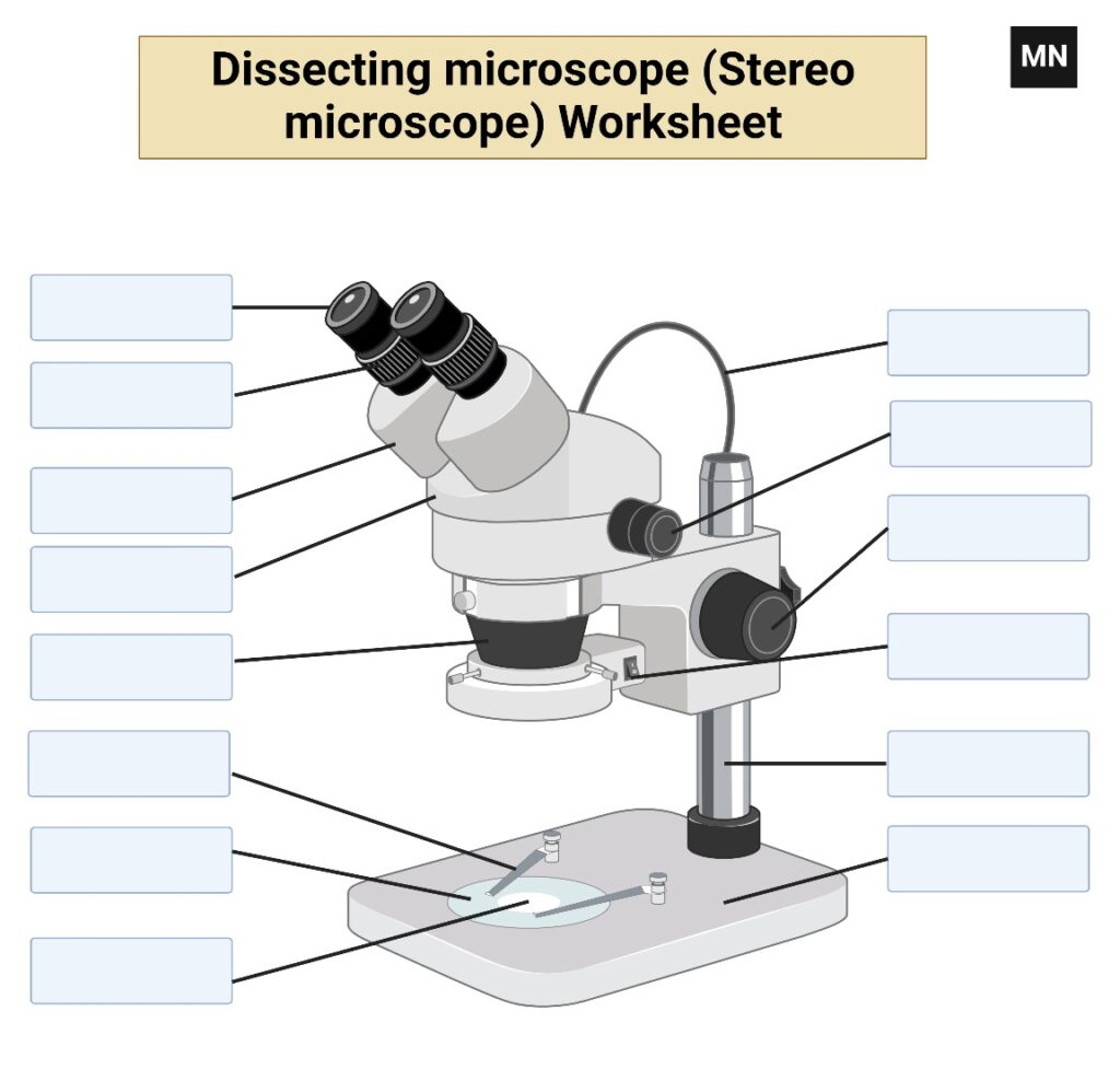
Dissecting Microscope Worksheet

Dissecting microscope images
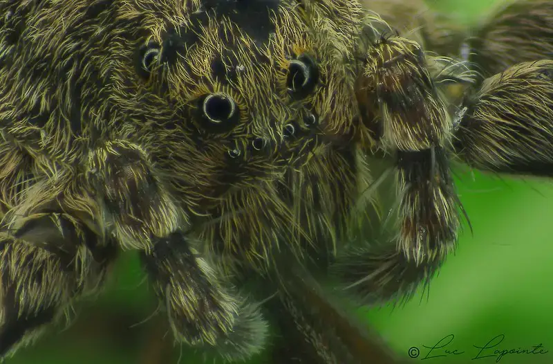
Dissecting microscope images – araignée poilue, 2 substacks de 21 images. Method=B (R=1,S=1) 90D sur stereo microscope
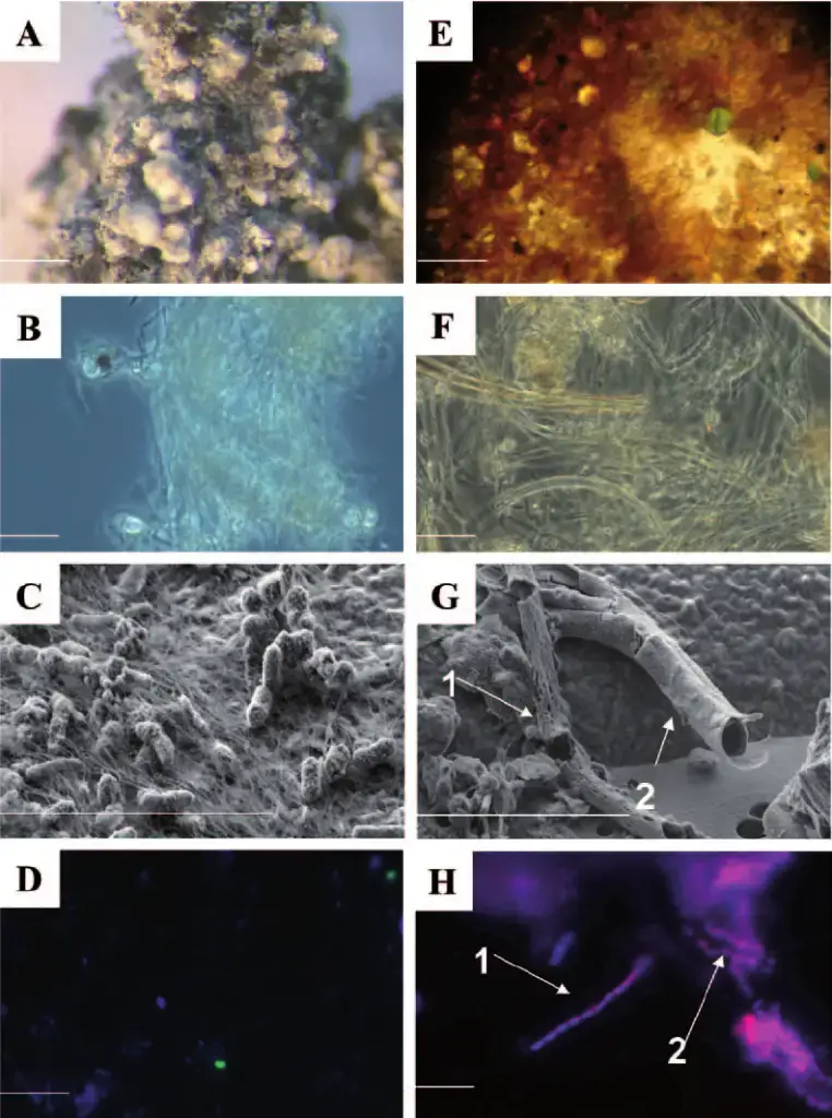
Dissecting microscope images – (A) Picture of a S aggregate under a dissecting microscope (A) on a white mat. The bar represents 1 mm on the scale. (B)  Picture of S filaments on a white mat in phase contrast. The bar represents 10 m in actual size. A high-resolution scanning electron micrograph (SEM) image showing filaments and related cells in the white mat (C). Arc94-targeted cells, as seen in fluorescent in situ hybridization (FISH) image (D) (green). A photograph of flakes of iron oxide (Fe oxide) found in an orange carpet. The bar represents 1 mm on the scale. An orange flake as seen using a light microscope in (F). (G) A magnified SEM picture of a flake with broken sheaths. The arrows denote two types of hidden microorganisms (the bacteria themselves are not visible,
Dissecting microscope images – (A) Picture of a S aggregate under a dissecting microscope (A) on a white mat. The bar represents 1 mm on the scale. (B) Picture of S filaments on a white mat in phase contrast. The bar represents 10 m in actual size. A high-resolution scanning electron micrograph (SEM) image showing filaments and related cells in the white mat (C). Arc94-targeted cells, as seen in fluorescent in situ hybridization (FISH) image (D) (green). A photograph of flakes of iron oxide (Fe oxide) found in an orange carpet. The bar represents 1 mm on the scale. An orange flake as seen using a light microscope in (F). (G) A magnified SEM picture of a flake with broken sheaths. The arrows denote two types of hidden microorganisms (the bacteria themselves are not visible,
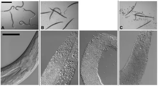
Dissecting microscope images – Nematodes cultivated in different environments, as seen under a dissecting microscope. Images in the top row have a 500 m scale bar. Cultured nematodes, seen under a compound microscope (bottom row). The anterior of the worm is toward the top right in all photographs in the bottom row, and the scale bar is 50 m. Soil-grown C. elegans (A) and B. thuringiensis (B) lacking Cry5B in a well-culture setting. In the top row, not a single one of the six nematodes has been infected. Everyone here is perfectly fine. Some of the worms on the top row appear blurry because they are swimming around in the well. The pharynx and gut of C. elegans that have been fed B. thuringiensis without Cry5B are normal and unharmed (bottom row). B) C. elegans co-cultured with B. thuringiensis and Cry5B in a well environment. In the top row, five of the six worms are fully infected (they lack normal colouring and internal structures) whereas one is not. Infected animals in the bottom row have internal structures that have been completely or nearly completely digested by the bacteria. These fatally infected animals harbour both vegetative and sporulated forms of the bacterium. (C) Similar photos to (B), but this time Bacillus anthracis has been cultivated with the nematodes.

Reference

Latest Questions

Start Asking Questions

This site uses Akismet to reduce spam. Learn how your comment data is processed.

⚠️
  1. Click on your ad blocker icon in your browser's toolbar
  2. Select "Pause" or "Disable" for this website
  3. Refresh the page if it doesn't automatically reload