

Hematopoiesis is a continuous physiological process by which all mature blood cells and bone marrow cells are formed. It is an essential process for maintaining normal blood cell number in the body.
This process starts from pluripotent hematopoietic stem cells (HSCs). These stem cells are mainly present in the soft spongy tissue of bone marrow in adult. They have the capacity of self-renewal and also form different progenitor cells.
The progenitor cells gradually differentiate and mature into the main blood cells. These include red blood cells (erythrocytes), white blood cells (leukocytes) and platelets. Erythrocytes carry oxygen to different body tissues. Leukocytes help in immune defence and fight against infection. Platelets help in blood clotting and wound healing.
Every day the body produces very large number of blood cells. More than 500 billion cells may be formed daily to maintain normal requirement of the body. This continuous production is needed because many blood cells have short life span.
The whole process is regulated by different chemical signals. These include cytokines and growth factors. The bone marrow niche also provides the supporting environment for the stem cells and helps to form correct type and number of blood cells according to body demand.
Location of Hematopoiesis
The location of hematopoiesis changes according to the age and developmental stage. It starts in embryo outside the bone marrow. Later it shifts to bone marrow.
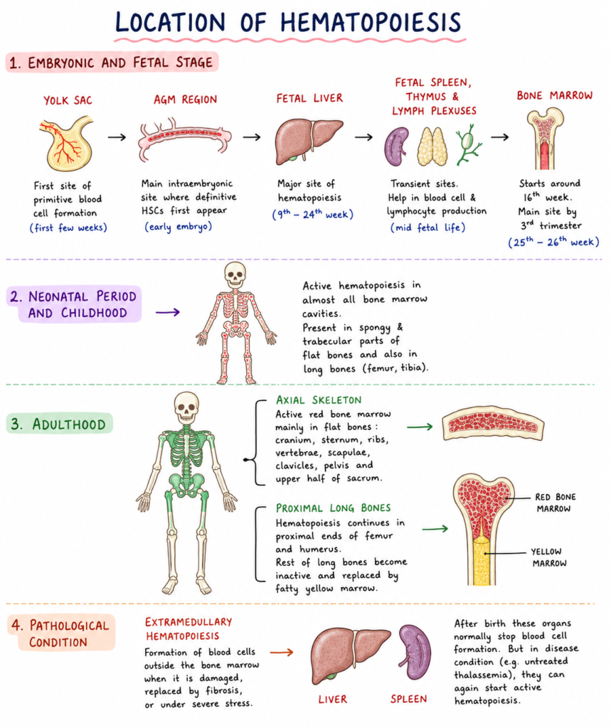
1. Embryonic and fetal stage
Yolk sac
It is the first site of primitive blood cell formation. It starts within first few weeks of gestation.
Aorta-gonad-mesonephros (AGM) region
It is the main intraembryonic site where definitive hematopoietic stem cells (HSCs) first appear. The placenta, embryonic head, vitelline and umbilical arteries also take part in this stage.
Fetal liver
It becomes the major site of hematopoiesis from 9th to 24th week of gestation. During this period most of the blood cell production occurs in the liver.
Fetal spleen, thymus and lymph plexuses
These are transient sites of blood cell formation. They help in production of blood cells and lymphocytes during mid fetal life.
Bone marrow
It starts hematopoietic activity around 16th week of gestation. It becomes the main site of blood cell formation by third trimester, mostly around 25th to 26th week.
2. Neonatal period and childhood
Entire bone marrow space
In newborn and children, active hematopoiesis occurs in almost all bone marrow cavities. It is present in spongy and trabecular parts of flat bones and also long bones like femur and tibia.
3. Adulthood
Axial skeleton
In adult, active red bone marrow is restricted mainly in flat bones. These include cranium, sternum, ribs, vertebrae, scapulae, clavicles, pelvis and upper half of sacrum.
Proximal long bones
Some hematopoiesis also continues in proximal ends of femur and humerus. The remaining parts of long bones become inactive and are replaced by fatty yellow marrow.
4. Pathological condition
Extramedullary hematopoiesis
It is the formation of blood cells outside the bone marrow. It occurs when bone marrow is damaged, replaced by fibrosis, or under severe stress.
Liver and spleen
After birth these organs normally stop blood cell formation. But in disease condition like untreated thalassemia, they can again start active hematopoiesis.

Step by step detail process of Hematopoiesis
The following are the steps of hematopoiesis–
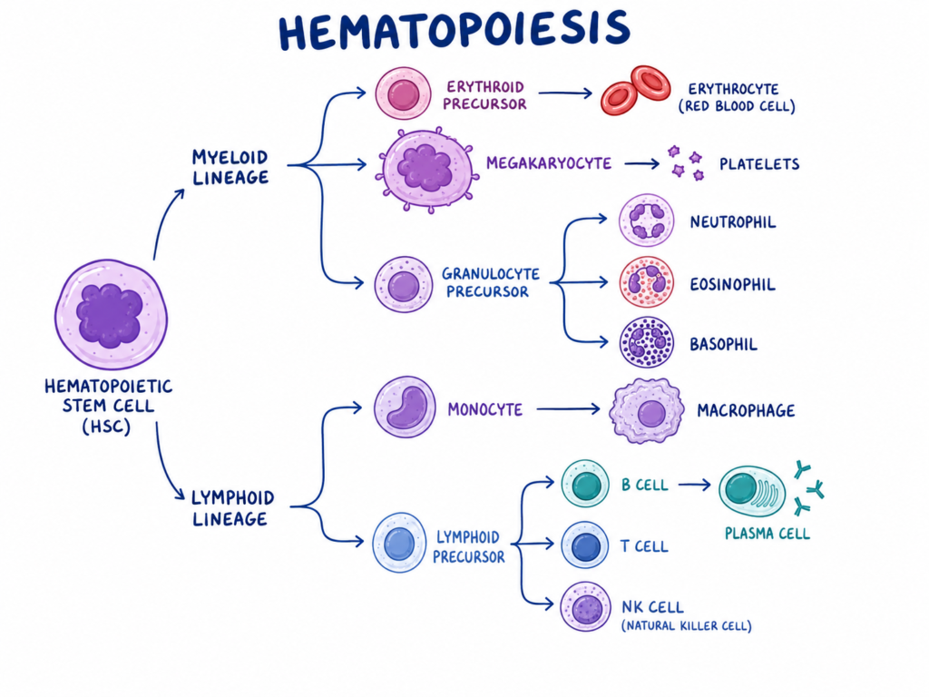
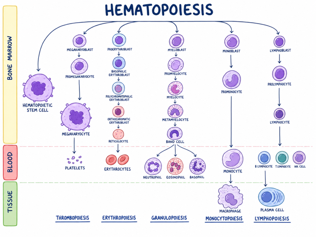
- The first step of hematopoiesis is started from hematopoietic stem cells (HSCs) present in the bone marrow. These cells are multipotent cells and can form all types of blood cells. Some of the HSCs divide to form same type of stem cells, so that the number of stem cells is maintained in the marrow. This process is called self-renewal.
- After this, some hematopoietic stem cells enter into the differentiation pathway. They divide and form multipotent progenitor cells (MPPs). These cells are still able to form different blood cells, but they do not have the same self-renewal power like the stem cells.
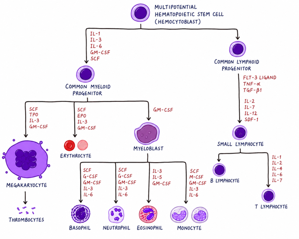
- In the next step, the multipotent progenitor cells become more restricted. They are not completely mature, but their future cell line become fixed. This is referred to as lineage commitment. Mainly two progenitor lines are formed, which are common myeloid progenitor (CMP) and common lymphoid progenitor (CLP).
- The common myeloid progenitor gives rise to different myeloid precursor cells. These cells are known as colony forming units (CFUs). Some cells become CFU-E, which are committed for formation of erythrocytes. Some cells become CFU-Meg, which are committed for formation of megakaryocytes.
- The megakaryocytes formed from myeloid line remain in the bone marrow and produce platelets by breaking small cytoplasmic fragments. In the same myeloid line, other precursor cells form neutrophils, eosinophils, basophils, monocytes and mast cells.
- The common lymphoid progenitor gives rise to lymphoid precursor cells. These cells are mainly concerned with formation of immune cells. They form B lymphocytes, T lymphocytes and Natural Killer (NK) cells. The T lymphocyte precursor goes to the thymus, where further maturation takes place.
- During terminal maturation, the immature precursor cells gradually change into mature blood cells. The nucleus, cytoplasm, granules and surface characters change according to the type of cell formed. The cells now become suitable for doing their normal function in blood and tissues.
- Finally, the mature cells are released from the bone marrow into blood circulation. Erythrocytes are used for oxygen transport. Leukocytes are used for body defence. Platelets are used in clotting of blood. In this way, the process of hematopoiesis continuously maintains blood cell formation.
Significance of Hematopoiesis
The following are the significance of hematopoiesis–
- Hematopoiesis is important for continuous formation and replacement of blood cells. Blood cells are destroyed after certain time, so new cells are formed again. In adult human body more than 500 billion new blood cells are formed every day, which includes about 175 billion erythrocytes, 175 billion platelets and 70 billion granulocytes. Thus it maintains normal blood cell number and homeostasis.
- It forms red blood cells (erythrocytes) which contain hemoglobin. These cells carry oxygen from lungs to different tissues of the body. If erythrocyte formation is reduced, then oxygen supply to tissue become poor and normal body function is affected.
- It forms different white blood cells (leukocytes) which are concerned with defence mechanism of body. These include lymphocytes, neutrophils and monocytes. They recognize foreign antigen and fight against bacterial and viral infection. During severe infection, more defensive cells are produced by emergency myelopoiesis.
- It forms megakaryocytes in the bone marrow, which give rise to platelets. These platelets stick together at the injured site and stop bleeding. They also help in wound healing process.
- Hematopoiesis is also important during embryonic life. In early embryo, primitive blood cell formation occurs along with formation of vascular channels. This is needed for early circulation and survival of embryo. Failure of primitive erythrocyte formation may cause embryonic death.
Properties of Hematopoietic Stem Cells
The following are the properties of hematopoietic stem cells (HSCs)–
- Self-renewal– HSCs have the capacity to divide and form the same type of stem cells again. This maintains the stem cell number in the bone marrow. By this property the stem cell pool is not depleted throughout life.
- Multipotency– HSCs are multipotent cells. They can give rise to all types of mature blood cells of myeloid and lymphoid lineages. Thus a single HSC can finally form erythrocytes, leukocytes and platelets.
- Morphology– HSCs are small round non-adherent cells. They have a rounded nucleus and the cytoplasm is very less in amount. Due to this character they resemble mature lymphocytes under microscope.
- Rarity– HSCs are very rare cells in the bone marrow. They are present about 1 in 10,000 cells in the myeloid tissue of bone marrow. Though the number is very low, they are responsible for continuous blood cell formation.
- Quiescence– Most of the HSCs remain in resting condition for long time. This resting stage is referred to as quiescence. It protects the genetic material of stem cells and prevents early exhaustion of the stem cell population.
- Low oxygen survival– HSCs can survive in the hypoxic condition of bone marrow. They use altered metabolism for long time survival in this low oxygen environment. This helps them to remain protected and functional.
- Mobility– HSCs have high capacity to pass through the bone marrow barrier. They can enter into blood circulation and move to other bones. They may also reach organs like thymus, liver and spleen for further development.
- Surface markers– HSCs are identified by special surface markers because they cannot be isolated only by seeing their shape. They are lineage negative (Lin-) cells and human HSCs commonly express CD34. Other markers like c-Kit and Sca-1 are also used in laboratory identification.
- Low dye uptake– HSCs absorb very little amount of some vital stains and dyes. This property helps in separation of HSCs from other bone marrow cells. So surface marker and dye uptake character are important for their identification.
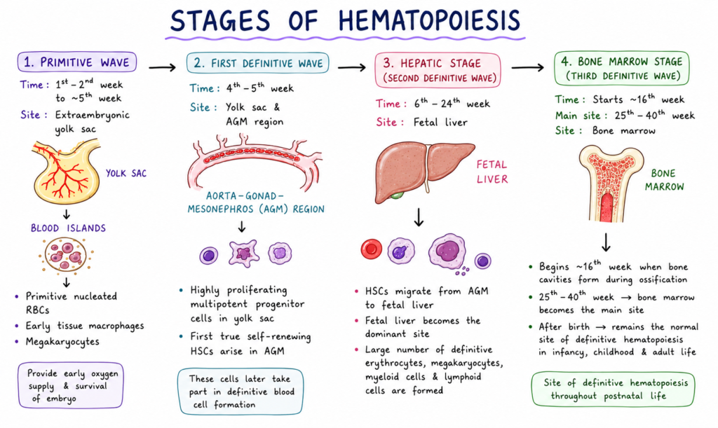
Stages of Hematopoiesis
The following are the stages of hematopoiesis–
- Primitive wave– Primitive hematopoiesis is the first blood forming stage of embryo. It takes place in the extraembryonic yolk sac from first two weeks to about 5th week of gestation. In this stage blood islands are formed. These blood islands give rise to primitive nucleated red blood cells, early tissue macrophages and megakaryocytes. The primitive red cells are needed for early oxygen supply of embryonic tissue and survival of embryo.
- First definitive wave– This wave occurs around 4th to 5th week of gestation. In this stage highly proliferating multipotent progenitor cells are formed in the yolk sac. At the same time first true self-renewing hematopoietic stem cells (HSCs) arise inside the embryo. The main site of this formation is aorta-gonad-mesonephros (AGM) region. These cells later take part in definitive blood cell formation.
- Hepatic stage– This is also called second definitive wave. It extends from 6th week to 24th week of gestation. During this stage HSCs migrate from the AGM region and colonize the fetal liver. So the fetal liver becomes the dominant site of blood cell formation. Here large number of definitive erythrocytes, megakaryocytes, myeloid cells and lymphoid cells are formed.
- Bone marrow stage– This is also called third definitive wave. It starts around 16th week of gestation when skeletal ossification forms bone cavities. Then the bone marrow gradually becomes active for blood cell production. Between 25th to 40th week, bone marrow becomes the main site of hematopoiesis. After birth, it remains the normal site of definitive hematopoiesis in infancy, childhood and adult life.
Growth Factors and Cytokines Regulating Hematopoiesis
The following are the important growth factors and cytokines regulating hematopoiesis–
- Stem cell factor (SCF)– It is produced by bone marrow stromal cells and endothelial cells. It is a broad acting growth factor. It is required for survival and proliferation of early hematopoietic stem cells (HSCs) and multipotent progenitor cells. Maintenance of stem cell pool is also done by this factor.
- Erythropoietin (EPO)– It is mainly produced by renal peritubular cells of kidney. It acts on erythroid progenitor cells. These cells are stimulated to form mature erythrocytes. Thus EPO is the main factor for red blood cell formation.
- Thrombopoietin (TPO)– It is mainly produced by hepatocytes of liver. It acts on megakaryocytic lineage. Formation of megakaryocytes is stimulated by this factor. Later these megakaryocytes produce platelets.
- GM-CSF– Granulocyte macrophage colony stimulating factor is mostly produced by T-cells. It acts on myeloid progenitor cells. Growth and differentiation of these cells are increased. It forms granulocytes and macrophages.
- G-CSF– Granulocyte colony stimulating factor acts mainly on neutrophil line. It stimulates growth and maturation of neutrophils. It is more active in infection condition. It also helps in movement of HSCs from bone marrow to blood.
- Interleukin-3 (IL-3)– It is produced by T-cells. It is a broad acting cytokine. It acts with other growth factors. Progenitor cells of erythrocytes, megakaryocytes, granulocytes and macrophages are stimulated by IL-3.
- Interleukin-4 (IL-4)– It is concerned with formation of basophils, mast cells and B-lymphocytes. It also takes part in allergic type immune reaction. Development of these cells are helped by IL-4.
- Interleukin-5 (IL-5)– It acts mainly on eosinophil lineage. Proliferation and differentiation of eosinophils are stimulated by this cytokine. So it is important in eosinophilic response.
- Interleukin-6 (IL-6)– It is a multifunctional cytokine. It acts on multipotent stem cells. It also helps in formation of platelets from mature megakaryocytes. It has role in acute phase reaction also.
- Interleukin-7 (IL-7)– It is important for lymphoid lineage. It helps in survival and development of T-cells and B-cells. Normal lymphocyte formation is maintained by this cytokine.
- Inhibitory cytokines– These cytokines suppress hematopoiesis. Important examples are tumor necrosis factor (TNF), interferon-gamma (IFN-γ) and transforming growth factor-beta (TGF-β). They are produced by bone marrow stromal cells and macrophages. In chronic inflammation, bone marrow suppression may occur due to these cytokines.
Regulation of Hematopoiesis
The following are the regulation of hematopoiesis–
- Bone marrow niche– It is the physical microenvironment of bone marrow. It is made by different supporting cells. These cells remain close to hematopoietic stem cells (HSCs) and regulate their survival, division, differentiation and resting condition.
- Osteoblasts– These are bone forming cells of the bone marrow niche. They regulate the HSC pool by producing angiopoietin-1, thrombopoietin and Jagged-1. They also produce inhibitory factor like osteopontin. Thus both support and control of HSCs is done by these cells.
- Endothelial cells– These cells line the blood vessels present in bone marrow. They secrete important maintenance factors like stem cell factor (SCF) and CXCL12. They also activate Notch and AKT signalling, by which HSCs are stimulated for proliferation and differentiation.
- Perivascular stromal cells– These cells are present near the blood vessels. They include mesenchymal stem cells also. They produce high amount of CXCL12, SCF and VCAM1. These factors help in retention and regulation of HSCs inside the bone marrow.
- Megakaryocytes– These are mature platelet forming cells and they are found near HSCs. They secrete CXCL4 and TGF-β1. These factors keep HSCs in dormant condition. This resting condition is important because it prevents exhaustion of stem cell population.
- Adipocytes– These are fat cells present in bone marrow. They produce adipokines like adiponectin and leptin. These factors can help in proliferation and differentiation of HSCs.
- Sympathetic nervous system– Nerve fibres are also present in the bone marrow. They regulate daily release of HSCs into the blood. This is done by changing the expression of CXCL12, which is an important retention molecule.
- Macrophages– These immune cells interact with stromal cells of bone marrow. They help to maintain normal level of CXCL12. Thus HSCs remain retained in the bone marrow niche.
- Stimulatory cytokines and growth factors– These factors increase survival, proliferation and maturation of blood cell precursors. Important examples are SCF, erythropoietin (EPO), thrombopoietin (TPO), G-CSF, M-CSF, IL-1, IL-3, IL-6 and IL-11. They act on different cell lines and promote formation of mature blood cells.
- Inhibitory cytokines– Some cytokines suppress hematopoiesis. These include tumor necrosis factor (TNF), interferon-gamma (IFN-γ) and transforming growth factor-beta (TGF-β). In chronic inflammation these factors may reduce bone marrow activity and cause marrow suppression.
- Adhesion molecules– Adhesion molecules keep HSCs attached in the bone marrow niche. The most important system is CXCL12-CXCR4 axis. Here CXCL12 binds with CXCR4 receptor and retains HSCs inside marrow. Other molecules like integrins, selectins and DEL-1 also help in cell attachment and lineage priming.
- Transcription factors– These are genetic regulators which control early development and lineage choice of blood cells. GATA1 and PU.1 regulate primitive red and white blood cell development. RUNX1 and GATA2 help in myelopoiesis. The HOX gene family controls early stem cell development and stage wise differentiation.
Regulation of Hematopoiesis
The regulation of hematopoiesis can be divided into two main part-
A. Cellular regulation by Bone Marrow Niche
- Bone marrow niche– It is the special microenvironment of bone marrow. It is made by different supporting cells. These cells remain around hematopoietic stem cells (HSCs) and control their survival, division, differentiation and resting stage.
- Osteoblasts– These are bone forming cells present in the bone marrow niche. They regulate the HSC pool by producing angiopoietin-1, thrombopoietin and Jagged-1. They also produce inhibitory factor like osteopontin. So support and control both are given by these cells.
- Endothelial cells– These cells form the lining of blood vessels in bone marrow. They secrete stem cell factor (SCF) and CXCL12. They also activate Notch and AKT signalling. By this, HSCs are stimulated for proliferation and differentiation.
- Perivascular stromal cells– These cells are located near blood vessels. Mesenchymal stem cells are also included in this group. They produce high amount of CXCL12, SCF and VCAM1. These proteins help in retention and regulation of HSCs inside marrow.
- Megakaryocytes– These are mature platelet forming cells and they remain close to HSCs. They secrete CXCL4 and TGF-β1. These factors keep HSCs in quiescent or dormant stage. So stem cell exhaustion is prevented by this regulation.
- Adipocytes– These are fat cells of bone marrow. They produce adipokines like adiponectin and leptin. These factors can promote proliferation and differentiation of HSCs.
- Sympathetic nervous system– Nerve fibres present in bone marrow regulate daily release of HSCs into blood. This regulation is done by controlling CXCL12 expression. So movement of HSCs from marrow to blood is also under nervous control.
- Macrophages– These cells interact with stromal cells of bone marrow. They maintain normal level of CXCL12. By this HSCs are retained properly in the bone marrow niche.
B. Molecular and genetic regulation
- Stimulatory cytokines and growth factors– These factors stimulate survival, proliferation and maturation of blood cell precursors. The important examples are SCF, erythropoietin (EPO), thrombopoietin (TPO), G-CSF, M-CSF, IL-1, IL-3, IL-6 and IL-11. They act on different cell line and help in formation of mature blood cells.
- Inhibitory cytokines– These cytokines suppress hematopoiesis. Important examples are tumor necrosis factor (TNF), interferon-gamma (IFN-γ) and transforming growth factor-beta (TGF-β). These factors may reduce bone marrow activity during chronic inflammation.
- Adhesion molecules– These molecules keep HSCs attached with the bone marrow niche. The most important system is CXCL12-CXCR4 axis. In this system CXCL12 binds with CXCR4 receptor and retains HSCs in marrow. Other molecules like integrins, selectins and DEL-1 also help in attachment and lineage priming.
- Transcription factors– These are genetic regulators of blood cell development. GATA1 and PU.1 regulate primitive red and white blood cell development. RUNX1 and GATA2 help in myelopoiesis. HOX gene family controls early stem cell development and stage wise differentiation.
Factors Affecting Hematopoiesis
The following are the factors affecting hematopoiesis–
- Growth factors and cytokines– Growth factors and cytokines are important chemical regulators of hematopoiesis. The important examples are stem cell factor (SCF), erythropoietin (EPO), thrombopoietin (TPO), granulocyte colony stimulating factor (G-CSF) and interleukins like IL-1, IL-3, IL-6 and IL-7. By these factors survival, proliferation and differentiation of blood progenitor cells are stimulated.
- Inhibitory cytokines– Some cytokines act in opposite way and suppress hematopoiesis. These include tumor necrosis factor-alpha (TNF-α), interferon-gamma (IFN-γ) and transforming growth factor-beta (TGF-β). During chronic inflammation and autoimmune attack, these factors may cause dormancy of stem cells or death of progenitor cells.
- Bone marrow niche– Bone marrow niche is the surrounding environment of hematopoietic stem cells (HSCs). It is formed by osteoblasts, endothelial cells, mesenchymal stem cells, macrophages, adipocytes and megakaryocytes. These cells provide local signals to HSCs. By these signals stem cells may remain resting, or they may divide and form blood cells.
- Adhesion molecules– Adhesion molecules are used to hold HSCs inside the bone marrow niche. The important molecules are CXCL12, CXCR4, DEL-1, integrins and selectins. These molecules attach stem cells with stromal cells and protect them in marrow. They also help in proper lineage development.
- Transcription factors– Transcription factors are internal genetic regulators of blood cell formation. The important factors are GATA1, GATA2, PU.1, RUNX1 and HOX gene family. These factors control early stem cell development and lineage commitment. Formation of red cell, white cell and myeloid cells are regulated by them.
- Sympathetic nervous system– Sympathetic nerve fibres are present in the bone marrow. They affect the release of hematopoietic stem cells from marrow into blood. This is done by controlling the level of CXCL12. Thus daily movement of HSCs is also regulated by nervous system.
- Metabolic and nutritional state– Hematopoiesis is affected by metabolic condition and nutrition of body. In obesity, fat cells may increase in the bone marrow and inflammatory cell formation may be increased. Deficiency of vitamin B12, folate and copper causes defective maturation of blood cells. So proper nutrition is needed for normal blood cell production.
- External toxins and treatment– Ionizing radiation, benzene, pesticides and some chemotherapeutic drugs damage the stem cells of bone marrow. Alkylating agents are important example. These agents may damage stem cell DNA. Due to this bone marrow failure or myelodysplastic syndrome may occur.
- Viral infection and autoimmunity– Some viral infections suppress hematopoiesis. The important examples are hepatitis virus, Epstein-Barr virus and parvovirus. In autoimmune disease, body’s own cytotoxic T-cells may attack hematopoietic stem cells. This may lead to severe marrow suppression and acquired aplastic anemia.
Disorders of Hematopoiesis
The following are the disorders of hematopoiesis–
- Aplastic anemia– It is a life threatening condition of bone marrow. In this condition the bone marrow cannot produce sufficient blood cells. So red blood cells, white blood cells and platelets are decreased together. This is called pancytopenia. It may be acquired due to autoimmune reaction, drugs, environmental toxins and viral infection. It may also be inherited.
- Myelodysplastic syndrome (MDS)– It is a clonal disorder of bone marrow stem cells. In this disease abnormal blood cells are produced from defective stem cells. The cells are formed but they are not properly functional. So it is called ineffective hematopoiesis. It has high chance to convert into acute myeloid leukemia (AML).
- Inherited bone marrow failure syndrome (IBMFS)– These are genetic disorders in which bone marrow failure occurs due to inherited defect. One or more blood cell line may be affected in these disorders.
- Fanconi anemia– It causes genomic instability, bone marrow failure and physical abnormalities. Short stature and absent thumb may be found.
- Dyskeratosis congenita– It occurs due to defective telomere maintenance. Due to this defect hematopoietic stem cells are exhausted early.
- Blackfan-Diamond anemia– It mainly causes failure of red blood cell formation. Defective ribosome biogenesis is present in this condition.
- Shwachman-Diamond syndrome– It causes neutropenia, exocrine pancreatic dysfunction and skeletal abnormalities.
- Congenital amegakaryocytic thrombocytopenia– It causes selective loss of megakaryocytes. So severe platelet deficiency occurs.
- Reticular dysgenesis– It is a severe combined immune deficiency. Marked leukopenia and neutropenia are present.
- Myeloproliferative neoplasms– These are disorders in which mature blood cells are produced in excess amount. The bone marrow becomes overactive and one or more blood cell line is increased.
- Polycythemia vera– In this disease red blood cells are produced in excess amount. The erythroid precursors over respond to erythropoietin due to genetic mutation.
- Essential thrombocythemia– In this condition excessive and uncontrolled production of platelets occurs.
- Primary myelofibrosis– In this condition scarring and fibrosis occur in the bone marrow. So normal blood cell formation is disturbed and hematopoiesis may shift to liver and spleen.
- Leukemia– It is a malignant disease of blood forming tissue. In this disease immature or abnormal leukocytes are produced in large number. These abnormal cells occupy the bone marrow and suppress normal blood cell formation.
- Acute myeloid leukemia (AML)– It is malignant increase of immature myeloid cells.
- Acute lymphoblastic leukemia (ALL)– It is malignant increase of immature lymphoid cells.
- Chronic myeloid leukemia (CML)– It is chronic increase of myeloid cells.
- Chronic lymphocytic leukemia (CLL)– It is chronic increase of lymphoid cells.
- Hemoglobinopathies– These are genetic disorders of hemoglobin and red blood cells. In these conditions normal erythropoiesis is affected and red cells are destroyed early.
- Beta-thalassemia– In this disease beta-globin chain formation is reduced or absent. Excess alpha-globin chain gets accumulated. Due to this erythroid precursors die early and ineffective erythropoiesis occurs.
- Sickle cell anemia– In this disease red blood cells become sickle shaped in low oxygen condition. These cells are destroyed early and they may block small capillaries.
- Paroxysmal nocturnal hemoglobinuria (PNH)– It is a rare disorder of red blood cells. In this condition abnormal red cells break down inside blood vessels. Hemoglobin is released into blood and may pass in urine. It may also develop after aplastic anemia.
- Myelophthisic syndrome– It is a condition in which normal hematopoietic bone marrow is replaced by abnormal tissue. This abnormal tissue may be metastatic tumor, hematological malignancy or fibrosis. Due to this replacement normal blood cell formation becomes decreased.
Clinical Significance of Hematopoiesis
The following are the clinical significance of hematopoiesis–
- Stem cell transplantation– Hematopoietic stem cell transplantation (HSCT) is based on the self-renewal and blood forming capacity of hematopoietic stem cells (HSCs). In this method defective or damaged bone marrow is replaced by normal stem cells. It is used in leukemia, aplastic anemia and inherited bone marrow failure syndrome. By this process normal blood cell formation may be restored.
- Blood cancers– Abnormal regulation of hematopoiesis may cause malignant blood cell formation. It may occur due to genetic mutation, radiation and toxic chemicals. The abnormal stem or progenitor cells produce abnormal blood cells. This is seen in myelodysplastic syndrome (MDS), myeloproliferative neoplasm and acute leukemia. So study of hematopoiesis is used in diagnosis and treatment of these diseases.
- Bone marrow failure– Failure of hematopoiesis causes failure of blood cell production. In aplastic anemia, RBC, WBC and platelets are reduced together. This condition is called pancytopenia. The cause may be autoimmune, genetic, drug induced or toxin induced. According to the cause, immunosuppressive treatment and supportive care are given.
- Hemoglobinopathies– Hematopoiesis has importance in sickle cell anemia and β-thalassemia. In these disorders normal hemoglobin production and erythropoiesis are affected. The change from fetal hemoglobin (HbF) to adult hemoglobin (HbA) is important in treatment. Hydroxyurea is used to increase HbF and it reduces disease symptoms.
- Congenital disorders– Early embryonic and fetal hematopoiesis is useful for understanding congenital anemia and inherited immune deficiency. Defect in early blood formation may cause abnormal blood condition in newborn. It also helps in early diagnosis and management of high risk pregnancy.
- Growth factor therapy– Some cytokines and growth factors are used as medicine. Erythropoietin (EPO) is used for stimulation of red blood cell production in anemia. Granulocyte colony stimulating factor (G-CSF) is used to increase neutrophils, especially after chemotherapy. So a particular blood cell line can be stimulated by giving these factors.
References
- Nimmana, B. K., & Penney, S. W. (2025, July 7). Aplastic anemia. In StatPearls. StatPearls Publishing. https://www.ncbi.nlm.nih.gov/books/NBK534212/
- UT Health San Antonio MD Anderson Mays Cancer Center. (n.d.). Aplastic anemia and myelodysplastic syndromes (MDS). Retrieved June 24, 2026, from https://cancer.uthscsa.edu/cancer-care/conditions/cancer-types/aplastic-anemia-MDS
- Barrett, D. (2024, October 18). Aplastic anemia and myelodysplastic syndromes: Similarities and differences. HealthTree for Myelodysplastic Syndromes.
- Aplastic anemia vs. myelodysplastic syndrome – Moffitt Cancer Center. (n.d.). Moffitt Cancer Center.
- Aplastic anemia: Etiology, molecular pathogenesis, and emerging concepts – PubMed. (n.d.). PubMed.
- Moore, C. A., & Krishnan, K. (2023, July 10). Bone marrow failure. In StatPearls. StatPearls Publishing. https://www.ncbi.nlm.nih.gov/books/NBK459249/
- Chapter 6: Hematology of the fetus and newborn – AccessMedicine. (n.d.). AccessMedicine.
- National Institute of Diabetes and Digestive and Kidney Diseases. (2020, July). Definition & facts for aplastic anemia & myelodysplastic syndromes. U.S. Department of Health and Human Services.
- Liang, X. (2024). Development of hematopoiesis in the fetus, at birth, and after birth. In X. Liang, B. Siegele, & J. Picarsic (Eds.), Pediatric pathology of hematopoietic and histiocytic disorders (pp. 1-4). Cambridge University Press.
- Liang, X. (2024). Development of hematopoiesis in the fetus, at birth, and after birth (Chapter 1). In X. Liang, B. Siegele, & J. Picarsic (Eds.), Pediatric pathology of hematopoietic and histiocytic disorders. Cambridge University Press. https://doi.org/10.1017/9781009105002.001
- Singh, R., & Sugumar, K. (2025, May 3). Embryology, hematopoiesis. In StatPearls. StatPearls Publishing. https://www.ncbi.nlm.nih.gov/books/NBK544245/
- Hematopoiesis – PMC – NIH. (n.d.). PubMed Central.
- Thermo Fisher Scientific. (n.d.). Hematopoiesis from pluripotent stem cells. Thermo Fisher Scientific.
- Hematopoietic stem cell niches and signals controlling immune cell development and maintenance of immunological memory – PMC. (n.d.). PubMed Central.
- Wikipedia contributors. (2026, June 8). Hematopoietic stem cell. In Wikipedia, The Free Encyclopedia. Retrieved from https://en.wikipedia.org/w/index.php?title=Hematopoietic_stem_cell&oldid=1358408084
- Hematopoietic stem cell discovery: Unveiling the historical and future perspective of colony-forming units assay – PMC. (n.d.). PubMed Central.
- Chapman, J., & Zhang, Y. (2023, May 1). Histology, hematopoiesis. In StatPearls. StatPearls Publishing. https://www.ncbi.nlm.nih.gov/books/NBK534246/
- Lymphoid and myeloid lineage commitment in multipotent … (n.d.).
- Dotson, J. L., & Lebowicz, Y. (2022, July 18). Myelodysplastic syndrome. In StatPearls. StatPearls Publishing. https://www.ncbi.nlm.nih.gov/books/NBK534126/
- Myelodysplastic syndrome: Clinical characteristics and significance of preclinically detecting biallelic mutations in the TET2 gene – PMC. (n.d.). PubMed Central.
- Emadi, A., & Law, J. Y. (2026, February). Myelodysplastic syndromes (MDS). Merck Manual Professional Edition.
- Novel combinations to improve hematopoiesis in myelodysplastic syndrome – PMC. (n.d.). PubMed Central.
- Bowers, E., & Singer, K. (2021). Obesity-induced inflammation: The impact of the hematopoietic stem cell niche. JCI Insight, 6(3), Article e145295. https://doi.org/10.1172/jci.insight.145295
- Pathogenesis of acquired aplastic anemia and the role of the bone … (n.d.).
- Quantitative analysis and spatiotemporal dynamics of vertebrate hematopoiesis: Ontogeny, niche architecture, and pathophysiological failure states. (n.d.).
- Mitroulis, I., Kalafati, L., Bornhäuser, M., Hajishengallis, G., & Chavakis, T. (2020). Regulation of the bone marrow niche by inflammation. Frontiers in Immunology, 11, Article 1540. https://doi.org/10.3389/fimmu.2020.01540
- Regulation of the bone marrow niche by inflammation – PMC – NIH. (n.d.). PubMed Central.
- T and B lymphocyte differentiation from hematopoietic stem cell – PMC – NIH. (n.d.). PubMed Central.
- The complex pathophysiology of acquired aplastic anaemia – PMC – NIH. (n.d.). PubMed Central.
- Purton, L. E., & Scadden, D. T. (2008, November 15). The hematopoietic stem cell niche. In StemBook. Harvard Stem Cell Institute. https://doi.org/10.3824/stembook.1.28.1
- Mobley, I. (2021, October 4). Understanding prenatal haematopoiesis in bone marrow. Front Line Genomics.