Major Histocompatibility Complex II (MHC II) molecules are protein molecules present on the surface of some immune cells. These are mainly found on professional antigen presenting cells (APCs). The cells include dendritic cells, macrophages and B cells.
MHC II molecules are different from MHC I molecules because MHC I is present on almost all nucleated cells. But MHC II is mainly present on those cells which take antigen and show it to immune cells. In human, these molecules are encoded by Human Leukocyte Antigen (HLA) gene complex.
The important HLA genes for MHC II are HLA-DR, HLA-DQ and HLA-DP. These genes form the molecules which are used in antigen presentation. It has important role in adaptive immunity.
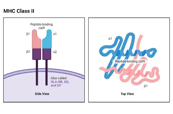
Structurally, MHC II molecule is heterodimer. It is composed of two different protein chains. One is alpha (α) chain and another is beta (β) chain. These two chains are joined together.
The two chains form a cleft like space. This is called peptide binding groove. This groove is open from both ends. Due to this open groove, long peptide fragment can bind with it.
The peptide bind with MHC II molecule is generally long. It contains about 13 to 25 amino acids. These peptide fragments are formed from extracellular foreign proteins.
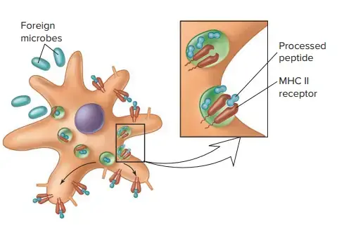
The function of MHC II molecules is to present extracellular antigen. These antigens may be from bacteria, viruses, fungi or other foreign particles. The APC first engulf the foreign protein and digest it.
The digestion occur inside acidic compartments of the cell. These compartments are called endosomes and lysosomes. In this place the antigen is broken into small peptide fragments.
Then MHC II molecule bind with one peptide fragment. This MHC II-peptide complex is carried to the cell surface. On the surface it is presented to CD4+ helper T cells.
When CD4+ helper T cell recognize this antigen, it becomes activated. Then it release signals for immune response. It also helps activation of B cells for antibody formation.
Distribution of Major Histocompatibility Complex II (MHC II molecules)
MHC II molecules are distributed mainly on professional antigen presenting cells (APCs). These cells normally express MHC II molecules on cell surface. These are as follows-
Dendritic cells– These cells contain high amount of MHC II molecules. It take antigen from tissue and present the processed antigen to CD4+ helper T cells.
Macrophages– Macrophages also express MHC II molecules. The expression is more after activation. It present engulfed antigen to helper T cells.
B cells– B cells have MHC II molecules on their surface. It bind processed antigen and show it to CD4+ T cells. This help in antibody response.
Langerhans cells– These are antigen presenting cells of skin. They also contain MHC II molecules. It take antigen from skin region and present it.
Thymic epithelial cells– MHC II molecules are present on epithelial cells of thymus. It has role in selection of developing T cells inside thymus.
Other immune cells– Group 3 innate lymphoid cells (ILC3) also show MHC II molecules. Some activated T cells may also express it. These include T peripheral helper cells, memory regulatory T cells and some CD8+ T cells.
Inducible cells– Many cells normally do not express MHC II molecules. But during inflammatory condition these cells can express it. This induction occur mainly by interferon-gamma (IFN-γ).
The inducible cells are endothelial cells, fibroblasts, mesenchymal stromal cells, epithelial cells and enteric glial cells.
Fibroblast-like synoviocytes of joint may also express MHC II molecules during inflammation.
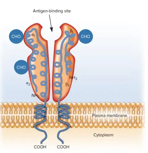
Structure of Major Histocompatibility Complex II (MHC II molecules)
- Heterodimeric composition- MHC II molecule is a heterodimer. It is made up of two different polypeptide chains. One is alpha (α) chain and another is beta (β) chain. These two chains are attached by non-covalent interaction.
- Molecular weight- The α chain is larger protein and it is about 32-35 kDa. The β chain is smaller and it is about 26-32 kDa.
- Domain architecture- Both α chain and β chain have three main regions.
- Two extracellular domains are present outside the cell membrane.
- One hydrophobic transmembrane segment is present in plasma membrane.
- One short cytoplasmic tail is present inside the cell.
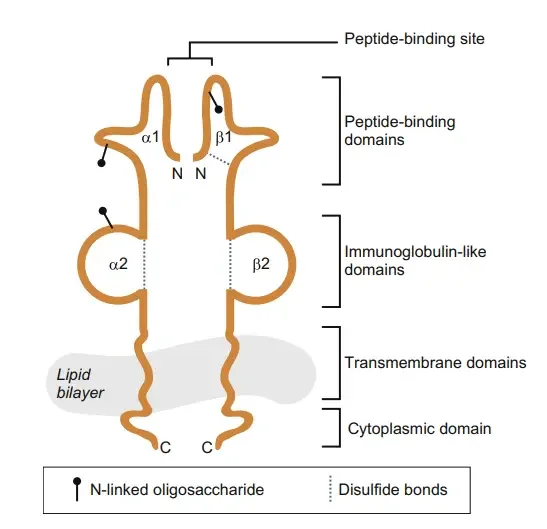
- Extracellular domains- The α chain has α1 and α2 domains. The β chain has β1 and β2 domains. These domains form the outer part of MHC II molecule.
- Transmembrane segment- It is hydrophobic part. It anchors the MHC II molecule into the plasma membrane. So the molecule remain fixed on cell surface.
- Cytoplasmic tail- It is short intracellular part of both chains. It extends into the cell. It may take part in signal transduction.
- Membrane proximal domains- The α2 and β2 domains are present close to cell membrane. These domains show conserved immunoglobulin-like fold. The β2 domain act as binding site for CD4 co-receptor of helper T cells.
- Peptide-binding cleft- This cleft is formed by α1 and β1 domains. These domains are membrane distal part. It forms the antigen binding site of MHC II molecule.
- The floor is made up of eight-stranded antiparallel β-sheet.
- The walls are made up of two parallel segmented α-helices.
- The cleft is open at both ends.
- Open-ended groove- The groove is open on both side. So longer peptide can remain attached in it. The peptide is usually 12 to 25 amino acids long and may extend outside the margin of groove.
- Anchor pockets- The cleft has polymorphic pockets called P1, P4, P6 and P9. These pockets interact with side chains of peptide. This help in deciding binding specificity.
- Binding forces- The peptide is held in the cleft by hydrogen bonds and van der Waals forces. These forces act between MHC II helical walls and peptide backbone.
Mechanism of action of Major Histocompatibility Complex II (MHC II)
- Antigen uptake- Professional antigen presenting cells (APCs) take the extracellular antigen first. These cells are dendritic cells, macrophages and B cells. The antigen may be from bacteria, fungi, virus or other foreign protein. The uptake occur by phagocytosis or endocytosis.
- Antigen processing- After uptake, the foreign protein enter into endocytic pathway. It is carried into acidic compartments like endosomes and lysosomes. In this place antigen remain for digestion.
- Peptide formation- The antigen is cleaved by cathepsins and other acid proteases. These enzymes cut large protein into small peptide fragments. These peptide fragments are later used for binding with MHC II molecule.
- MHC II synthesis- At the same time MHC II molecule is formed in rough endoplasmic reticulum (RER). The alpha (α) chain and beta (β) chain are synthesized here. These two chains join and form one MHC II molecule.
- Invariant chain binding- The newly formed MHC II molecule has open peptide binding groove. Invariant chain (Ii) bind with this groove. It block the groove and prevent binding of self protein inside ER.
- Transport to MIIC- The MHC II-invariant chain complex leaves the ER. It pass through Golgi apparatus. Then it is directed into acidic compartment called MHC class II compartment (MIIC).
- CLIP formation- Inside MIIC, most part of invariant chain is digested. Mainly cathepsin S and cathepsin L are involved. A small part of invariant chain remain inside the groove. This part is called CLIP (Class II-associated Invariant chain Peptide).
- CLIP removal- HLA-DM interact with MHC II-CLIP complex. It remove CLIP from the binding groove. After this the groove become empty for antigenic peptide.
- Peptide loading- The processed foreign peptide bind in the empty groove of MHC II molecule. HLA-DM help in loading of proper peptide. Weakly bound peptide are removed and stable peptide remain attached. In B cells, HLA-DO regulate the activity of HLA-DM.
- Complex formation- After stable peptide binding, peptide-MHC II complex is formed. This complex dissociate from HLA-DM. It is now ready for movement to cell surface.
- Surface transport- The peptide-MHC II complex moves to plasma membrane of APC. It is displayed on the cell surface. The antigenic peptide remain exposed outside.
- T cell recognition- CD4+ helper T cell recognize the peptide. T cell receptor (TCR) bind with peptide-MHC II complex. CD4 co-receptor also bind with MHC II molecule.
- T cell activation- If the peptide is recognized as foreign, CD4+ helper T cell become activated. It release cytokines. It help B cells for antibody production and activate macrophages for killing of pathogen.
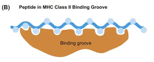
How are self and non-self peptides deposited in the MHC II binding pocket?
- Protein source- Non-self peptides are obtained from extracellular pathogen proteins. These proteins may come from bacteria, viruses and fungi. They are taken up by APCs through endocytosis or phagocytosis.
- Self protein source- Self peptides are obtained from the body’s own proteins. Some self proteins are taken from extracellular space. Some are brought from cytoplasm to endosomal pathway by autophagy.
- Self peptide amount- In normal healthy condition, many peptides shown by MHC II molecules are self peptides. About 70-90% of presented peptides may be self peptides.
- Protein degradation- The collected self and non-self proteins are carried into acidic compartments. These are late endosomes, lysosomes or phagolysosomes. In this place proteins are cut into small peptide fragments.
- Protease action- The degradation is done by cathepsins and other acid proteases. These enzymes break large protein into smaller peptide. These peptides become available for MHC II binding pocket.
- MHC II formation- New MHC II molecules are formed in endoplasmic reticulum (ER). The alpha (α) chain and beta (β) chain join there and form the peptide binding groove.
- Invariant chain binding- The groove of new MHC II molecule is blocked by invariant chain (Ii). It prevent binding of unwanted native protein in ER. So the binding pocket remain protected.
- Transport to MIIC- The MHC II-invariant chain complex move from ER to Golgi apparatus. Then it reach acidic compartment called MHC class II compartment (MIIC). Here the degraded peptides are also present.
- CLIP formation- Inside MIIC, most part of invariant chain is digested. Mainly cathepsin S and cathepsin L act on it. Only a small fragment remain in the pocket. This fragment is called CLIP (Class II-associated Invariant chain Peptide).
- CLIP removal- HLA-DM bind with MHC II-CLIP complex. It helps in removing CLIP from the binding pocket. After this the groove become free for peptide binding.
- Peptide deposition- Available self peptides and non-self peptides now enter into the empty MHC II binding pocket. The peptide which fit properly remain attached. Weakly attached peptide are removed.
- Peptide editing- HLA-DM act as peptide editing molecule. It remove unstable peptide again and again. Stable peptide with tight binding is finally deposited in the groove.
- Stable complex- After proper peptide binding, stable peptide-MHC II complex is formed. The peptide may be self or non-self. It is held inside the groove by weak bonds and pocket interaction.
- Surface display- The stable peptide-MHC II complex move to plasma membrane. It is displayed on the surface of APC. Then it can be recognized by CD4+ helper T cells.
Functions of MHC II
- Exogenous antigen presentation- MHC II molecules present exogenous antigen on the cell surface. These antigens are taken from outside the cell. It may be from bacteria, fungi, virus and other foreign protein.
- CD4+ T cell activation- MHC II-peptide complex is recognized by CD4+ helper T cells. The TCR of helper T cell bind with this complex. After recognition, CD4+ T cell become activated.
- Self and non-self recognition- MHC II molecules show both self and non-self peptide. By this process immune system can identify own harmless protein and foreign harmful protein.
- B cell activation- B cells present antigen with MHC II molecules to helper T cells. After this, B cells undergo clonal expansion. It also help in somatic hypermutation and antibody class switching.
- Antibody production- Activated helper T cell give signal to B cells. Then B cells form plasma cells and produce specific antibody. This antibody act against the same antigen.
- Macrophage activation- Macrophages present engulfed antigen through MHC II molecules. Th1 cells recognize this antigen. Then Th1 cells release interferon-gamma (IFN-γ) which increase killing activity of macrophage.
- Inflammatory response- MHC II presentation help in forming proper type of immune response. It help in differentiation of naive T cells into Th1, Th2, Th17 or regulatory T cells (Tregs) according to type of pathogen.
- Positive selection- In thymus, thymic epithelial cells present self peptide with MHC II molecules. Developing thymocytes which bind with low or moderate affinity survive. These cells become CD4+ T cell lineage.
- Negative selection- MHC II molecules also help in removal of strongly self reactive T cells. In thymus, medullary thymic cells present self antigen. Strongly reactive T cells undergo apoptosis.
- Regulatory T cell formation- Some thymocytes bind with MHC II-self peptide complex with intermediate to high affinity and proper costimulatory signal. These cells differentiate into regulatory T cells (Tregs).
- Autoimmunity control- Tregs formed by this process suppress unwanted immune response. It help in preventing autoimmune reaction in peripheral tissues.
References
- 2. Central & peripheral tolerance. (n.d.). Immunopaedia.
- 4. MHC & antigen presentation. (n.d.). Immunopaedia.
- Associations and interactions between bare lymphocyte syndrome factors. (n.d.). PubMed Central (PMC).
- Bare lymphocyte syndrome type II. (2017). MedlinePlus.
- Bare lymphocyte syndrome type II. (2017). MedlinePlus Genetics.
- Central tolerance. (n.d.). Wikipedia.
- CIITA and its dual roles in MHC gene transcription. (n.d.). PubMed Central (PMC).
- CLIP (protein). (n.d.). Wikipedia.
- Comparison of peptide conformations presented by MHC-I and MHC-II. (n.d.). ResearchGate.
- Expression of MHC II genes. (n.d.). ResearchGate.
- Ferrante, A. (2013). Thermodynamics of peptide-MHC class II interactions: Not all complexes are created equal. Frontiers in Immunology, 4, 308.
- For many but not for all: How the conformational flexibility of the peptide/MHCII complex shapes epitope selection. (n.d.). PubMed Central (PMC).
- HLA-DM and HLA-DO, key regulators of MHC-II processing and… (n.d.). PubMed Central (PMC).
- HLA-DR risk variants in rheumatoid arthritis: What we know and still do not know. (2026). BMJ.
- Imbachí-Salamanca, A., & Vásquez, G. (2024). Central tolerance in T cells, what’s new?. Revista Colombiana de Reumatología, 31(4). SciELO.
- Interactions of CD4 with MHC class II molecules, T cell receptors and p56lck. (n.d.). PubMed.
- Interactions of CD4 with MHC class II molecules, T cell receptors and p56lck. (n.d.). ResearchGate.
- Janeway, C. A., Jr., Travers, P., Walport, M., & Shlomchik, M. J. (2001). The major histocompatibility complex and its functions. In Immunobiology: The immune system in health and disease (5th ed.). Garland Science. NCBI Bookshelf.
- Kerry, J. (2025, February 13). Major histocompatibility complex. Rapid Novor.
- Larid, G., Pancarte, M., Offer, G., Clavel, C., Martin, M., Pradel, V., Auger, I., Lafforgue, P., Roudier, J., Serre, G., & Balandraud, N. (2021). In rheumatoid arthritis patients, HLA-DRB1*04:01 and rheumatoid nodules are associated with ACPA to a particular fibrin epitope. Frontiers in Immunology, 12, 692041.
- Leddon, S. A., & Sant, A. J. (2010). Generation of MHC class II:peptide ligands for CD4 T cell allorecognition of MHC class II molecules. Current Opinion in Organ Transplantation, 15(4), 505–511. PMC.
- León Machado, J. A., & Steimle, V. (2021). The MHC class II transactivator CIITA: Not (quite) the odd-one-out anymore among NLR proteins. International Journal of Molecular Sciences, 22(3), 1074. MDPI.
- Lu, X.-B., Wang, Z.-X., Liu, S.-B., Zhang, X.-Y., Lu, L.-F., Li, S., Chen, D.-D., Nie, P., & Zhang, Y.-A. (2019). Interferon regulatory factors 1 and 2 play different roles in MHC II expression mediated by CIITA in grass carp, Ctenopharyngodon idella. Frontiers in Immunology, 10, 1106.
- Major histocompatibility complex. (n.d.). Wikipedia.
- McCracken, R. (2006). HLA-DM. Davidson College.
- MHC class II deficiency. (n.d.). Immunodeficiency Search.
- MHC class II. (n.d.). Wikipedia.
- MHC I vs II peptide binding – Understanding T cell presentation. (n.d.). peptides & elephants.
- Molecular architecture, catalytic pathways, and immunological functions of major histocompatibility complex class II molecules. (n.d.).
- National Center for Biotechnology Information. (2026). CIITA class II major histocompatibility complex transactivator [Homo sapiens (human)]. NCBI.
- Nesmiyanov, P. (n.d.). Antigen processing and presentation. British Society for Immunology.
- New insights into the functional role of the rheumatoid arthritis shared epitope. (n.d.). PubMed Central (PMC).
- Promoter-specific functions of CIITA and the MHC class II… (n.d.). PubMed Central (PMC).
- Regulation of class II MHC expression in APCs: Roles of types I, III, and IV class II transactivator. (n.d.). PubMed.
- Self-association of CIITA and its transactivation potential. (n.d.). PubMed Central (PMC).
- Species-specific binding of CD4 to the beta 2 domain of major histocompatibility complex class II molecules. (n.d.). PubMed Central (PMC).
- Structural insights into HLA-DM mediated MHC II peptide exchange… (n.d.). PubMed Central (PMC).
- Structural insights into human MHC-II association with invariant… (n.d.). PNAS.
- The activation threshold of CD4+ T cells is defined by TCR/peptide-MHC class II interactions in the thymic medulla. (n.d.). PubMed Central (PMC).
- The rheumatoid arthritis HLA-DRB1 shared epitope. (n.d.). PubMed Central (PMC).
- Type 1 diabetes associated HLA-DQ2 and DQ8 molecules are relatively resistant to HLA-DM mediated release of invariant chain-derived CLIP peptides. (n.d.). PubMed Central (PMC).
- Use of eluted peptide sequence data to identify the binding characteristics of peptides to the insulin-dependent diabetes susceptibility allele HLA-DQ8 (DQ 3.2). (n.d.). PubMed.
- Vignali, D. A., Moreno, J., Schiller, D., & Hämmerling, G. J. (1992). Species-specific binding of CD4 to the beta 2 domain of major histocompatibility complex class II molecules. Journal of Experimental Medicine, 175(4), 925-932. PubMed.
- Wang, J. H., Meijers, R., Xiong, Y., Liu, J. H., Sakihama, T., Zhang, R., Joachimiak, A., & Reinherz, E. L. (2001). Crystal structure of the human CD4 N-terminal two-domain fragment complexed to a class II MHC molecule. RCSB PDB.
- What is the evidence behind the shared epitope – calreticulin axis in rheumatoid arthritis? (2025). PatSnap.
- Yamano, T., Steinert, M., & Klein, L. (2015). Thymic B cells and central T cell tolerance. Frontiers in Immunology, 6, 376.