Type I hypersensitivity reaction is an immediate hypersensitivity reaction. It is also called anaphylactic hypersensitivity. It is produced against some harmless foreign substances called allergens.
The allergens may be pollen, food, drugs, dust and insect venom. These substances enter into the body and the immune system reacts against them. The reaction is very fast and occurs within few minutes.
This reaction is mainly due to IgE antibody. The cells involved are mast cells and basophils. These cells contain granules inside their cytoplasm.
During first exposure, the allergen enters into the body. It stimulates the production of IgE antibody. This condition is called sensitization.
The IgE antibodies formed are attached on the surface of mast cells and basophils. So these cells become sensitized. Usually severe symptoms are not seen in this first exposure.
During second exposure, the same allergen again enters into the body. The allergen binds with the IgE antibody present on the mast cell surface. It causes cross linking of IgE molecules.
After cross linking, the mast cells and basophils are activated. They release their granules outside the cell. This process is called degranulation.
The granules contain chemical mediators. The important mediators are histamine, leukotrienes and prostaglandins. These substances produce the allergic reaction.
Histamine causes dilatation of blood vessels. It also increases vascular permeability. Due to this fluid comes out from blood vessels and swelling is produced.
It also causes itching, redness and wheal formation. In bronchi, it causes contraction of smooth muscles. So bronchoconstriction and difficulty in breathing occurs.
The symptoms may be local or systemic. Local symptoms are sneezing, itching, watery eyes, skin rashes and urticaria.
When the reaction occurs in whole body, it is called anaphylaxis. It is a severe condition. It produces severe bronchoconstriction, fall of blood pressure, respiratory difficulty and shock.
Mechanism of Type I Hypersensitivity Reaction
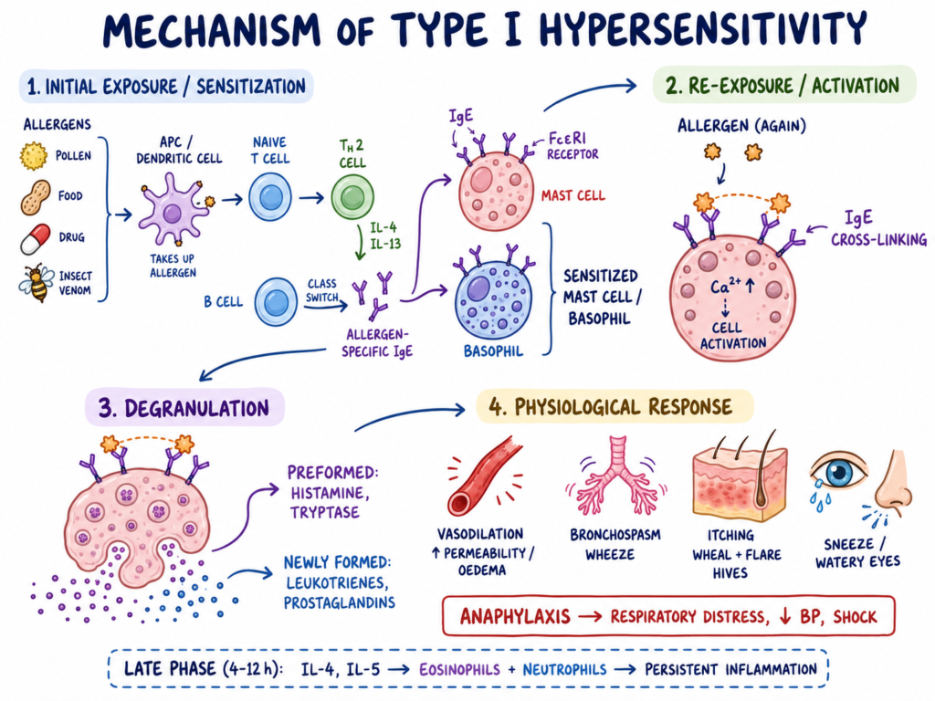
Phase 1: Initial exposure and sensitization
In this phase, the allergen first enters into the body. The allergen may be pollen, food, drug or insect venom. It is harmless normally, but in allergic person it acts as antigen.
The allergen is taken by antigen presenting cells, mainly dendritic cells. These cells process the allergen and present it to naive T lymphocytes. Then the naive T cells change into T helper 2 cells (Th2 cells).
The Th2 cells release cytokines mainly Interleukin-4 (IL-4) and Interleukin-13 (IL-13). These cytokines stimulate B lymphocytes. Then B cells undergo class switching and produce allergen specific IgE antibodies.
The produced IgE antibodies bind with high affinity FcεRI receptors on the surface of mast cells and basophils. In this way the mast cells and basophils become sensitized. This stage is called sensitization or priming of immune system.
Phase 2: Re-exposure and cellular activation
In this phase, the same allergen again enters into the body. The allergen now binds with the IgE antibodies already attached on mast cells and basophils.
One allergen binds with more than one IgE molecule. This causes cross linking of IgE antibodies and their receptors on the cell surface. This cross linking is the main signal for activation of mast cells and basophils.
After cross linking, intracellular reaction starts inside the cell. There is rapid biochemical signalling. Calcium level increases inside the cell and the granules become ready for release.
Phase 3: Degranulation and chemical release
In this phase, the internal granules of mast cells move toward the cell membrane. These granules fuse with the outer cell membrane. This fusion is helped by fusion proteins like SNAREs.
After fusion, the granule contents are released outside the cell. This process is called degranulation. It occurs rapidly after activation of mast cells and basophils.
The released mediators are of two types. Preformed mediators include histamine and tryptase. Newly formed mediators include leukotrienes and prostaglandins.
Phase 4: Physiological response and symptoms
In this phase, the released mediators act on different tissues. Histamine causes vasodilation and increases capillary permeability. Due to this, fluid comes out into tissues and oedema is produced.
The mediators also act on smooth muscles. In the respiratory tract, smooth muscle contraction causes bronchospasm. So breathing difficulty, wheezing and chest tightness may occur.
In skin, it produces itching, redness and wheal and flare reaction. Local symptoms include sneezing, itching, watery eyes, hives and urticaria.
In severe condition, the reaction becomes systemic. This is called anaphylaxis. It produces respiratory distress, fall in blood pressure and shock.
After 4 to 12 hours, late phase reaction may occur. Cytokines such as IL-4 and IL-5 attract eosinophils and neutrophils at the site. These cells produce persistent inflammation and second wave of allergic symptoms.
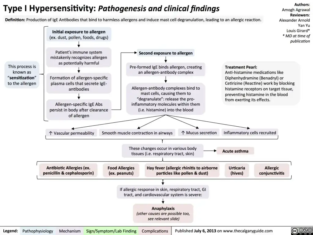
Clinical Manifestation of Type I Hypersensitivity Reaction
The following are the clinical manifestation of Type I hypersensitivity reaction–
- Skin and mucous membrane– Urticaria, intense pruritus, erythema, cutaneous flushing and local wheal and flare reaction are seen. Angioedema may occur in lips, tongue, uvula and around the eyes. Swelling around eye is called periorbital edema.
- Upper respiratory tract– Sneezing, running nose (rhinorrhea) and nasal congestion are common. These are produced due to irritation and swelling of nasal mucosa.
- Throat and larynx– Hoarseness of voice, fullness in throat, lump like feeling and persistent throat clearing may occur. Stridor is seen in laryngeal swelling. Severe swelling may cause airway obstruction.
- Lower respiratory tract– Shortness of breath (dyspnea), wheezing, persistent cough and bronchoconstriction are seen. In severe condition, oxygen level in blood decreases and it is called hypoxemia.
- Cardiovascular system– Rapid heart rate (tachycardia) and abnormal heart rhythm (tachyarrhythmia) may occur. Fall in blood pressure (hypotension), dizziness, syncope and cardiovascular collapse may be seen in severe reaction.
- Gastrointestinal tract– Nausea, vomiting, abdominal cramps and diarrhea may occur. These are due to action of mediators on smooth muscle of intestine.
- Other systemic manifestation– Loss of muscle tone (hypotonia) and loss of bladder or bowel control (incontinence) may occur in severe systemic reaction.
- Anaphylaxis– Anaphylaxis is severe systemic reaction. It produces respiratory distress, severe bronchoconstriction, laryngeal edema, hypotension and shock.
- Late phase manifestation– In allergic rhinitis, post nasal drip, loss of smell and Eustachian tube dysfunction may occur. These are delayed symptoms after early allergic reaction.
- Biphasic reaction– Symptoms may occur again after initial recovery. It may peak after 8 to 11 hours and occurs without further exposure to allergen.
Diagnosis of Type I Hypersensitivity Reaction
The following are the diagnosis of Type I hypersensitivity reaction–
- Clinical diagnosis– Diagnosis is mainly done by observing the symptoms. In anaphylaxis, the reaction occurs very fast. Respiratory distress, fall in blood pressure, skin and mucosal changes are important findings.
- Symptom assessment– The patient may show itching, urticaria, swelling, wheezing, difficulty in breathing and hypotension. These symptoms appearing rapidly after exposure to allergen helps in diagnosis.
- Emergency diagnosis of anaphylaxis– Anaphylaxis is diagnosed clinically. Laboratory test is not necessary for immediate diagnosis. Treatment should not be delayed for doing test.
- Skin prick test (SPT)– In this test, a small amount of specific allergen is introduced into the skin by pricking. If allergen specific IgE is present on skin mast cells, then local wheal and flare reaction appears within 15 to 20 minutes.
- Precaution before skin prick test– Antihistamine drugs should be stopped before testing. Usually it is stopped for 3 to 7 days. Otherwise false negative result may occur.
- Intradermal test– In this test, minute amount of allergen is injected under the superficial layer of skin. It is used when skin prick test is negative but allergy is still suspected. It is not used for food allergy.
- Allergen specific IgE blood test– This test detects IgE antibodies against particular allergen in blood. It is used when skin test cannot be done. It is useful in extensive skin disease, pregnancy and when patient cannot stop antihistamine.
- Serum tryptase test– Tryptase is an enzyme released from mast cells during degranulation. Increased serum tryptase helps to confirm systemic mast cell activation after suspected anaphylaxis.
- Timing of tryptase sample– Acute blood sample is taken ideally after 1 to 2 hours of symptom onset. Baseline sample is taken after at least 24 hours when symptoms are fully resolved. The two values are compared for diagnosis.
Treatment / Management of Type I Hypersensitivity Reaction
The following are the treatment and management of Type I hypersensitivity reaction–
- Removal of allergen– The offending allergen is removed first. Insect stinger, food, drug or other triggering substance should be stopped. It prevents further exposure to same allergen.
- Airway management– Airway is checked immediately in anaphylaxis. If airway is blocked or swelling is present, airway support is given. Severe laryngeal oedema may need definitive airway.
- Epinephrine– Epinephrine is the first line drug in systemic anaphylaxis. It is given by intramuscular (IM) injection in anterolateral thigh. Adult dose is commonly 0.3 to 0.5 mg and child dose is 0.01 mg/kg, according to emergency protocol.
- Action of epinephrine– Epinephrine acts on α and β receptors. It increases blood pressure, dilates the airway and decreases further mast cell degranulation. So it reverses shock and breathing difficulty.
- Intravenous fluid– IV fluid resuscitation is given when hypotension and shock is present. Isotonic crystalloids are used. It helps to restore blood volume and blood pressure.
- Antihistamines– H1 blockers such as diphenhydramine and H2 blockers such as famotidine are used as supportive drugs. They reduce itching, hives and skin manifestations. They are not replacement of epinephrine in anaphylaxis.
- Bronchodilators– Inhaled bronchodilator such as albuterol is used when wheezing and bronchospasm continue. It relaxes bronchial smooth muscle and improves breathing.
- Corticosteroids– Corticosteroids such as methylprednisolone may be given. It helps to reduce prolonged inflammation and late phase or biphasic reaction.
- Glucagon– Glucagon is used in refractory anaphylaxis. It is useful when patient is taking beta-blocker drug and not responding properly to epinephrine.
- Allergen avoidance– Avoidance of known allergen is the main preventive management. It is used in food allergy, allergic asthma, allergic rhinitis and skin allergy. The patient should avoid the triggering food, drug, dust, pollen or insect sting.
- Allergic rhinitis management– Allergic rhinitis is managed by allergen avoidance, nasal saline irrigation, intranasal glucocorticoids and antihistamines. Intranasal corticosteroids are used as first line treatment in many allergic rhinitis cases.
- Urticaria and angioedema management– Second generation H1 antihistamines such as cetirizine are used. They reduce itching and wheal formation with less sedation. Short course of systemic corticosteroid may be used when symptoms persist.
- Atopic dermatitis management– In atopic dermatitis, irritating factors are removed. Harsh soap, detergent and low humidity are avoided. Skin emollients, topical corticosteroids and calcineurin inhibitors are used during flare.
- Food allergy management– In food allergy, strict avoidance of trigger food is the main treatment. Accidental exposure is treated quickly by required medicine. Oral immunotherapy may be used in selected cases to increase tolerance.
- Allergic asthma management– In allergic asthma, allergen avoidance is done. Inhaled bronchodilators and inhaled corticosteroids are used according to asthma severity. It reduces bronchospasm and airway inflammation.
- Epinephrine auto-injector– Patient with severe allergy should carry epinephrine auto-injector. Patient and family members should know how to use it. Emergency action plan is also needed.
- Allergen immunotherapy (AIT)– Allergen immunotherapy is given by subcutaneous or sublingual route. It is a disease modifying treatment and can produce long term tolerance. It increases regulatory T cells (Treg) and blocking antibodies like IgG4 and IgA.
- Targeted biologics– Monoclonal antibodies are used in severe and resistant allergic disease. Omalizumab binds free circulating IgE and prevents its attachment to mast cells. Dupilumab blocks IL-4 and IL-13 signalling, and Tezepelumab also decreases allergic inflammatory pathway.
Examples of Type I Hypersensitivity Reaction
The following are the examples of Type I hypersensitivity reaction–
- Allergic rhinitis– It is also called hay fever. It occurs due to inhaled allergens like pollen and dust.
- Allergic asthma– It occurs due to allergen mediated narrowing of bronchi. Wheezing and difficulty in breathing are seen.
- Anaphylaxis– It is a severe systemic allergic reaction. It occurs rapidly and may become life threatening.
- Urticaria– It is also called hives. Raised itchy patches are formed on the skin.
- Angioedema– It is localized swelling of tissue. It commonly occurs in lips, eyelids, tongue and face.
- Atopic dermatitis– It is also called eczema. It produces itchy inflamed skin lesion.
- Allergic conjunctivitis– It is allergic reaction of conjunctiva. Redness, itching and watering of eyes are seen.
- Food allergy– It may occur due to peanut, tree nuts, fish, shellfish, milk, egg, wheat and soy.
- Drug allergy– It may occur due to drugs like penicillin, cephalosporins and other beta-lactam antibiotics.
- Insect venom allergy– It occurs after bee sting, wasp sting and fire ant bite.
- Latex allergy– It occurs due to exposure to latex products. Skin itching, rash or systemic reaction may occur.
- Alpha-gal anaphylaxis– It is allergic reaction against alpha-gal sugar present in mammalian meat.
- Eosinophilia– Increased number of eosinophils may be seen in allergic type reaction.
- Sweet itch– It is an allergic skin reaction due to insect bite. Itching and skin irritation are present.
References
- Adrenaline in the Acute Treatment of Anaphylaxis. (n.d.). PMC.
- Allergy and Asthma Center of Western Colorado. (2019-2025). Allergy Skin Testing.
- McLendon, K., & Sternard, B. T. (2023). Anaphylaxis. In StatPearls. StatPearls Publishing.
- Antigen-Specific Tolerance in Immunotherapy of Th2-Associated Allergic Diseases. (n.d.). PMC.
- Clinical and Molecular Landscapes of Type I Hypersensitivity: From Intracellular Signaling to Advanced Therapeutic Paradigms. (n.d.).
- Current Strategies to Inhibit High Affinity FcεRI-Mediated Signaling for the Treatment of Allergic Disease. (n.d.). Frontiers.
- Dalal, R., & Grujic, D. (2024). Epinephrine. In StatPearls. StatPearls Publishing.
- Epinephrine in anaphylaxis. (n.d.). PMC – NIH.
- Petersen, L. J., Church, M. K., & Skov, P. S. (1997). Histamine is released in the wheal but not the flare following challenge of human skin in vivo: a microdialysis study. Clinical and Experimental Allergy, 27(3), 284-295. https://doi.org/10.1046/j.1365-2222.1997.d01-502.x
- IgE and non-IgE food allergy: A review of immunological mechanisms. (n.d.). PMC.
- IgE and non-IgE-mediated pathways in anaphylaxis. (n.d.). PMC.
- IgE receptor signaling in food allergy pathogenesis. (n.d.). PMC – NIH.
- AMBOSS: Medical Knowledge Distilled. (n.d.). Immediate response: Type I Hypersensitivity Reaction [Video]. YouTube.
- National Clinical Guideline Centre (UK). (2014). Measuring serum tryptase after suspected anaphylaxis. In Drug Allergy: Diagnosis and Management of Drug Allergy in Adults, Children and Young People (NICE Clinical Guidelines, No. 183). National Institute for Health and Care Excellence (NICE).
- Mechanisms of Allergen Immunotherapy and Potential Biomarkers for Clinical Evaluation. (n.d.). PMC.
- Mechanisms of action that contribute to efficacy of omalizumab in chronic spontaneous urticaria. (n.d.). PMC.
- Mechanisms of allergen-specific immunotherapy and immune tolerance to allergens. (n.d.). PMC.
- Mechanobiology of Type 1 hypersensitivity: Elucidating the impacts … (n.d.).
- Omalizumab-induced reductions in mast cell Fc Epsilon RI expression and function. (n.d.).
- Omalizumab: A Monoclonal Anti-IgE Antibody. (n.d.). PMC.
- Godse, K., Mehta, A., Patil, S., Gautam, M., & Nadkarni, N. (2015). Omalizumab—A Review. Indian Journal of Dermatology, 60(4), 381–384. https://doi.org/10.4103/0019-5154.160490
- Canadian Agency for Drugs and Technologies in Health. (2024). Retesting Intervals for Tryptase: Rapid Review.
- Retesting Intervals for Tryptase. (n.d.). CDA-AMC – Canada’s Drug Agency.
- SNARE complex-mediated degranulation in mast cells. (n.d.). PMC.
- SNARE complex‐mediated degranulation in mast cells. (n.d.). ResearchGate.
- Asthma and Allergy Foundation of America. (n.d.). Skin Testing for Allergies.
- Yang, Y., Kong, B., Jung, Y., Park, J.-B., Oh, J.-M., Hwang, J., Cho, J. Y., & Kweon, D.-H. (2018). Soluble N-Ethylmaleimide-Sensitive Factor Attachment Protein Receptor-Derived Peptides for Regulation of Mast Cell Degranulation. Frontiers in Immunology, 9, 725. https://doi.org/10.3389/fimmu.2018.00725
- Jutel, M., & Akdis, C. (2008). T-Cell Regulatory Mechanisms in Specific Immunotherapy. In K. Blaser (Ed.), T Cell Regulation in Allergy, Asthma and Atopic Skin Diseases. Chemical Immunology and Allergy (Vol. 94, pp. 1–20). S. Karger AG.
- Table 4, Summary of Recommendations for Patients With Confirmed or Suspected Anaphylaxis. (2024). In Retesting Intervals for Tryptase: Rapid Review. Canadian Agency for Drugs and Technologies in Health. NCBI Bookshelf.
- Lorentz, A., Baumann, A., Vitte, J., & Blank, U. (2012). The SNARE Machinery in Mast Cell Secretion. Frontiers in Immunology, 3, 143. https://doi.org/10.3389/fimmu.2012.00143
- The skin prick test – European standards. (n.d.). PMC.
- Abbas, M., & Goldin, J. (2025). Type I Hypersensitivity Reaction. In StatPearls. StatPearls Publishing.
- Type I hypersensitivity. (2025). In Wikipedia. Wikimedia Foundation.
- Saavedra Torres, J. S., & Annamaraju, P. (2025). Type III Hypersensitivity Reaction. In StatPearls. StatPearls Publishing.
- Anderson, L. A. (2023). What’s the mechanism of action for epinephrine? Drugs.com.
- Robinson, D. (n.d.). Wheal and flare. Ent Clinics.