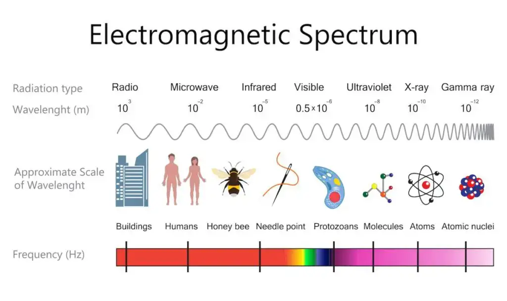
What is X-Ray Spectroscopy?
- X-Ray Spectroscopy can define as technique used for study of interaction between X-rays and matter, mainly for determination of elemental composition or chemical state.
- It’s based on emission / absorption of X-rays by atoms when they are excited by high-energy sources.
- The spectrum produced is analyzed for getting information about structure and properties of sample materials.
- In general overview, X-ray spectroscopy involve the measurement of characteristic radiation that comes out from material after excitation.
- The energy levels of atoms are affected by inner shell transitions, so unique fingerprints of each element are observed.
- Many different methods are existed like XRF (X-ray fluorescence), XPS (X-ray photoelectron spectroscopy), and EDS (Energy dispersive spectroscopy) etc.
- It has been widely utilized for qualitative and quantitative analysis, surface study, and elemental mapping by scientists from various fields.
- The importance of X-ray spectroscopy is very high by analytical and material science viewpoint.
- It has been applied for determination of atomic arrangement, oxidation states, bonding conditions, impurities, etc.
- It helps in studying metals, semiconductors, biological tissues, geological samples, and so on.
- Industrial quality control, corrosion study, thin film analysis, forensic and environmental testing all benefited by this technique.
- Because it is nondestructive, the original sample mostly remain safe after testing which make it very useful and economical in research and industrial work.
- The discovery of X-rays by Wilhelm Conrad Röntgen in 1895 had started the basis of this spectroscopy.
- Later on, scientists like Henry Moseley in 1913 used X-ray spectra to establish relation between atomic number and frequency (now called Moseley’s law).
- Through early 20th century, more instruments were developed for X-ray diffraction and fluorescence methods.
- By 1950s–1960s, with arrival of electron microscopes and solid-state detectors, X-ray spectroscopy techniques were refined and automated.
- Even today, they keep being advanced with digital detectors, synchrotron sources and micro-beam analysis, making it still one of the most sturdy and hardy analytical tools in science.
History of X-ray spectroscopy
- The groundwork was laid when Wilhelm Conrad Röntgen discovered X-rays in 1895 while experimenting with cathode‐ray tubes, he noticed a fluorescent screen glowing though it was covered, and that was first sign of new rays.
- Early investigations showed that when inner electron shells were excited, they emitted characteristic X‐rays, and this phenomenon was used for first forms of X‐ray analysis.
- Around 1913, Henry G. J. Moseley used X-ray spectra of metals to derive a relation between frequency of emitted X-rays and atomic number (now called “Moseley’s law”).
- In the years following, the method of diffraction of X‐rays by crystals (by William Henry Bragg & William Lawrence Bragg) was applied to X-ray spectroscopy, and it was established that X-rays are electromagnetic waves and that their spectra could be measured precisely.
- The term “X-ray spectroscopy” gradually came to stand for various techniques (fluorescence, absorption, emission) using X-rays to characterise materials.
- In 1924, Manne Siegbahn was awarded the Nobel Prize in Physics for his research in X-ray spectroscopy, which marked a mature stage in its development.
- Later mid-20th century, the instrumentation improved (better detectors, crystals, excitation sources) and industrial / analytical applications of X-ray spectroscopy expanded widely.
- More recently, with synchrotron sources and micro‐analysis, X-ray spectroscopy has become very refined and is used for studying electronic structure, materials, etc.
X Ray Spectroscopy Principle – Principle of x ray spectroscopy
- A high-energy beam (like X-rays or electrons) is directed onto a sample, and the atoms in the sample are excited by the incident radiation.
- An inner-shell electron (for example from K or L shell) is knocked out or raised to a higher energy level, leaving a “core-hole” in the atom.
- When that core-hole is filled by an outer electron falling into the vacancy, energy is released in form of a photon (an X-ray) whose energy (or wavelength) is characteristic of the element.
- The emitted X-ray photons are measured (either by energy-dispersive detector or wavelength-dispersive crystal) and the spectrum obtained shows peaks that correspond to specific elements.
- The intensity (number of photons) of a given characteristic line is roughly proportional to the concentration of that element in the sample, allowing quantitative analysis (with calibration).
- In absorption-type X-ray spectroscopy (like X‑Ray Absorption Spectroscopy, XAS) the incident X-ray energy is swept and the absorption by core level electrons is monitored, giving information on electronic/structural environment.
- Both emission and absorption versions depend on the fact that inner-shell electrons have discrete binding energies, so transitions produce discrete X-ray energies, and that gives elemental/chemical specificity
- Detector geometry, sample matrix, excitation source, absorption effects, scattering etc all influence the measurement and must be considered (for correct results).

How X-ray spectroscopy works
- The process is initiated when a sample is irradiated with a beam of high-energy X-rays (or sometimes electrons/charged particles) that strike atoms and excite them.
- Upon excitation an inner‐shell electron (for example in K or L shell) is ejected or promoted to a higher energy level leaving a “core‐hole”.
- The atom is left unstable and then an outer‐shell electron falls into the core‐hole, and this transition releases energy in form of a characteristic X-ray photon (or sometimes an Auger electron, depending on method).
- The energy (or wavelength) of the emitted photon is characteristic for the element (because it depends on energy difference between specific shells) so we can identify what elements are present.
- In some methods the absorption of X-rays is measured instead: the incident X-ray energy is scanned and the absorption coefficient (µ) is measured just above a core‐level binding energy, giving information on electronic structure and local environment.
- In a typical energy‐dispersive setup (EDS) the detector measures directly the energy of incoming photons, whereas in a wavelength‐dispersive setup (WDS) a crystal diffracts the photons and the wavelength is measured via Bragg’s law.
- The intensity of emitted or absorbed X-rays is linked to concentration of the elements (quantitative) and the peak positions give qualitative identity.
- A sample can remain mostly unchanged (non-destructive) when the method is properly chosen, which makes this work useful for wide range of materials.
Instrumentation of X-Ray Spectroscopy
1. X–ray Source – It is mainly used for production of X–rays. Usually a X–ray tube or sometimes a synchrotron is used. In the tube, high energy electrons are accelerated toward a metal target (like Cu, Mo etc.), where the X–rays are generated by bombardment process.
2. Sample Holder – The specimen which is to be analyzed is placed here. It is usually made adjustable so the sample can be correctly oriented with the incident beam, the alignment is quite important for precise result.
3. Collimator – The beam of X–rays is directed and narrowed by the collimator, it helps to reduce scattering and noise in data. Various types of collimators (pinhole/slit) are used depends on system.
4. Monochromator – It is employed for selection of desired wavelength of X–rays from the polychromatic beam. Usually a crystal (like LiF or NaCl) is used to diffract the X–rays at specific angle according to Bragg’s law (nλ = 2d sinθ).
5. Detector – The detector is used for measuring the intensity of emitted or diffracted X–rays. There are different types – proportional counter, scintillation detector, semiconductor detector etc. The choice depends on energy range and resolution needed.
6. Goniometer – It is the device used for precise measurement of angle between incident and diffracted beam, it controls movement of sample and detector simultaneously.
7. Data Processing / Recording Unit – The electronic system or computer interface that collects, amplifies and stores data from detector. Spectra are displayed and analyzed by software for identifying elements and structure.
Applications of X-Ray Spectroscopy
- Elemental and chemical characterisation of materials are commonly done by X-ray spectroscopy, the technique is used to identify what elements are present and their chemical state.
- Within materials science it is applied for micro-/nano-structure analysis of alloys, thin films, batteries, fuel cells etc.
- In geology / mineral exploration the method is used for analysing rocks, ores, soils etc., which helps in mineral identification and resource assessment.
- In environmental monitoring trace or major elements in water, soil, air particulates are measured and mapped by X-ray spectroscopy setups.
- In forensic science many unknown samples (metal fragments, glass, paint chips) are analysed by this technique for composition and provenance which supports investigation work.
- The technique is used in cultural heritage and archaeology for non-destructive analysis of artifacts, paintings, ancient tools etc., to find elemental make-up and conservation state.
- In astrophysics / space science the method is used to study remote materials (for example planetary surfaces, cosmic dust) by instrumentation onboard spacecraft to obtain elemental signatures.
- In chemistry and biochemistry advanced X-ray absorption/emission spectroscopy (XAS/XES) applications are used to study electronic & geometric structure of atom in molecules or catalysts, dynamic reactions etc.
Advantages of X-Ray Spectroscopy
- A major advantage is that non-destructive analysis is possible, the sample often remains intact and unchanged after measurement.
- Multi-element detection is allowed, many elements (major, minor and trace) can be identified and quantified in one go.
- The technique is rapid, with measurement times often very short (for instance 2-5 minutes for some setups) which makes it suited for high throughput / industrial work.
- Minimal or simple sample preparation is required, solid, powder, liquid forms can be used which saves time and effort.
- Good for surface / near surface analysis as well as bulk in some variants, so versatile from viewpoint of depth of study.
- High analytical precision and reproducibility are provided (when instrumentation is well calibrated) which helps in quality control and research.
- The technique is element-specific, meaning that precise fingerprint of each element’s characteristic X-rays is used so identification is reliable.
Limitations of X-Ray Spectroscopy
- A major limitation is that light elements (for example atomic number Z < 11 or Z < 12) are often very poorly detected especially by standard X-ray fluorescence (XRF) methods, so their quantification is difficult.
- In some cases matrix effects (that is effects from surrounding material / sample composition) strongly influence the emitted or absorbed X-rays, so the result are skewed or less accurate, for example attenuation or enhancement by neighbouring atoms.
- Depth-penetration is limited, meaning that only surface or near-surface layers are studied well, so bulk composition may not be well represented especially when surface is coated or altered.
- Very low concentration (trace / ultra-trace) detection is often not possible or requires long measurement time and special set-ups; for example concentrations below parts-per-billion are outside routine reach.
- Sample preparation / geometry / surface condition issues must be handled carefully, for the errors due to sample heterogeneity, surface contamination or shape effects are large and they degrade reliability.
- The instrumentation cost / complexity for higher resolution methods (for example wavelength dispersive spectrometers) can be higher and footprint larger, making routine application harder in some settings.
- In certain spectroscopies the chemical state (for example oxidation states) or bonding environment of element may not be well distinguished by simple elemental X-ray methods; isotopes cannot be distinguished either.
FAQ
What is X-ray spectroscopy?
X-ray spectroscopy is a technique used to study the interaction between X-rays and matter. It involves analyzing the emitted or scattered X-rays to obtain information about the elemental composition, chemical bonding, and structural characteristics of a material.
How does X-ray spectroscopy work?
X-ray spectroscopy works by irradiating a sample with an intense X-ray beam. When the X-rays interact with the atoms in the sample, they cause the emission or scattering of X-rays with characteristic energies and wavelengths. By analyzing the emitted or scattered X-rays, scientists can determine the elemental composition and other properties of the material.
What are the different types of X-ray spectroscopy techniques?
Some common types of X-ray spectroscopy techniques include X-ray fluorescence (XRF), X-ray photoelectron spectroscopy (XPS), X-ray absorption spectroscopy (XAS), and X-ray emission spectroscopy (XES).
What are the applications of X-ray spectroscopy?
X-ray spectroscopy has a wide range of applications, including materials science, chemistry, geology, environmental analysis, pharmaceuticals, forensic science, and archaeology. It is used for elemental analysis, identification of unknown substances, structural characterization, quality control, and many other purposes.
What are the advantages of X-ray spectroscopy?
X-ray spectroscopy offers several advantages, including its non-destructive nature, high sensitivity to trace elements, ability to provide both qualitative and quantitative analysis, and its capability to analyze a wide range of materials, from solids to liquids and gases.
What are the limitations of X-ray spectroscopy?
One limitation of X-ray spectroscopy is that it requires access to a pure crystalline sample for certain techniques like X-ray crystallography. Additionally, X-ray spectroscopy techniques may not be suitable for analyzing light elements such as hydrogen and helium.
What are the different components of an X-ray spectroscopy setup?
An X-ray spectroscopy setup typically consists of an X-ray source (e.g., X-ray tube), sample holder, detectors, collimators, monochromators, and data analysis software.
How is data analysis done in X-ray spectroscopy?
Data analysis in X-ray spectroscopy involves comparing the measured X-ray signals with reference spectra and using mathematical algorithms to determine the elemental composition, bonding information, and other relevant properties of the sample.
Is X-ray spectroscopy safe?
X-ray spectroscopy techniques are generally safe when conducted by trained professionals and with proper safety precautions. However, it is important to follow radiation safety guidelines and minimize unnecessary exposure to X-rays.
How has X-ray spectroscopy contributed to scientific advancements?
X-ray spectroscopy has played a crucial role in various scientific advancements. It has helped in understanding the structure of crystals, identifying unknown compounds, developing new materials, studying the electronic properties of materials, and advancing fields such as materials science, chemistry, and solid-state physics.
- Balci, Metin (2005). Basic 1H- and 13C-NMR Spectroscopy Volume 446 || Introduction. , (), 3–8. doi:10.1016/b978-044451811-8.50001-2
- Feng, X., Zhang, H. & Yu, P. (2020). X-ray fluorescence application in food, feed, and agricultural science: a critical review. Crit Rev Food Sci Nutr, 1-11.10.1080/10408398.2020.1776677
- Smyth, M. S. & Martin, J. H. (2000). x-ray crystallography. Mol Pathol, 53, 8-14.10.1136/mp.53.1.8
Franklin, R. E. & Gosling, R. G. (1953). Molecular Configuration in Sodium Thymonucleate. Nature, 171, 740-741.https://doi.org/10.1038/171740a0 - https://www.iucr.org/__data/assets/pdf_file/0013/733/chap16.pdf
- https://www.azolifesciences.com/article/X-Ray-Spectroscopy-An-Overview.aspx
- http://instructor.physics.lsa.umich.edu/adv-labs/X-Ray_Spectroscopy/x_ray_spectroscopy_v2.pdf
- https://new.bhu.ac.in/Content/Syllabus/Syllabus_3006312820200416111005.pdf
- https://www.iaea.org/topics/x-ray-spectrometry
- http://www.issp.ac.ru/ebooks/books/open/X-Ray_Spectroscopy.pdf
- http://websites.umich.edu/~jphgroup/XAS_Course/Harbin/Lecture1.pdf
- Text Highlighting: Select any text in the post content to highlight it
- Text Annotation: Select text and add comments with annotations
- Comment Management: Edit or delete your own comments
- Highlight Management: Remove your own highlights
How to use: Simply select any text in the post content above, and you'll see annotation options. Login here or create an account to get started.