What is Vertebra?
- A vertebra (plural: vertebrae) is a fundamental bone component of the vertebral column, or spine, present in all vertebrate species. The term “vertebra” originates from the Latin word “vertere,” meaning to turn, reflecting the spine’s function in providing flexibility and movement.
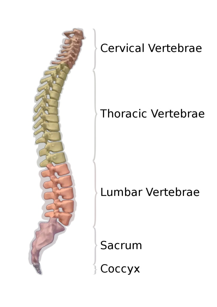
- In humans, the vertebral column consists of 33 vertebrae extending from the base of the skull to the coccyx, or tailbone. These vertebrae are divided into five distinct regions, each with a specific number of bones: the cervical region (7 vertebrae), thoracic region (12 vertebrae), lumbar region (5 vertebrae), sacral region (1 fused vertebra), and coccygeal region (1 fused vertebra). This configuration not only supports the body’s structure but also allows for a range of movements and flexibility.
- Each vertebra is an irregularly shaped bone with a complex structure, comprising a combination of bone and hyaline cartilage. The central portion of the vertebra is known as the centrum, which is crucial for bearing the body’s weight. Attached to the centrum are intervertebral discs that act as cushions, providing shock absorption and protecting the vertebrae during movement.
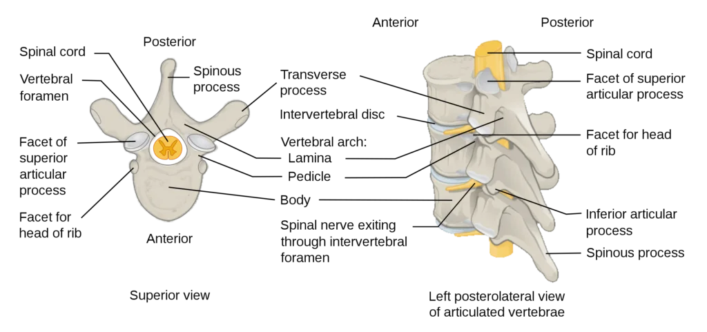
- The posterior part of a vertebra forms the vertebral arch, which consists of eleven parts: two pedicles, two laminae, and seven processes. These structures provide attachment points for muscles and ligaments, such as the ligamenta flava, which contribute to the spine’s stability and flexibility. The pedicles and laminae create a protective enclosure for the spinal cord, forming the vertebral foramen—a large, central opening that houses the spinal canal.
- Vertebrae also feature vertebral notches, which, when aligned with adjacent vertebrae, form the intervertebral foramina. These foramina serve as entry and exit points for spinal nerves, facilitating communication between the brain and the rest of the body.
- The overall shape and structure of vertebrae vary slightly across different species, reflecting adaptations to their environments and modes of movement. For instance, land-dwelling animals have vertebrae that support weight-bearing and enable a range of movements necessary for walking and running, while aquatic animals have vertebrae adapted for swimming.
- The vertebral column’s design—comprising the vertebrae and intervertebral discs—provides a balance of strength, flexibility, and protection. This intricate system allows for movement while safeguarding the spinal cord, a critical component of the central nervous system.
- In summary, vertebrae are integral to the structure and function of the vertebral column in vertebrates. Their unique design, featuring a blend of bone and cartilage, supports a wide range of movements, protects the spinal cord, and serves as a defining characteristic of vertebrate species. Understanding the anatomy and function of vertebrae offers valuable insights into the complexities of vertebrate biology and the evolutionary adaptations that enable diverse modes of life.
Definition of Vertebra
A vertebra is an individual bone that forms part of the vertebral column (spine) in vertebrate animals. It is an irregularly shaped bone composed of a central body, a vertebral arch, and various processes, providing structural support, flexibility, and protection for the spinal cord.
Parts of Vertebrae
1. Vertebral Body
- Centrum: The main anterior part of the vertebral body, composed of cancellous (spongy) bone, which is characterized by a porous structure. This cancellous bone is covered by a layer of cortical bone, which is denser and harder.
- Posterior Vertebral Arch (Neural Arch): Extends posteriorly from the vertebral body and forms the vertebral foramen, which houses and protects the spinal cord.
- Intervertebral Disc Attachment: The vertebral body has roughened surfaces for the attachment of intervertebral discs, which provide cushioning and flexibility to the spine.
2. Vertebral Arch
- Pedicles: Two short, thick processes extending from the sides of the vertebral body, connecting it to the vertebral arch.
- Laminae: Broad plates that extend backward and medially from each pedicle to complete the vertebral arch, forming the posterior part of the vertebral foramen.
3. Processes
- Spinous Process: Projects posteriorly from the vertebral arch, serving as an attachment point for muscles and ligaments.
- Transverse Processes: Extend laterally from the vertebral arch and also serve as attachment points for muscles and ligaments. In the thoracic vertebrae, they articulate with the ribs.
- Articular Processes: Include two superior and two inferior processes that restrict the range of movement and help stabilize the spine. These processes articulate with adjacent vertebrae to form facet joints, which are part of the vertebral arch.
4. Vertebral Foramen
- Central Opening: The vertebral foramen is formed by the vertebral body and arch, creating a canal through which the spinal cord passes.
5. Intervertebral Foramina
- Notches and Foraminal Openings: Shallow depressions called vertebral notches, located above and below the pedicles, align with adjacent vertebrae to form intervertebral foramina. These openings allow for the passage of spinal nerves and blood vessels.
Structure of Vertebra
The vertebrae in the human vertebral column vary in size and shape according to their location and the specific demands of stress and mobility in different regions of the spine. Each vertebra is classified as an irregular bone due to its complex structure. Below is a detailed explanation of the anatomy and structure of a typical vertebra:

1. Vertebral Body (Centrum)
- Centrum: The central part of the vertebral body, or centrum, forms the large, anterior portion of the vertebra. It is composed primarily of cancellous bone, also known as spongy bone, which is characterized by a porous, honeycomb-like structure. This cancellous bone is covered by a thin layer of cortical bone, the hard and dense type of osseous tissue.
- Endplates: The upper and lower surfaces of the vertebral body are flattened and rough, known as vertebral endplates. These endplates provide attachment for the intervertebral discs and are formed from a thickened layer of cancellous bone, with the top layer being denser. The endplates serve several functions:
- Containing adjacent discs
- Evenly distributing applied loads
- Anchoring collagen fibers of the discs
- Acting as a semi-permeable interface for the exchange of water and solutes
2. Vertebral Arch (Neural Arch)
- Pedicles: Two short, thick processes called pedicles extend posteriorly from the sides of the vertebral body. They connect the body to the arch and form the sides of the vertebral foramen.
- Laminae: From each pedicle, a broad plate called a lamina projects backward and medially, joining to complete the vertebral arch. The laminae form the posterior border of the vertebral foramen.
- Ligamenta Flava: The upper surfaces of the laminae are rough to provide attachment points for the ligamenta flava, ligaments that connect the laminae of adjacent vertebrae along the spine.
3. Vertebral Foramen and Canal
- Vertebral Foramen: The space enclosed by the vertebral arch and the vertebral body forms the vertebral foramen. When the vertebrae are stacked, these foramina align to create the vertebral canal, which houses and protects the spinal cord.
4. Processes and Notches
- Spinous and Transverse Processes: Various processes extend from the vertebral arch, including the spinous process (extending posteriorly) and transverse processes (extending laterally). These serve as attachment points for muscles and ligaments.
- Vertebral Notches: Above and below the pedicles are shallow depressions called vertebral notches (superior and inferior). When adjacent vertebrae articulate, these notches align to form intervertebral foramina, through which spinal nerves and blood vessels pass.
5. Intervertebral Discs and Articulation
- Intervertebral Discs: These discs are located between the vertebrae, providing cushioning and shock absorption. They consist of a tough outer layer called the annulus fibrosus and a gel-like core known as the nucleus pulposus.
- Articulation: Vertebrae articulate with each other at the intervertebral discs and facet joints, forming a strong yet flexible column that supports the body and allows a range of movements.
| Component | Description |
|---|---|
| Vertebral Body | – Centrum: The central part, composed of cancellous (spongy) bone covered by a thin layer of cortical (compact) bone. |
| – Endplates: Flattened, rough surfaces on the upper and lower parts of the vertebral body, providing attachment for intervertebral discs and distributing loads. | |
| Vertebral Arch | – Pedicles: Short, thick processes extending from the sides of the vertebral body to connect it to the arch. |
| – Laminae: Broad plates projecting backward and medially from each pedicle to complete the vertebral arch. | |
| – Ligamenta Flava: Ligaments attaching to the rough upper surfaces of the laminae, connecting adjacent vertebrae. | |
| Vertebral Foramen | – The space enclosed by the vertebral arch and body, forming the vertebral canal that houses and protects the spinal cord. |
| Processes and Notches | – Spinous Process: Extends posteriorly from the vertebral arch, serving as an attachment point for muscles and ligaments. |
| – Transverse Processes: Extend laterally from the vertebral arch, serving as attachment points for muscles and ligaments. | |
| – Vertebral Notches: Shallow depressions above and below the pedicles that align with adjacent vertebrae to form intervertebral foramina for spinal nerves. | |
| Intervertebral Discs | – Located between vertebrae, providing cushioning and shock absorption, composed of the annulus fibrosus (outer layer) and nucleus pulposus (gel-like core). |
| Articulation | – Vertebrae articulate with each other at intervertebral discs and facet joints, forming a strong and flexible column that supports the body and allows movement. |
Types of Vertebrae
Vertebrae are the individual bones that make up the vertebral column, or spine, and they are categorized into several types based on their location and characteristics. Understanding these types is crucial for comprehending the structure and function of the vertebral column. Here, we delve into the various types of vertebrae found in most vertebrates:
- Cervical Vertebrae (C1-C7):
- Location: Found at the base of the skull, forming the neck region.
- Characteristics:
- There are seven cervical vertebrae, numbered C1 through C7.
- C1 is known as the atlas, and C2 is called the axis. These two vertebrae have unique shapes to support the skull and enable neck movement.
- They allow for the full range of motion of the neck.
- Example: Despite their long necks, even giraffes have only seven cervical vertebrae, like humans.
- Thoracic Vertebrae (T1-T12):
- Location: Situated below the cervical vertebrae, in the upper and middle back.
- Characteristics:
- There are twelve thoracic vertebrae.
- These vertebrae articulate with the ribs, contributing to the protection of the chest cavity containing vital organs like the heart and lungs.
- Example: Thoracic vertebrae are larger than cervical vertebrae.
- Lumbar Vertebrae (L1-L5):
- Location: Located below the thoracic vertebrae, in the lower back.
- Characteristics:
- There are five lumbar vertebrae, making them the largest of the vertebrae.
- They support the most weight and allow for significant flexion, extension, and side-bending.
- Example: Chimpanzees have only three lumbar vertebrae.
- Sacrum and Coccyx:
- Location: The sacrum is a triangular bone located at the base of the vertebral column, formed by the fusion of five sacral vertebrae (S1-S5).
- The coccyx, or tailbone, is formed by the fusion of three to five coccygeal vertebrae.
- Characteristics:
- These vertebrae do not have intervertebral discs.
- They are sometimes referred to as the caudal vertebrae.
- There is significant variation in the number of caudal vertebrae among species, ranging from a few to as many as 50.

Development of Vertebrae
The development of vertebrae is a complex process that begins in the early stages of embryonic development and continues through puberty. Here, we explore the key stages and factors involved in the development of vertebrae:
- Embryonic Development:
- Vertebrae develop around the notochord, a flexible rod-like structure present in the early embryo.
- Each vertebra forms from three primary ossification centers:
- An anterior midline centrum, which forms most of the vertebral body.
- Two bilateral posterolateral centers, which form the two halves of the neural arch and the posterior portion of the vertebral body.
- The neurocentral joint is where these ossification centers meet in the posterior vertebral body.
- Ossification of these centers typically begins in the latter half of the embryonic period, with fusion starting around the time of birth.
- Occasionally, the two neural arch centers do not fuse in the midline, resulting in an unfused spinous process.
- Pubertal Development:
- During puberty, five secondary ossification centers develop in each vertebra.
- The secondary centers on the tips of the transverse and spinous processes contribute to their length.
- In the developing vertebral body, two ring or annular epiphyses form, one above and one below the ossifying centrum.
- Failure of a ring epiphysis to unite may result in a limbus vertebra.
- Fusion of these secondary centers is typically complete by 25 years of age.
- Sclerotome Formation and Migration:
- Somites, which are early structures in the embryo, develop into sclerotomes.
- Sclerotomes give rise to the vertebrae, rib cartilage, and part of the occipital bone.
- Sclerotome cells migrate medially toward the notochord and meet cells from the other side of the paraxial mesoderm.
- The lower half of one sclerotome fuses with the upper half of the adjacent one to form each vertebral body.
- Sclerotome cells then move dorsally to surround the developing spinal cord, forming the vertebral arch.
- Other cells move distally to the costal processes of thoracic vertebrae to form the ribs.
Vertebrae Function
The vertebrae play crucial roles in the skeletal system, providing support, protection, and facilitating movement. Here are the key functions of vertebrae:
- Support:
- Vertebrae form the vertebral column, also known as the spine, which supports the weight of the body.
- The vertebral column provides a stable base for the attachment of muscles and ligaments, aiding in posture and movement.
- Protection:
- The vertebrae enclose and protect the spinal cord, a vital part of the central nervous system.
- The vertebral foramen, a hole in each vertebra, forms the spinal canal, through which the spinal cord passes, safeguarding it from damage.
- The sturdy structure of vertebrae provides a protective barrier for the spinal cord against external forces and injuries.
- Movement:
- The vertebrae, along with the intervertebral discs, facilitate controlled movement and flexibility of the spine.
- Intervertebral foramina, openings between adjacent vertebrae, allow spinal nerves to exit and enter the spinal cord, enabling communication between the brain and the rest of the body.
- The surfaces of the vertebrae and their processes provide attachment points for muscles and ligaments, allowing for coordinated movement and stability of the spine.
- Feeding of Intervertebral Discs:
- The vertebrae support the intervertebral discs, which act as cushions between the vertebrae and absorb shock during movement.
- The reflex (hyaline ligament) plate, which separates the cancellous bone of the vertebral body from each disc, supports the discs’ nutrition and maintenance.
FAQ
How many lumbar vertebrae are there?
There are five lumbar vertebrae in the human spine.
How many cervical vertebrae are there?
There are seven cervical vertebrae in the human spine.
How many thoracic vertebrae are there?
There are twelve thoracic vertebrae in the human spine.
How many vertebrae are there?
There are typically thirty-three vertebrae in the human vertebral column, including seven cervical vertebrae, twelve thoracic vertebrae, five lumbar vertebrae, five fused sacral vertebrae forming the sacrum, and four coccygeal vertebrae forming the coccyx.
How many sacral vertebrae are there?
There are five sacral vertebrae in the human spine, which fuse to form the sacrum.
How many vertebrae are in the lumbar spine?
There are five vertebrae in the lumbar spine.
What is a vertebra?
A vertebra is an individual bone that forms part of the vertebral column, also known as the spine. Each vertebra provides structural support and protects the spinal cord.
How many vertebrae do humans have?
Humans typically have thirty-three vertebrae, consisting of seven cervical, twelve thoracic, five lumbar, five fused sacral, and four coccygeal vertebrae.
How many vertebrae are in the thoracic spine?
There are twelve vertebrae in the thoracic spine.
How many coccyx vertebrae are there?
There are usually four coccygeal vertebrae, but the number can vary.
How many thoracic vertebrae are there?
There are twelve thoracic vertebrae.
How many cervical vertebrae are there?
There are seven cervical vertebrae.
How many lumbar vertebrae are there?
There are five lumbar vertebrae.
Where is the lumbar vertebrae located?
The lumbar vertebrae are located in the lower back, between the thoracic vertebrae and the sacrum.
Where is the thoracic vertebrae located?
The thoracic vertebrae are located in the mid to upper back region, between the cervical vertebrae and the lumbar vertebrae.
Where is the cervical vertebrae located?
The cervical vertebrae are located in the neck region, between the skull and the thoracic vertebrae.
Where is the sacral vertebrae located?
The sacral vertebrae are located at the base of the spine, below the lumbar vertebrae and above the coccyx.
Where is the coccyx vertebrae located?
The coccyx vertebrae are located at the very bottom of the vertebral column, below the sacrum.
What is vertebrae?
Vertebrae are the individual bones that make up the vertebral column, or spine. They provide support, protect the spinal cord, and allow for movement.
How many vertebrae in the spine?
There are thirty-three vertebrae in the human spine.
References
- https://biologydictionary.net/vertebrae/#parts-of-vertebrae
- https://radiopaedia.org/articles/vertebra
- https://courses.lumenlearning.com/suny-ap1/chapter/the-vertebral-column/
- https://teachmeanatomy.info/back/bones/vertebral-column/
- https://www.cancer.gov/publications/dictionaries/cancer-terms/def/vertebral-column
- https://www.ncbi.nlm.nih.gov/books/NBK459200/
- https://www.sciencedirect.com/topics/immunology-and-microbiology/vertebra
- https://www.sciencedirect.com/topics/neuroscience/vertebral-column
- Text Highlighting: Select any text in the post content to highlight it
- Text Annotation: Select text and add comments with annotations
- Comment Management: Edit or delete your own comments
- Highlight Management: Remove your own highlights
How to use: Simply select any text in the post content above, and you'll see annotation options. Login here or create an account to get started.