What is Trichonympha?
- Trichonympha, a mesmerizing genus of parabasalid protists, has captured the attention of researchers and nature enthusiasts alike due to its remarkable symbiotic relationship with lower termite species and wood-feeding cockroaches. These tiny organisms, residing in the hindgut of their hosts, play a crucial role in breaking down cellulose found in wood, enabling their hosts to access valuable nutrients. In return, the host provides a conducive living environment for Trichonympha to thrive.
- One of the most striking features of Trichonympha is their appearance—numerous flagella adorn their surface, making them some of the most visually captivating single-celled organisms. These delicate hair-like structures grant them impressive motility, helping them navigate the complex microenvironment of their host’s gut.
- Early observations suggested that Trichonympha depended on specific symbiotic bacteria for digesting cellulose. However, recent research has revealed their surprising ability to digest cellulose even in the absence of these bacteria, demonstrating the remarkable versatility of these protists.
- Beyond their interactions with their host insects, Trichonympha also appears to engage in a symbiotic relationship with certain bacterial species found in their habitat. This intricate web of interactions highlights the complexity of the microecosystems within the hindguts of termites and wood-feeding cockroaches.
- Trichonympha belongs to the domain Eukaryota, signifying the presence of membrane-bound organelles, including a nucleus enclosed within a nuclear membrane. Within the Eukaryota domain, they fall under the phylum Metamonada, a group of anaerobic flagellates with diverse relationships with various animals, some of which can be pathogenic.
- Categorized under the class Parabasalia within the super-group Excavata, Trichonympha is part of a larger family of single-celled organisms known for their flagella-driven motility and their propensity for forming symbiotic relationships with diverse animals.
- Specifically, Trichonympha resides in the order Hypermastigida, composed of heavily flagellated protozoa primarily found in the guts of termites and wood roaches. This order’s association with their host insects highlights the crucial role they play in the digestion of cellulose, facilitating the breakdown of wood materials that would otherwise remain indigestible to their hosts.
- Within the family Trichonymphidae, these intriguing organisms are characterized by their short, broad body and relatively short flagella. They boast a distinctive ribbon-shaped body, and their putative nucleus is located in the upper third part of their body. These features are well-suited to their habitat in the gut of termites and wood-feeding cockroaches, where they fulfill their vital ecological role.
- Trichonympha’s inclusion in the super-group Excavata emphasizes their membership in a diverse assemblage of single-celled organisms found in various habitats worldwide. The Excavata group encompasses both free-living and parasitic organisms, underscoring the importance of Trichonympha and its relatives in the intricate tapestry of life on Earth.
- In conclusion, Trichonympha stands as a captivating example of nature’s ingenuity, demonstrating the fascinating interplay between microscopic life forms and their hosts. As these protists continue to captivate researchers, they remind us of the intricate relationships that underpin the delicate balance of ecosystems, revealing the hidden wonders that exist even in the tiniest corners of our world.
Morphology/Structure of Trichonympha Species
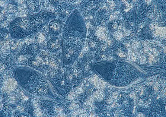
- Trichonympha species exhibit a captivating morphology, boasting large dimensions that range from 30 to 110 micrometers in length and 21 to 90 micrometers in width. This generous size allows them to stand out among other single-celled organisms, catching the eye of researchers and microscopists.
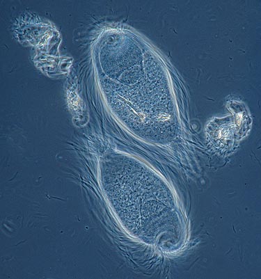
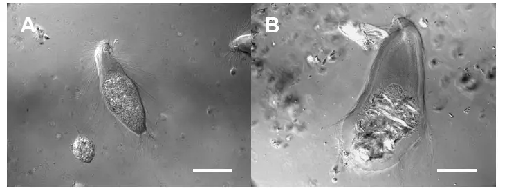
- At the heart of their unique structure lies a rounded, bell-shaped body. This characteristic shape, coupled with their intriguing features, sets them apart from their single-celled counterparts. However, it is the sheer abundance of flagella that truly defines the visual spectacle of Trichonympha. In awe-inspiring numbers, these protists possess as many as ten thousand or more flagella, which play a crucial role in enabling them to navigate through the highly viscous environment of their host’s hindgut. Such a vast array of flagella grants them impressive motility and maneuverability within this challenging microenvironment.
- Remarkably, the anterior part of the cell, known as the rostral cap, protrudes distinctly, lending a unique profile to Trichonympha’s structure. Unlike the broader posterior region of the cell, the rostral cap showcases a more slender appearance, further distinguishing these intriguing organisms.
- Microscopic examinations have unveiled the internal architecture of Trichonympha, revealing two distinct regions: the ectoplasm and the endoplasm. Most of the cell’s organelles are confined within the endoplasm, highlighting the specialized roles of these cellular compartments.
- One of the most astonishing features of Trichonympha is the absence of mitochondria. Unlike the vast majority of eukaryotic cells, these flagellates have evolved to thrive without this essential organelle. The fascinating adaptations that enable Trichonympha to function effectively in the absence of mitochondria remain a subject of ongoing scientific interest.
- Moreover, the flagella pattern of Trichonympha is unique in that it does not cover the rostral cap of the cell. This distinctive arrangement contributes to their efficient movement and navigation within the complex gut environment of their host.
- Another striking aspect of Trichonympha’s structure is the exceptionally long basal body. These basal bodies, responsible for flagella production, vary in length depending on their location within the cell. Those located in the anterior region of the cell tend to be longer, while those in the posterior part of the cell are comparatively shorter. The mechanisms behind this variation in basal body length are yet to be fully elucidated, presenting an exciting avenue for further research.
- In summary, the morphology of Trichonympha species stands as a testament to the ingenuity of nature, showcasing a plethora of unique features that have enabled them to thrive in the specialized niche of their host’s hindgut. Their large size, multitude of flagella, absence of mitochondria, and intriguing basal body variations all contribute to their captivating structure, making them a captivating subject of study for scientists seeking to unravel the mysteries of the microscopic world.
Requirements
- Clean Glass Slide: A clean glass slide serves as the foundation for microscopic investigations. It provides a smooth and clear surface on which to place the specimens, ensuring optimal visibility and accuracy during observation.
- Clean Coverslip: Placing a clean coverslip over the specimen on the glass slide is crucial for creating a secure and uniform viewing platform. The coverslip prevents the specimen from getting compressed and maintains the integrity of its structure for detailed examination.
- Termites: To study Trichonympha, access to termites (or cockroaches that feed on wood) is imperative. These host insects harbor the intriguing symbiotic relationship with Trichonympha, making them a fundamental element of any investigation.
- Petri Dish: A Petri dish provides a controlled and confined environment for the manipulation and study of specimens. It facilitates the careful handling of termites and Trichonympha while maintaining a stable environment for observation.
- Forceps: Fine-tipped forceps offer a delicate and precise method for handling delicate specimens like Trichonympha and for positioning them appropriately on the glass slide. They minimize damage and ensure accurate placement during the preparation process.
- Dissecting Needles: Dissecting needles play a critical role in separating and manipulating tissues or organisms under low magnification. They aid in isolating Trichonympha from the host gut or assisting in the study of specific structures.
- Compound Microscope: A compound microscope is an indispensable tool for examining Trichonympha at high magnification. It enables researchers to observe the intricate details of these fascinating protists, such as their unique flagella arrangement and internal structures.
- Stereomicroscope: A stereomicroscope, also known as a dissecting microscope, complements the compound microscope by providing a three-dimensional view of specimens. This instrument aids in the initial examination and handling of larger specimens like termites before more detailed analysis under higher magnification.
- Pipette: A pipette facilitates the precise handling and transfer of small volumes of liquid. It proves invaluable for collecting and manipulating Trichonympha samples, allowing researchers to study these protists under various conditions.
Procedure
- Prepare the Specimen:
- Using a pipette, place a single drop of isotonic saline solution onto a clean glass slide or a small Petri dish. The saline solution maintains a suitable environment for the Trichonympha and allows for better observation.
- Secure the Host Insect:
- With the aid of forceps, gently hold the termite by its head to ensure a stable and controlled grip. Position the termite on the drop of saline solution on the glass slide or Petri dish.
- Dissect the Gut:
- For a detailed examination of Trichonympha, place the slide or Petri dish on a stereomicroscope. This magnified view enables precise gut dissection while minimizing damage to the Trichonympha.
- Using a dissecting needle under the stereomicroscope, carefully remove the gut of the termite. The stereomicroscope provides enhanced visibility, ensuring accurate manipulation and isolation of the gut.
- Prepare the Sample:
- Gently pull apart the dissected gut to obtain smaller pieces. This step aims to create an accessible sample containing the Trichonympha, ready for observation.
- Set Up the Slide:
- Transfer the pieces of the gut onto a glass slide containing a drop of the saline solution. Alternatively, you can utilize the original slide used for dissection and add the gut pieces to it.
- Cover the Sample:
- Place a clean coverslip over the gut pieces, ensuring a secure and uniform viewing surface. The coverslip prevents compression and preserves the integrity of the sample during observation.
- Microscopic Observation:
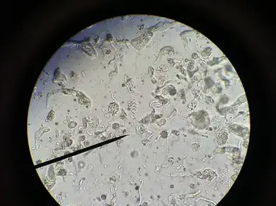
- Now, place the prepared slide under a compound microscope for detailed observation of Trichonympha.
- Start with the low-power objective to locate the gut pieces and potential Trichonympha clusters.
- Gradually switch to the high-power objective to obtain a clear and magnified view of the intricate flagellates. This higher magnification allows for the examination of Trichonympha’s unique features and behaviors.
By following this well-structured procedure, researchers can unlock the microscopic wonders of Trichonympha and gain valuable insights into their symbiotic relationships with termites and wood-feeding cockroaches. As scientists continue to refine their observations, this captivating journey into the microcosm contributes to our understanding of the complexities that exist within the tiniest corners of the natural world.
Observation
- Flagellar Extravaganza: When the compound microscope unveils the world of Trichonympha, one of the most striking features is the abundant array of flagella adorning their surface. These delicate hair-like structures emerge in astounding numbers, granting these single-celled organisms a breathtaking appearance. The flagella’s sheer quantity is unparalleled among other microorganisms, accentuating the significance of these protists in their host’s ecosystem.
- The Bell-Shaped Wonder: As we continue our observation, Trichonympha reveals its distinctive body structure. Its form may be likened to a bell, with a gracefully protruding anterior part. This unique profile sets Trichonympha apart, capturing the curiosity of scientists and enthusiasts alike. The elegant bell shape serves a vital purpose, aiding the protists in navigating through the intricate, viscous environment of the termite or wood-feeding cockroach’s hindgut.
- A Tale of Two Ends: The body of Trichonympha exhibits an intriguing contrast between its anterior and posterior regions. The anterior, or rostral cap, protrudes in a manner that sets it apart from the broader posterior. This dissimilarity adds to the protists’ visual allure, emphasizing their specialized adaptations to the microenvironments they inhabit.


FAQ
What is Trichonympha, and why is it studied under the microscope?
Trichonympha is a genus of parabasalid protists found in the hindgut of termites and wood-feeding cockroaches. They are studied under the microscope because of their intriguing symbiotic relationship with their host insects and their unique features, such as a high number of flagella.
How does Trichonympha appear under the microscope?
Under the microscope, Trichonympha is characterized by its bell-shaped body with a protruding anterior and broad posterior. The surface is covered with numerous flagella, creating a visually striking spectacle.
What is the role of flagella in Trichonympha?
Flagella in Trichonympha are crucial for its motility and navigation through the highly viscous environment of the termite or cockroach hindgut. The abundance of flagella sets Trichonympha apart from other single-celled organisms.
What is the significance of Trichonympha’s symbiotic relationship with termites and wood-feeding cockroaches?
Trichonympha aids its host insects in breaking down cellulose from wood, which is a challenging task for the insects themselves. In return, the insects provide a conducive environment for Trichonympha to thrive.
How do researchers prepare Trichonympha samples for observation under the microscope?
Researchers typically use an isotonic saline solution to place the gut of the termite or cockroach on a glass slide. The dissected gut pieces are then covered with a clean coverslip before observation.
What tools are essential for observing Trichonympha under the microscope?
Tools like a compound microscope, stereomicroscope, clean glass slides, coverslips, pipettes, forceps, and dissecting needles are necessary for a successful observation.
Can Trichonympha be observed using both low and high-power objectives of the microscope?
Yes, researchers start with a low-power objective to locate the gut pieces and potential Trichonympha clusters. They then switch to the high-power objective for a more detailed and magnified view of the protists.
Are there variations in Trichonympha’s basal body length?
Yes, Trichonympha’s basal bodies vary in length depending on their location within the cell. Basal bodies in the anterior region tend to be longer, while those in the posterior region are shorter.
How do researchers handle Trichonympha samples during observation?
Researchers use fine-tipped forceps and dissecting needles to handle the gut pieces containing Trichonympha under the stereomicroscope. This delicate manipulation ensures minimal damage to the protists.
What makes Trichonympha a fascinating subject of study?
Trichonympha’s unique morphology, symbiotic relationship with termites and wood-feeding cockroaches, and its ability to digest cellulose make it a captivating subject for researchers studying microorganisms and their ecological interactions.
- Text Highlighting: Select any text in the post content to highlight it
- Text Annotation: Select text and add comments with annotations
- Comment Management: Edit or delete your own comments
- Highlight Management: Remove your own highlights
How to use: Simply select any text in the post content above, and you'll see annotation options. Login here or create an account to get started.