What is Thymus Gland?
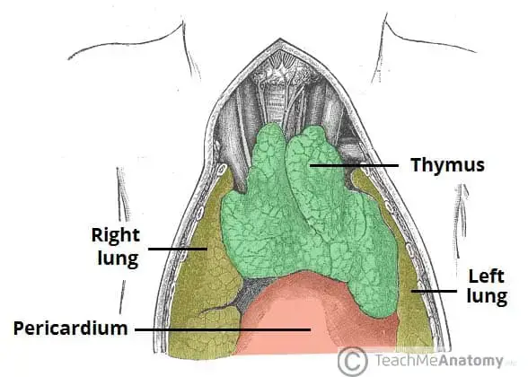
- The thymus gland is a vital primary lymphoid organ located in the anterior mediastinum of the chest, positioned behind the sternum and in front of the heart. This specialized gland plays an indispensable role in the development and maturation of T lymphocytes, commonly referred to as T cells, which are crucial components of the adaptive immune system. The thymus typically consists of two lobes, each characterized by a central medulla surrounded by an outer cortex and encased in a protective capsule.
- Within the thymus, immature T cells, known as thymocytes, undergo a complex maturation process facilitated by specialized epithelial cells. These epithelial cells are essential for the development of thymocytes, enabling them to appropriately interact with major histocompatibility complex (MHC) molecules through a process called positive selection. This process ensures that only those T cells capable of recognizing foreign antigens while being tolerant to self-proteins survive. Conversely, T cells that react against the body’s own proteins are eliminated during negative selection, which further emphasizes the organ’s critical role in maintaining immune tolerance.
- The thymus is most active during the neonatal and pre-adolescent stages of life, when the demand for T cells is high. Its size and activity peak around puberty, after which a gradual involution occurs, leading to a significant reduction in its size and functionality as it becomes increasingly replaced by adipose tissue. Nevertheless, T cell development continues, albeit at a diminished rate, throughout adulthood.
- Abnormalities within the thymus can have serious implications for immune function. A decreased production of T cells may lead to immune deficiencies and can contribute to the development of autoimmune diseases, such as autoimmune polyendocrine syndrome type 1 and myasthenia gravis. These conditions are often associated with thymoma, a tumor arising from thymic tissue, or lymphomas originating from immature lymphocytes, including T cells. The surgical removal of the thymus, known as thymectomy, may be indicated in certain pathological conditions.
- The significance of the thymus extends beyond its role in T cell maturation; it is also a hub for various immune cells, including macrophages, neutrophils, and dendritic cells. Cytokines, such as tumor necrosis factor (TNF) and interferon, regulate the proliferation and secretory activity of thymic epithelial cells, further illustrating the organ’s complex interactions within the immune system.
- Historically, the thymus has been recognized since ancient Greek times, with its name derived from the Greek word for “soul.” However, it was not until the 1960s that its functional importance in immune response became evident. Today, the thymus is acknowledged as a critical organ in defending the body against pathogens, tumors, and antigens, solidifying its place in the study of immunology and medicine.
Definition of Thymus Gland
The thymus gland is an important organ of the lymphatic system located in the chest behind the breastbone. It plays a crucial role in the development and maturation of immune cells known as T cells, which are essential for the body’s immune response. The thymus gland is most active during childhood and gradually diminishes in size and function after puberty.
Characterisitcs of Thymus Gland
The thymus gland is a unique organ with distinct characteristics. Here are the key characteristics of the thymus gland:
- Location: The thymus gland is located in the upper chest, behind the sternum (breastbone), in front of the heart, and above the superior vena cava. It is part of the mediastinum, which is the central region of the chest cavity.
- Structure: The thymus gland has a lobular structure, consisting of two lobes connected by a central portion called the isthmus. Each lobe is further divided into smaller lobules. It is composed of both glandular tissue and lymphoid tissue.
- Function: The primary function of the thymus gland is the maturation and development of T cells, which are a type of white blood cell involved in cell-mediated immunity. The thymus provides a microenvironment where T cells differentiate, mature, and acquire their immunological functions.
- Maturation Site: The thymus is the primary site for T cell maturation. Immature T cells, known as thymocytes, migrate from the bone marrow to the thymus. Within the thymus, they undergo a process of maturation, selection, and education to become functional T cells capable of recognizing foreign antigens and distinguishing them from self-antigens.
- Hormonal Influence: The thymus gland produces various hormones that play a role in T cell development and maturation. These hormones include thymosin, thymopoietin, and thymulin, among others. They help regulate the differentiation and function of thymocytes.
- Size and Involution: The size of the thymus gland changes throughout life. It is relatively large in infants and children, reaching its maximum size during puberty. However, with age, the thymus undergoes a process called involution, where it gradually decreases in size and becomes replaced by fatty tissue. This involution is a natural part of aging.
- Immune-Endocrine Interaction: The thymus gland serves as an interface between the immune and endocrine systems. It functions as both a lymphoid organ and an endocrine gland. It interacts with other components of the immune system, such as lymph nodes and spleen, as well as secretes hormones that influence T cell development and immune responses.
- Role in Autoimmunity: The thymus gland plays a crucial role in preventing autoimmune diseases. Through processes like positive and negative selection, the thymus eliminates T cells that could potentially attack the body’s own tissues, promoting self-tolerance and preventing autoimmune responses.
These characteristics make the thymus gland a vital organ for the development and regulation of the immune system, particularly in the maturation of T cells and the establishment of immune tolerance.
Structure of Thymus Gland
The thymus gland is a bilobed organ essential for the development and maturation of T cells, which are critical components of the immune system. Located in the superior mediastinum, beneath the sternum, this gland undergoes significant structural changes throughout a person’s life. The thymus is organized into distinct components that facilitate its complex functions.
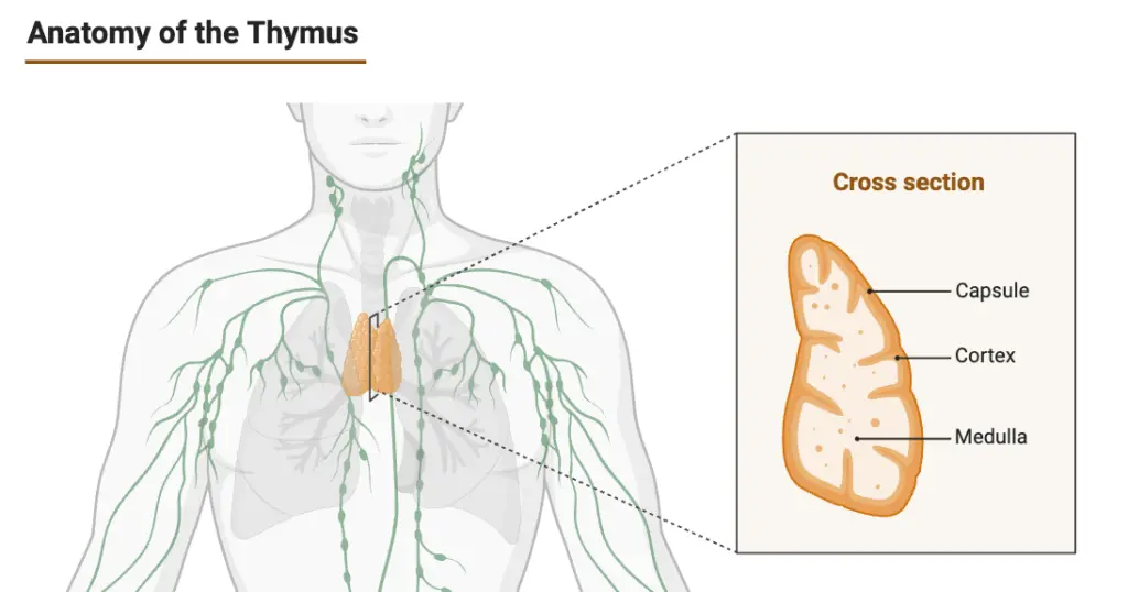
- The thymus is bilobed, consisting of two pyramid-shaped lobes, each exhibiting a lobulated surface.
- Each lobe is encapsulated by a dense layer of connective tissue, which provides structural integrity, and is internally divided by connective tissue septa that create distinct compartments.
- The majority of the thymic mass is composed of a three-dimensional network of reticular cells, which support the thymocytes and contribute to the organ’s overall architecture.
- Within the thymus, there are two main subcomponents: the cortex and the medulla.
- The outer cortex contains loosely packed lymphocytes, primarily thymocytes, that are involved in T cell maturation.
- The inner medulla consists of reticulocytes, which are rich in cytoplasm and play a role in the final maturation of T cells.
- Epithelial cells within the thymus are classified into four distinct subtypes based on various characteristics, including antigenic expression and ultrastructure.
- These subtypes include subcapsular cortical, inner cortical, medullary, and Hassall’s corpuscles.
- Hassall’s bodies, unique structures within the thymus, are concentric arrays of squamous cells that aid in the maturation of thymocytes and in clearing apoptotic cells, making them vital for lymphopoiesis.
- The thymus reaches its largest size during puberty, reflecting its peak activity in T cell production.
- Following this period, the gland gradually undergoes involution, during which its organized structure is replaced by adipose tissue, leading to a decrease in functional capacity.
- The blood supply to the thymus is provided by several arteries, including the inferior thyroid, internal thyroid, and intercostal arteries, ensuring that the gland receives adequate nutrients and oxygen.
- Additionally, the thymus is anchored to the sternum by bilateral sternohyoid and sternothyroid muscles, which assist in maintaining its position within the thoracic cavity.
- The structural integrity and cellular composition of the thymus are crucial for its function in the immune system.
- The positive and negative selection processes that T cells undergo in this organ ensure that only those cells capable of responding appropriately to foreign antigens and tolerant of self-antigens are released into the circulation.
- Approximately 95% of thymocytes fail to survive this rigorous selection process, emphasizing the thymus’s role in maintaining immune tolerance.
Detail Anatomy of Thymus Gland
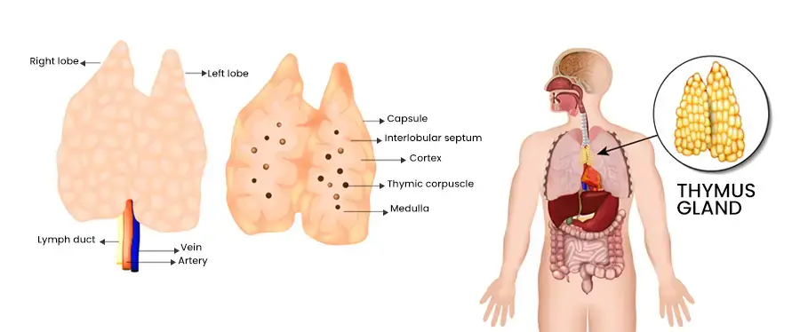
The thymus gland is a critical component of the immune system, primarily responsible for the maturation of T cells, which play a crucial role in adaptive immunity. Its anatomical structure supports its function and adapts throughout an individual’s life, particularly during childhood and adolescence.
- The thymus is anatomically located at the upper part of the sternum, nestled between the sternum and the aortic arch, close to the collarbone.
- At birth, it measures approximately 1 to 2 inches in width and about half an inch in thickness, growing larger throughout childhood before beginning to involute during adolescence.
- The thymus is encased in a collagen-type connective tissue capsule that provides structural support and protection.
- Internally, it is divided into two main lobes, which consist of irregular lobules or sub-lobes. Each lobe contains several distinct structures and cell types that contribute to its functionality.
- The cortex is the outer region of each lobe, located nearest to the capsule. It serves as the site for developing T cell lymphocytes, where the initial stages of T cell maturation occur.
- The medulla, situated centrally within each lobule, contains fully developed T cells ready for release into the peripheral immune system.
- Epithelioreticular cells play a pivotal role in thymic architecture. They create a lattice-like framework within the thymus that delineates compartments for both developing and mature T cells, ensuring an organized environment for T cell maturation.
- The thymus also contains blood vessels within its capsule and lobular walls. These vessels provide essential oxygen and nutrients to the thymic tissue, supporting cellular activities.
- Additionally, lymphatic vessels are present, facilitating the transport of lymphatic fluid throughout the thymus and the wider lymphatic system, which is crucial for immune surveillance.
- Within the thymus, macrophages function as guardians of T cell development. They are responsible for identifying and eliminating T cells that fail to develop correctly, ensuring that only functional T cells are released into circulation.
- Anatomical variations of the thymus gland may occur, especially in infants, where it can extend above the clavicle.
- In some cases, an enlarged thymus may exert pressure on surrounding structures, such as the trachea or heart. However, it is often deemed inadvisable to remove the thymus in these instances, as such actions could adversely affect immune system development.
Location of Thymus Gland
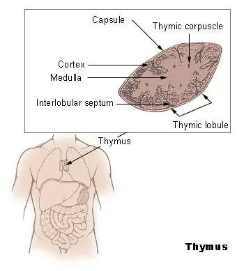
- The thymus gland is situated in the anterior (front) part of the chest, specifically behind the breastbone or sternum and between the lungs. It has a pinkish-grey color and is characterized by its lobed structure, consisting of two primary lobes and smaller lobes branching out from within. The size of the lobes can vary and they may be separate or fused together.
- The thymus gland is a soft, bilobed organ that is encapsulated. It is located near the pericardium in the superior mediastinum (the upper part of the middle compartment of the chest) and the anterior region of the inferior mediastinum (the lower part of the middle compartment of the chest). It lies deep within the sternum and anterior to the major arteries of the heart. It extends upward from the level of the inferior poles of the thyroid gland and reaches the fourth costal cartilage.
- On either side of the thymus gland, running parallel to it, are the phrenic nerves, which continue to supply the diaphragm. The two separate lobes of the thymus are connected in the middle by an isthmus.
Blood supply in Thymus Gland
The blood supply of the thymus gland is integral to its functions, particularly in supporting the development and maturation of T cells. The vascular architecture of the thymus allows for efficient delivery of nutrients and oxygen while also facilitating immune cell interactions.
- Arterial Supply
- The thymus receives its blood supply from several key arteries, which include branches of the internal thoracic artery, inferior thyroid artery, and occasionally the superior thyroid artery.
- These arteries navigate through the septa of the thymic capsule, ensuring that the blood reaches both the cortex and medulla of the gland.
- Upon entering the thymus, these arterial branches either penetrate the thymic tissue directly or extend into the outer capsule, distributing blood throughout the organ.
- Venous Drainage
- The venous blood from the thymus is collected by specific vessels known as thymic veins.
- These veins primarily drain into the left brachiocephalic vein, internal thoracic vein, and inferior thyroid veins. In certain instances, they may also empty directly into the superior vena cava.
- This drainage system is crucial for removing deoxygenated blood and metabolic waste products, ensuring that the thymus maintains optimal functioning.
- Lymphatic Drainage
- Accompanying the blood vessels, lymphatic vessels in the thymus are responsible for draining lymph away from the gland.
- These lymphatics do not enter the thymus but instead transport lymph to various lymph nodes, including the brachiocephalic, tracheobronchial, and parasternal lymph nodes.
- This drainage pathway is important for immune surveillance and the distribution of lymphocytes.
- Nervous Supply
- The thymus gland receives autonomic nervous innervation from the vagus nerve and the cervical sympathetic chain.
- Additionally, branches from the phrenic nerves reach the thymic capsule but do not infiltrate the gland itself. This innervation is essential for regulating blood flow and immune responses within the thymus.
- Blood-Thymus Barrier
- Within the thymus, the organization of blood flow is structured into intricate arcades, particularly in the cortex. This architecture contributes to the formation of the blood-thymus barrier.
- This barrier is characterized by non-fenestrated endothelium, a thick basal lamina, and reticular endothelial cells, along with associated lymphocytes and macrophages.
- Its primary function is to restrict the passage of proteins from the bloodstream into the thymic environment, thereby maintaining a specialized microenvironment crucial for T cell development.
Development of Thymus Gland
The development of the thymus gland is a complex process that involves the differentiation of various cell types and the establishment of intricate cellular structures. This process is essential for the formation of a functional immune system, particularly in generating T cells, which play a critical role in adaptive immunity.
- Embryonic Origin
- The thymus gland originates from the third pharyngeal pouch, with contributions from the fourth pharyngeal pouch in some cases.
- Initially, these pouches give rise to epithelial outgrowths that extend into surrounding mesoderm and neural crest-derived mesenchyme, establishing the basic structure of the thymus.
- Formation of Cellular Structures
- As the epithelial cells proliferate and differentiate, they interact with hematopoietic precursors migrating from the bone marrow. This interaction is crucial for the formation of thymocytes, the immature T cells that will mature within the thymus.
- The mesodermal tissue invades and surrounds the epithelial buds, leading to the organization of the thymus into lobules and a spongy architecture. This structure is characterized by cellular cords and clusters, which facilitate further development and organization.
- Importance of Iodine
- Iodine plays a significant role in the development and functional activity of the thymus. It is essential for the synthesis of thyroid hormones, which influence the growth and differentiation of thymic cells.
- Postnatal Growth and Activity
- Following birth, the thymus gland continues to grow, reaching its maximum size and functionality by puberty. At this stage, the thymus typically weighs between 20 to 50 grams and is highly active in generating T cells.
- The thymus is particularly active during fetal and neonatal periods, providing an essential environment for T cell maturation.
- Thymic Involution
- After puberty, a process known as thymic involution begins, characterized by a decrease in size and activity. This decline is attributed to hormonal changes, particularly increases in sex hormones.
- The production of T cells starts to decline after the first year of life, and over time, fat and connective tissue gradually replace the thymic volume.
- Thymic involution is a natural part of aging and can be influenced by external factors, such as severe illness or conditions like HIV infection, which further contribute to thymus shrinkage and reduced immune function.
- Aging and Evolutionary Perspective
- As individuals age, the thymus undergoes progressive involution, and fat cells increasingly infiltrate the gland. In older adults, the thymus may weigh between 5 to 15 grams and can become challenging to detect.
- Notably, thymic involution is a conserved evolutionary phenomenon observed in most vertebrate species possessing a thymus, indicating its fundamental role in immune system development and maintenance.
Hormones of Thymus Gland
The thymus gland plays a crucial role in the development and regulation of the immune system, primarily through the secretion of its hormones: thymosin, thymopoietin, and serum thymic factor. Each of these hormones has specific functions that contribute to the maturation and function of T lymphocytes, which are essential for adaptive immunity.
- Thymosin
- Source and Composition: Thymosin is secreted by the epithelial cells located in both the cortex and medulla of the thymus gland. It is primarily composed of proteins and exhibits thermal stability, maintaining its function even at temperatures up to 80°C.
- Functions:
- The principal role of thymosin is to facilitate the differentiation of T cells from precursor cells, an essential process for establishing a functional immune response.
- It enhances the immunological capabilities of various immune cells, thus strengthening the body’s ability to fight infections.
- Thymosin is instrumental in increasing the expression of phenotypic markers on lymphocytes, which are crucial for their identification and functionality within the immune system.
- Thymopoietin
- Source and Composition: Thymopoietin is a polypeptide hormone produced in the thymus, known for its relatively smaller molecular structure compared to thymosin.
- Functions:
- While thymopoietin has been shown to have neuromuscular functions, its significance in immunology is notable as well.
- Increased levels of thymopoietin are associated with the activation and differentiation of T cells, thereby contributing to the immune response.
- This hormone may influence the development of specific T cell subsets, playing a role in the adaptive immune system’s versatility.
- Serum Thymic Factor:
- Although less emphasized than thymosin and thymopoietin, serum thymic factor also contributes to immune system regulation.
- It has been implicated in the modulation of T cell activity and plays a supportive role in the maturation of T cells in conjunction with other thymic hormones.
T cell maturation by Thymus Gland
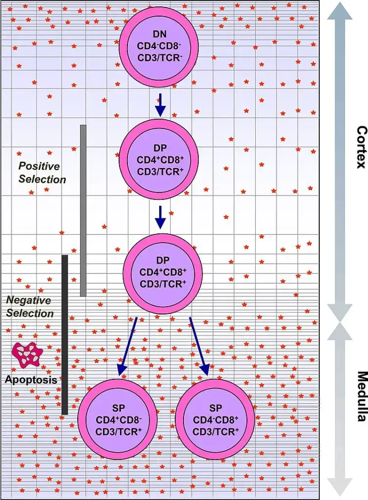
- The thymus gland plays a crucial role in the maturation of T cells, which are a vital component of the immune system responsible for cell-mediated immunity. T cells originate as hematopoietic precursors in the bone marrow and migrate to the thymus, where they are known as thymocytes. Within the thymus, a process of maturation takes place.
- During maturation, T cells undergo a selection process to ensure they can appropriately recognize antigens while avoiding reactions against self-antigens found on the body’s own tissues. This selection involves both positive and negative selection mechanisms. Positive selection ensures that T cells can bind to the major histocompatibility complex (MHC) molecules on the body’s cells. If a T cell receptor matches the specific antigen presented by the MHC, the T cell becomes activated. Negative selection, on the other hand, prevents T cells from reacting against self-antigens, thus avoiding autoimmune responses.
- The positive selection process occurs in the cortex of the thymus, where T cells that can properly bind to MHC molecules are selected. The negative selection process takes place in the medulla of the thymus, where T cells that would react against self-antigens are eliminated. After undergoing these selection processes, the mature T cells that have successfully passed leave the thymus, guided by a molecule called sphingosine-1-phosphate.
- While in the thymus, the maturation of T cells is influenced by various hormones and cytokines secreted by cells within the gland. These include thymulin, thymopoietin, and thymosins, which contribute to the development and functionality of T cells. Once T cells have left the thymus, further maturation occurs in the peripheral circulation.
- In summary, the thymus gland plays a vital role in T cell maturation by ensuring the proper selection and development of T cells capable of recognizing foreign antigens while avoiding self-reactivity. The thymus creates an environment for T cell maturation through a complex interplay of positive and negative selection processes, guided by various hormones and cytokines. The mature T cells that emerge from the thymus contribute to the effective functioning of the immune system in providing protection against pathogens and foreign substances.
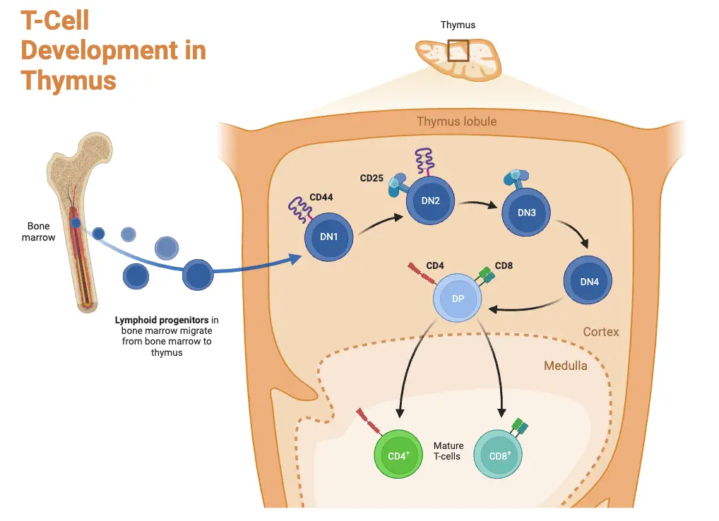
Positive selection by Thymus Gland
Positive selection is a crucial process that occurs in the thymus gland, ensuring the development of functional T cells with the ability to recognize and interact appropriately with antigens presented by major histocompatibility complex (MHC) molecules. Here are the key points about positive selection:
- T cells undergo V(D)J recombination gene rearrangement stimulated by RAG1 and RAG2 genes, resulting in the formation of distinct T cell receptors. This process introduces diversity in the T cell receptor repertoire.
- The V(D)J recombination process is error-prone, leading to the production of thymocytes with non-functional T cell receptors or receptors that may react against self-antigens (autoreactive).
- Thymocytes that successfully generate a functional T cell receptor begin to express both CD4 and CD8 cell surface proteins simultaneously.
- The fate of the thymocytes depends on their interaction with the surrounding thymic epithelial cells. The T cell receptor on thymocytes interacts with the MHC molecules displayed on the surface of epithelial cells.
- Thymocytes with T cell receptors that do not react or react weakly with the MHC molecules will undergo apoptosis and be eliminated from further development.
- Thymocytes with T cell receptors that strongly interact with MHC molecules will survive and undergo proliferation.
- The outcome of positive selection determines the nature of the mature T cell. A mature T cell expresses either CD4 or CD8, but not both, depending on the strength of binding between the T cell receptor and MHC class I or class II molecules.
- T cell receptors that primarily bind to MHC class I molecules tend to develop into mature “cytotoxic” CD8-positive T cells, which are involved in cell-mediated immune responses.
- T cell receptors that predominantly bind to MHC class II molecules tend to develop into CD4-positive T cells, which play a role in activating other immune cells and coordinating immune responses.
Negative selection by Thymus Gland
Negative selection is an important process that occurs in the thymus gland to eliminate T cells that could potentially attack the body’s own proteins, thus preventing autoimmune responses. Here are the key points about negative selection:
- Negative selection takes place in the medulla of the thymus, where epithelial cells and dendritic cells express a wide range of proteins from various tissues of the body.
- The negative selection process is stimulated by a gene called Autoimmune Regulator (AIRE), which plays a role in the expression of self-antigens in the thymus.
- Thymocytes, or developing T cells, undergo interaction with these self-antigens presented by thymic epithelial cells and dendritic cells.
- Thymocytes that strongly react to self-antigens, indicating a potential to recognize and attack the body’s own proteins, do not survive. They undergo apoptosis, a programmed cell death process.
- This process of eliminating autoreactive thymocytes helps to prevent the development of autoimmune diseases by removing T cells that could cause harm to the body’s own tissues.
- However, some CD4-positive T cells that are exposed to self-antigens can persist as a specialized subset of T cells called T regulatory cells (Tregs).
- T regulatory cells play a crucial role in maintaining immune tolerance by suppressing the activation of autoreactive T cells and preventing excessive immune responses against self-antigens.
- Tregs contribute to immune homeostasis and help prevent autoimmune diseases by regulating the immune system and promoting self-tolerance.
Disorders of Thymus Gland
The thymus gland, an essential organ for immune system development, can be affected by various disorders that compromise its function and the body’s immune response. Understanding these conditions is vital for recognizing their implications for overall health.
- Thymic Cysts
- Definition and Nature: Thymic cysts are fluid-filled sacs that may develop within the thymus gland, often originating congenitally. These cysts typically remain benign and can be surgically excised if necessary.
- Pathophysiology: The presence of these cysts can inhibit the proliferation of thymocytes, which are critical for T cell development. This inhibition can disrupt normal immune function.
- Symptoms: Individuals with thymic cysts may experience respiratory issues, such as a persistent cough, and are more prone to upper respiratory tract infections due to impaired immune capabilities.
- Thymic Involution
- Definition: Thymic involution refers to the natural shrinking of the thymus gland, which typically occurs with age but can be accelerated by various stressors.
- Causes: In infants, factors such as malnutrition, neglect, and abuse can lead to premature involution. Additionally, treatments like chemotherapy, radiation therapy, and prolonged steroid use can also induce involution, given the thymus’s sensitivity to stress.
- Implications: While involution is a normal part of development, premature involution in infants may signal underlying social or health issues, warranting further investigation into their care and environment.
- Thymic Hypoplasia
- Definition: Hypoplasia refers to the reduced number of cells within the thymus gland, resulting from developmental defects.
- Etiology: This condition is often associated with abnormalities in the third and fourth pharyngeal pouches during embryonic development. Factors such as maternal alcohol consumption or exposure to certain organic acids may interfere with neural crest cell differentiation, leading to hypoplasia.
- Clinical Manifestations: The clinical symptoms of thymic hypoplasia are contingent upon the severity of the cell reduction and the extent of genetic defects. Affected individuals may exhibit compromised immune function, leading to increased susceptibility to infections.
Functions of Thymus Gland
The thymus gland serves critical functions in the immune system, particularly in the development and maturation of T cells, which are essential for cellular immunity. Understanding these functions is vital for comprehending how the immune system protects the body from pathogens.
- Development and Differentiation of T Cells
- The primary function of the thymus gland is to promote the development, activation, and differentiation of T lymphocytes. This process is essential for establishing a robust immune response against pathogens.
- Thymosin and thymopoietin, the key hormones produced by the thymus, play a crucial role in stimulating precursor cells known as prothymocytes to differentiate into thymocytes, which eventually develop into functional T lymphocytes.
- Cytokine Release
- The thymus gland facilitates the release of various cytokines that are instrumental in regulating T cell development. These cytokines support the maturation process of T cells at multiple stages, ensuring that they acquire the necessary functions to combat infections effectively.
- This regulatory mechanism is vital for maintaining the balance of the immune response, promoting appropriate activation while preventing overactivity that could lead to autoimmune disorders.
- Role in Fetal and Childhood Immunity
- The thymus begins its role in the immune system as early as the 12th week of gestation, contributing to fetal immunity. This early development is crucial for establishing immune competence before birth.
- The gland remains active during childhood, continuing to produce T cells that are essential for a developing immune system. This activity underscores the thymus’s importance in ensuring a strong immune defense during the early years of life.
- Endocrine Functions
- Besides its immunological roles, the thymus also functions as an endocrine gland. It produces hormones such as human growth hormone (HGH), which are essential for the growth and development of various body systems.
- This endocrine function supports overall physical development and contributes to the body’s ability to respond to various physiological challenges.
Difference between Thyroid and Thymus
Thyroid and thymus are two distinct glands in the body with different characteristics and functions. Here is a comparison between the two:
Definition:
- Thyroid: The thyroid is a gland of the endocrine system responsible for producing thyroid hormones.
- Thymus: The thymus is an organ of the lymphatic system involved in the production and maturation of T cells of the immune system.
Anatomy:
- Thyroid: The thyroid consists of two lobes located in the neck region. It is composed of follicles surrounded by epithelial cells.
- Thymus: The thymus has an outer capsule and an inner medulla section. It consists of two lobules.
Changes in Size with Age:
- Thyroid: The size of the thyroid gland does not change significantly as you age.
- Thymus: The thymus tends to become smaller as you age. This process is known as thymic involution.
What It Produces:
- Thyroid: The thyroid gland produces two important hormones called thyroxine (T4) and triiodothyronine (T3), which regulate the metabolic rate and activity in the body.
- Thymus: The thymus produces and matures T cells, which are crucial for the cell-mediated immune response.
Functions:
- Thyroid: The thyroid gland plays a key role in controlling the metabolic rate and various physiological functions in the body, such as energy production, growth, and development.
- Thymus: The thymus is involved in the development and maturation of T cells, which are responsible for the cell-mediated immune response, including recognizing and attacking foreign substances and pathogens.
Disorders:
- Thyroid: Disorders of the thyroid gland include hypothyroidism (too few thyroid hormones) and hyperthyroidism (excess production of thyroid hormones).
- Thymus: Disorders associated with the thymus include myasthenia gravis, an autoimmune condition that affects the neuromuscular junction, and hypogammaglobulinemia, a condition characterized by low levels of immunoglobulins (antibodies).
| Characteristics | Thyroid | Thymus |
|---|---|---|
| Definition | A gland of the endocrine system | An organ of the lymphatic system |
| Anatomy | Two lobes comprised of follicles surrounded by epithelia | An outer capsule and inner medulla section, two lobules |
| Changes in size with age | Does not change in size as you age | Becomes smaller as you age |
| What it produces | Two thyroid hormones called thyroxine (T4) and triiodothyronine (T3) | T cells of the immune system |
| Functions | Controls metabolic rate and activity in the body | Cell-mediated immune response |
| Disorders | Too few hormones or too many hormones may be produced | The disorder, myasthenia gravis, and the condition, hypogammaglobulinemia |
FAQ
What is the thymus gland?
The thymus gland is a small organ located in the chest, near the heart. It is part of the immune system and plays a crucial role in the maturation of T cells, a type of white blood cell.
What is the function of the thymus gland?
The thymus gland is responsible for the development and maturation of T cells, which are essential for cell-mediated immunity. It helps educate T cells to recognize foreign antigens while preventing them from attacking the body’s own tissues.
When does the thymus gland reach its maximum size?
The thymus gland is relatively large during infancy and childhood. It reaches its maximum size during puberty and gradually decreases in size thereafter through a process called involution.
What is thymic involution?
Thymic involution is the natural shrinking and replacement of the thymus gland with fatty tissue that occurs as a person ages. It is a normal part of the aging process.
Can the thymus gland be affected by diseases?
Yes, the thymus gland can be affected by various diseases, including thymic tumors (thymomas), thymic carcinomas, and autoimmune disorders such as myasthenia gravis.
How is thymus cancer diagnosed?
Thymus cancer is diagnosed through a combination of imaging tests (such as CT scans), biopsy, and analysis of the tumor tissue. Medical professionals use these methods to determine the type and stage of the cancer.
Are there any symptoms associated with thymus cancer?
Symptoms of thymus cancer may include coughing, chest pain, difficulty swallowing, shortness of breath, and weight loss. However, symptoms can vary depending on the location and size of the tumor.
Can thymus cancer be treated?
Yes, treatment options for thymus cancer include surgery, radiation therapy, chemotherapy, targeted therapy, and immunotherapy. The choice of treatment depends on the type and stage of the cancer, as well as individual factors.
Can the removal of the thymus gland affect the immune system?
While the thymus gland is important for T cell maturation, its removal (thymectomy) is sometimes necessary in certain medical conditions, such as thymus tumors or myasthenia gravis. However, other lymphoid organs and tissues can compensate for its absence, and the immune system can still function effectively.
Can thymus gland disorders lead to immune system problems?
Yes, disorders of the thymus gland can impact the immune system. For example, thymic hypoplasia or dysfunction can lead to immune deficiencies, affecting the body’s ability to mount an adequate immune response. Additionally, autoimmune disorders such as myasthenia gravis can arise from dysfunction within the thymus gland.
- Lydyard, P.M., Whelan,A.,& Fanger,M.W. (2005).Immunology (2 ed.).London: BIOS Scientific Publishers.
- Brooks, G. F., Jawetz, E., Melnick, J. L., & Adelberg, E. A. (2010). Jawetz, Melnick, & Adelberg’s medical microbiology. New York: McGraw Hill Medical.
- Playfair, J., & Chain, B. (2001). Immunology at a Glance. London: Blackwell Publishing.
Owen, J. A., Punt, J., & Stranford, S. A. (2013). Kuby Immunology (7 ed.). New York: W.H. Freeman and Company. - Pearse G. Normal structure, function and histology of the thymus. Toxicol Pathol. 2006;34(5):504-14. doi: 10.1080/01926230600865549. PMID: 17067941.
- Askin DF, Young S. The thymus gland. Neonatal Netw. 2001 Dec;20(8):7-13. doi: 10.1891/0730-0832.20.8.7. PMID: 12144107.
- Thapa, Puspa, and Donna L Farber. “The Role of the Thymus in the Immune Response.” Thoracic surgery clinics vol. 29,2 (2019): 123-131. doi:10.1016/j.thorsurg.2018.12.001
- Bach JF. Thymic hormones. J Immunopharmacol. 1979;1(3):277-310. doi: 10.3109/08923977909026377. PMID: 233313.
- Zdrojewicz Z, Pachura E, Pachura P. The Thymus: A Forgotten, But Very Important Organ. Adv Clin Exp Med. 2016 Mar-Apr;25(2):369-75. doi: 10.17219/acem/58802. PMID: 27627572.
- Remien K, Jan A. Anatomy, Head and Neck, Thymus. [Updated 2021 Feb 9]. In: StatPearls [Internet]. Treasure Island (FL): StatPearls Publishing; 2021 Jan-. Available from: https://www.ncbi.nlm.nih.gov/books/NBK539748/
- https://teachmeanatomy.info/thorax/organs/thymus/
- https://www.news-medical.net/health/The-Structure-and-Function-of-the-Thymus.aspx
- https://www.verywellhealth.com/thymus-anatomy-4800309
- https://visualsonline.cancer.gov/details.cfm?imageid=9266
- Text Highlighting: Select any text in the post content to highlight it
- Text Annotation: Select text and add comments with annotations
- Comment Management: Edit or delete your own comments
- Highlight Management: Remove your own highlights
How to use: Simply select any text in the post content above, and you'll see annotation options. Login here or create an account to get started.