

Telophase is the last stage of mitosis, where separated chromosomes have already reached the two opposite poles of the cell. It is more or less the reverse process of prophase. During this stage, the chromosomes become uncoiled and gradually change into chromatin form. Around each group of chromosome, the nuclear envelope is again formed.
Two daughter nuclei are formed in this way. The spindle fibres also disappear. Cytokinesis generally starts in late anaphase or telophase, during which the cytoplasm is divided and two daughter cells are formed. At the end of this stage, the nuclear division becomes complete and interphase-like nuclei are present in the daughter cells.
Where Telophase Occurs in the Cell-Division Sequence
Telophase is the last stage of mitosis (nuclear division). It comes after anaphase. The sequence of mitosis includes prophase, prometaphase, metaphase, anaphase and telophase.
During anaphase, the sister chromatids are separated and move towards the opposite poles of the cell. When their movement is completed, the cell enters into telophase. At this stage, the chromosomes are already present at the two poles.
Cytokinesis (division of cytoplasm) can also start during late anaphase and continue in telophase. During this process, the cytoplasm undergoes division and two daughter cells are formed.
What Happens During Telophase?
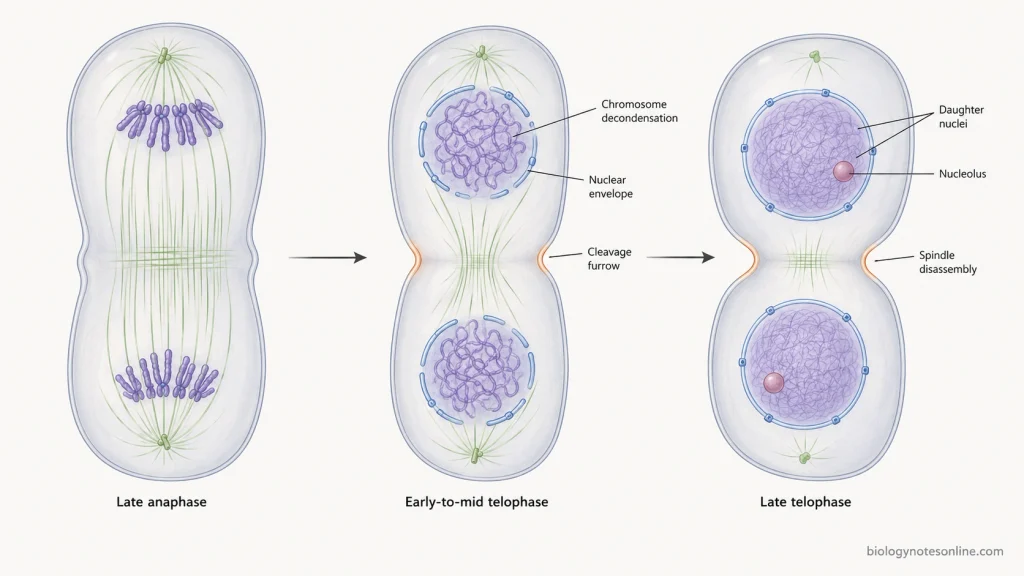
The following are the major events that take place during telophase–
- Chromosome movement- The daughter chromosomes which were separated during anaphase now reach the opposite poles. Two separate groups of chromosomes are formed, one at each end of the cell.
- Cdk1 inactivation- Cyclin B is degraded, resulting in decrease of Cdk1 activity. The different proteins which were phosphorylated during earlier mitosis are now gradually dephosphorylated. In this step, the cell starts moving out of the mitotic condition.
- Chromosome decondensation- The highly condensed chromosomes start becoming loose. They gradually change into the extended chromatin form and individual chromosomes become less clearly visible.
- Nuclear envelope formation- Around the two groups of chromosomes, nuclear membrane starts forming again. The membrane structures and nuclear envelope proteins are assembled around each chromosome group. Finally, two separate daughter nuclei are formed.
- Nuclear pore formation- Along with nuclear envelope, Nuclear Pore Complexes (NPCs) are also assembled in the newly formed membrane. Nuclear lamina is restored. The separation between nuclear and cytoplasmic contents is established again.
- Spindle disassembly- The mitotic spindle which was involved in separation of chromosomes is now no longer required. Most of its microtubules start disassembling. However, some microtubules remain in the central region of the cell for some time and are involved in the later process of cytokinesis.
- Nucleolus reappearance- The nucleolus again appears inside each daughter nucleus. During this process, chromosomes continue to decondense and transcriptional activities gradually start again. The newly formed nuclei return towards the interphase condition.
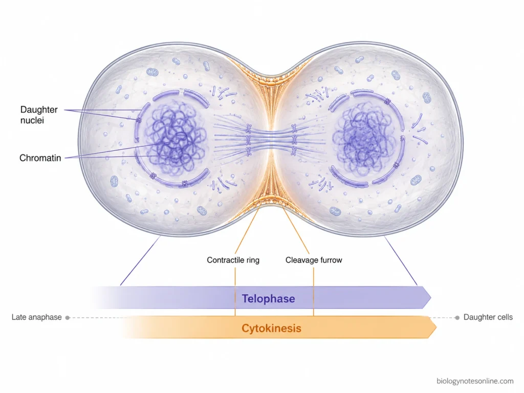
- Cytokinesis- Cytokinesis generally starts during late anaphase and continues during telophase. In animal cells, the actin-myosin contractile ring forms a cleavage furrow which moves inward, separating the cytoplasm. In plant cells, a cell plate is formed between the two daughter nuclei. The cytoplasm is finally separated into two daughter cells.
Telophase and Cytokinesis: What Is the Difference?
Telophase and cytokinesis occur near the end of cell division, but both are different processes. Telophase completes nuclear division, whereas cytokinesis divides the cytoplasm.
| Features | Telophase | Cytokinesis |
|---|---|---|
| Main process | Two daughter nuclei are formed. | Cytoplasm is divided into two parts. |
| Chromosomes | Chromosomes reach the poles and start decondensing. | Chromosome separation has already taken place. |
| Nuclear envelope | Nuclear envelope is formed again around each chromosome group. | It has no major role in nuclear-envelope formation. |
| Main structure involved | Chromosomes, nuclear envelope and spindle changes are involved. | Cleavage furrow in animal cells and cell plate in plant cells are involved. |
| Time of occurrence | It is the final stage of mitosis. | It generally starts during late anaphase or telophase and continues until cell separation. |
| Final result | Nuclear division is completed. | Two separate daughter cells are produced. |
Telophase in Mitosis
Telophase is the final stage of mitosis during which the separated daughter chromosomes reach the opposite poles and two new nuclei are formed. Many changes which occurred during prophase are reversed in this stage.
The following are the major events of telophase-
- Chromosomes at poles- The daughter chromosomes reach the two opposite poles of the spindle. Each pole now contains one complete set of chromosomes.
- M-Cdk inactivation- Cyclin B is degraded and M-Cdk activity decreases. The mitotic proteins which were phosphorylated during earlier stages are now dephosphorylated. This allows the cell to come out of the mitotic state.
- Chromosome decondensation- The chromosomes start losing their highly condensed structure. They become extended into chromatin and gradually the individual chromosomes are no longer clearly seen.
- Nuclear envelope formation- Nuclear membrane starts forming around each separated chromosome group. The membrane components associate with chromosomes and fuse with one another. In this process, two daughter nuclei are formed at the opposite ends of the cell.
- Nuclear pore formation- Nuclear pore complexes (NPCs) are assembled in the newly developing nuclear envelope. Dephosphorylated lamins again associate and the nuclear lamina is formed. Nuclear proteins can then be transported into the newly formed nucleus.
- Spindle disappearance- The spindle is no longer required for chromosome movement and its microtubules start disassembling. The microtubule system gradually returns towards its interphase arrangement.
- Nucleolus reappearance- The nucleolus which disappeared during the beginning of mitosis is formed again. Chromatin becomes less condensed and transcriptional activity can resume inside the daughter nuclei.
- Formation of daughter nuclei- At the end of telophase, two interphase-like nuclei are present. Each nucleus contains the chromosome set which was separated during anaphase. The nuclear division is now completed.
- Cytokinesis- Cytokinesis generally starts during late anaphase and continues through telophase. In animal cells, an actin-myosin contractile ring produces a cleavage furrow and divides the cytoplasm. In higher plant cells, division takes place by formation of a cell plate between the two daughter nuclei.
Telophase I and Telophase II in Meiosis
Telophase in meiosis occurs after the chromosomes have moved towards their respective poles. It occurs two times, telophase I at the end of meiosis I and telophase II at the end of meiosis II. The chromosome condition, however, is not the same in these two stages.
What Happens During Telophase I?
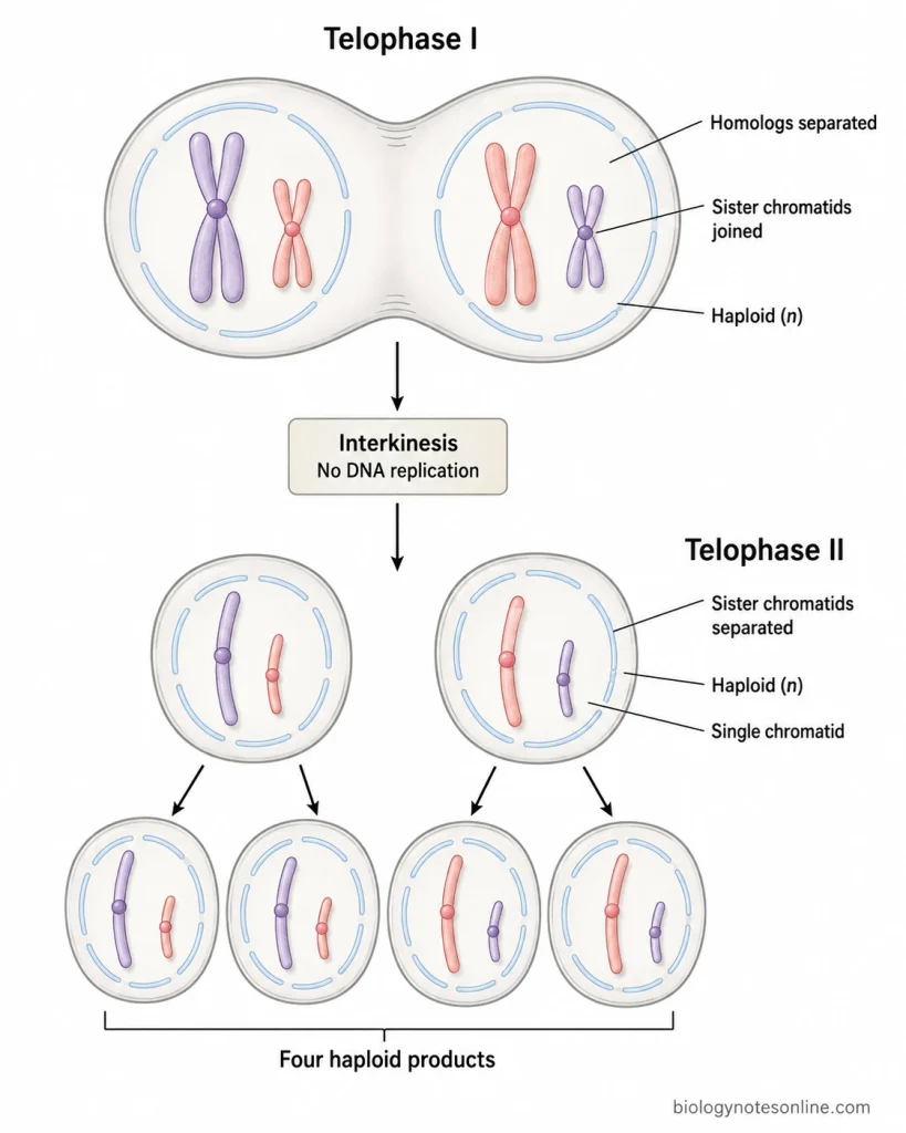
Telophase I follows anaphase I, where the homologous chromosomes are separated from each other. The chromosomes reaching each pole form a haploid set, but every chromosome is still composed of two sister chromatids.
The following are the events taking place during telophase I-
- The homologous chromosomes which were separated during anaphase I reach the opposite poles. Thus, one chromosome of each homologous pair is present at either pole.
- Sister chromatids remain attached with each other. They do not separate in the first meiotic division, and each chromosome is therefore still a duplicated chromosome with two chromatids.
- The chromosome set present at each pole is haploid (n). Only one member from each homologous pair is present, although the chromosomes themselves still contain two sister chromatids. This is the reduction in chromosome number produced by meiosis I.
- Nuclear membrane may start forming around the chromosomes. The chromosomes can also become less condensed and return towards a chromatin-like condition.
- Telophase I is not equally complete in all organisms. In some cells, the chromosomes remain condensed and formation of nuclear envelope is also absent before the next meiotic division starts.
- Cytokinesis can occur after meiosis I, producing two haploid cells. Both cells contain chromosomes having two chromatids each.
Interkinesis Between Meiosis I and Meiosis II
The short period present between meiosis I and meiosis II is called interkinesis. It may resemble an interphase in some cells, but an important difference is the absence of another DNA replication phase.
- DNA replication does not occur during this period. There is no S phase between the two meiotic divisions.
- The chromosomes have already been replicated before meiosis I. Hence, each chromosome entering meiosis II still consists of the two sister chromatids which were retained after meiosis I.
- If nuclear envelope had been formed, it disappears when meiosis II starts. Chromosomes that had partly decondensed can again become condensed during prophase II.
What Happens During Telophase II?
Telophase II is the last nuclear stage of meiosis. It follows anaphase II, in which the sister chromatids are finally separated and moved towards opposite poles. The condition is therefore different from telophase I.
- The chromosomes derived from separated sister chromatids reach their respective poles.
- These chromosomes begin to lose their condensed condition. Gradually, they become extended into chromatin.
- Nuclear envelope is formed around the chromosome groups present at each pole. New nuclei are thus established.
- Each chromosome now has only one chromatid because separation of sister chromatids has already occurred during anaphase II. In telophase I, on the other hand, the two sister chromatids were still present together.
- The newly formed nuclei contain a haploid chromosome set. Nuclear organization is restored as telophase II proceeds.
- Cytokinesis occurs with or after this stage. Following completion of meiosis II, one original diploid meiotic cell generally gives rise to four haploid products.
- Telophase II completes the nuclear divisions of meiosis. Crossing over does not take place here. It occurs during prophase I, while the chromosome arrangement producing independent assortment is established earlier during metaphase I.
Mitosis vs Telophase I vs Telophase II
Mitosis telophase and the two meiotic telophases occur after chromosome segregation, but the chromosomes arriving at the poles are not in the same condition. The main differences are as follows-
| Features | Mitosis Telophase | Telophase I | Telophase II |
|---|---|---|---|
| What was separated | Sister chromatids | Homologous chromosomes | Sister chromatids |
| Chromosome-set condition | Usually same chromosome-set number as the parent cell | Haploid | Haploid |
| Chromatids in each chromosome | One | Two sister chromatids remain together | One |
| Result after cytokinesis | Usually two daughter cells | Usually two haploid cells | Generally four haploid products after meiosis II |
| Following stage | Interphase in many dividing cells | Interkinesis or prophase II | Meiotic division ends |
Telophase and Cytokinesis in Animal and Plant Cells
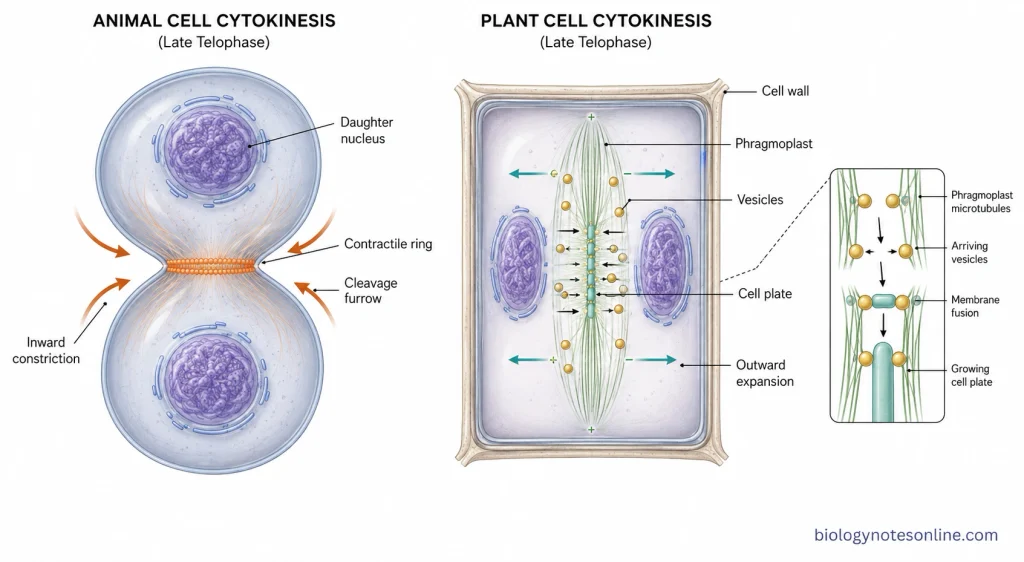
Telophase is followed closely by cytokinesis and in many cells both processes overlap with each other. During telophase, the separated chromosomes are enclosed into daughter nuclei, while cytokinesis divides the cytoplasm. The process of cytokinesis is different in animal and plant cells mainly because plant cells have a rigid cell wall.
Telophase and Cytokinesis in Animal Cells
The following are the events taking place in animal cells-
- During telophase, the daughter chromosomes have reached the opposite poles. The chromosomes now start becoming less condensed, and nuclear envelope is formed again around each chromosome group. Thus, two daughter nuclei are produced.
- Cytokinesis generally begins before telophase is fully completed. The site of division is formed around the equatorial region of the cell, between the two separating chromosome groups.
- A ring made mainly of actin filaments and myosin II is assembled just below the plasma membrane. This is known as the contractile ring. Its position is associated with the mitotic spindle, hence division occurs between the two daughter nuclei.
- Contraction of this ring pulls the plasma membrane towards the center. A depression is therefore produced on the cell surface, which is referred to as the cleavage furrow.
- The cleavage furrow becomes deeper as contraction continues. It moves inward from the outer surface, gradually constricting the cytoplasm into two parts. Animal cytokinesis is therefore an outside-to-inside type of division.
- During this process, membrane is also supplied to the region of the dividing cell. Finally, only a narrow connection remains between the daughter cells.
- This connection is removed during the final separation or abscission. Two individual daughter cells are thus formed, each having its own nucleus and cytoplasmic contents.
Telophase and Cytokinesis in Plant Cells
In plant cells, telophase also produces two daughter nuclei, but division of cytoplasm does not occur by pinching of the plasma membrane. Due to the presence of the rigid cell wall, a new partition is constructed inside the parent cell instead. The plant cell also has a very unique cell division process whereby there is formation of a phragmoplast during cytokinesis.
- After the chromosomes reach their respective poles, they start losing the condensed condition. Nuclear envelopes are formed around the two chromosome groups and daughter nuclei are established.
- Between these two daughter nuclei, remnants and newly organized microtubules form a structure called the phragmoplast. It is a plant-specific structure consisting mainly of microtubules together with actin filaments and other associated components.
- Vesicles are transported towards the central region of the phragmoplast. These membrane vesicles accumulate and fuse with each other at the middle of the cell.
- The fused vesicles gradually produce an initial cell plate. Unlike the cleavage furrow of animal cells, cell plate formation begins internally and expands towards the outer region of the parent cell.
- More vesicles continue to fuse with the developing plate. The cell plate therefore enlarges outward through the cytoplasm, while the phragmoplast also expands towards the cell cortex.
- The growing cell plate finally reaches and joins with the existing plasma membrane. The membranes of the cell plate become the new plasma membranes of the daughter cells, and cell wall material is deposited between them.
- The new partition separates the cytoplasm into two daughter cells. Primary plasmodesmata can remain at places where portions of endoplasmic reticulum are retained across the newly developing wall, allowing connection between adjacent plant cells.
How to Identify Telophase Under a Microscope or in a Diagram
Telophase can be identified mainly from the position of chromosomes and formation of two new nuclei. The chromosomes are already present at the opposite ends of the cell, therefore it looks quite different from metaphase or early anaphase. The following are some of the important characters used for identification-
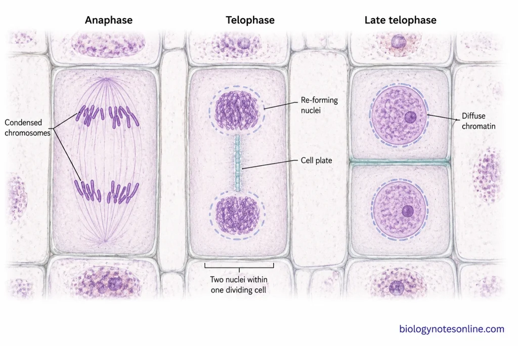
- Look for two groups of chromosomes at the opposite poles of the same cell. There will be no chromosome arrangement at the equatorial plate. Separation of sister chromatids has already occurred before this stage.
- The chromosome groups become less compact. During this process, the clearly condensed chromosomes start changing back into chromatin, therefore their outline may appear less sharp than the chromosomes of metaphase and anaphase.
- Two nuclear regions can be seen instead of one. Nuclear envelope starts forming around each group of chromosomes and finally two daughter nuclei are produced. This is one of the main characters of telophase.
- The spindle becomes less prominent and gradually disappears. So, a distinct spindle with chromosomes moving along it is generally not the major appearance of a telophase cell.
- Cytokinesis may also be seen because it overlaps with the later part of mitosis. In an animal cell, a constriction or cleavage furrow is present between the two developing daughter nuclei. The furrow moves inward as division proceeds.
- In a plant cell, the outer cell wall does not pinch inward. A developing cell plate may be visible across the middle of the cell, between the two daughter nuclei. It is the region where the new partition between daughter cells is being formed.
- While observing an onion root tip or another stained plant tissue, find a cell having two chromosome masses or two newly forming nuclei inside one original cell boundary. A cell plate in the middle gives further indication of late telophase or cytokinesis. Telophase cells can be directly recognized in stained onion root-tip preparations by this late mitotic appearance.
- Telophase should not be confused with anaphase. In anaphase, two chromosome groups are still moving apart and remain distinctly condensed. In telophase, they have reached the poles, chromosomes start decondensing and nuclei are formed again.
- It can also look close to two interphase nuclei when telophase becomes advanced. Check whether both nuclei are still present within a recently dividing cell and whether cytokinesis is incomplete. By the end of telophase, cytokinesis is usually nearly completed and two interphase daughter cells are formed.
What Controls the Transition Into Telophase?
The entry into telophase is controlled mainly by the fall of mitotic Cdk activity after chromosome separation. For this, several proteins are degraded and the phosphorylated proteins of mitosis are again dephosphorylated. The major control is based on APC/C, cyclin B and Cdk1 activity.
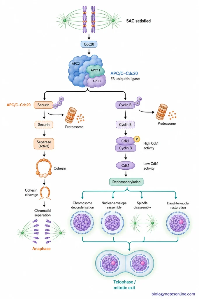
The following are the important events controlling this transition-
- Before chromosome separation, the spindle assembly checkpoint (SAC) checks the attachment of chromosomes with spindle microtubules. If one or more kinetochores remain unattached, Cdc20 is inhibited and the cell is prevented from progressing normally. Thus, premature chromosome separation is avoided.
- When proper chromosome attachment has been achieved, the checkpoint signal becomes inactive. Cdc20 is then available for activation of the anaphase-promoting complex/cyclosome (APC/C). It is an E3 ubiquitin ligase and controls anaphase as well as later exit from mitosis.
- The activated APC/C-Cdc20 causes ubiquitination and destruction of securin. Separase is released from its inhibition and cohesin joining the sister chromatids is cleaved. The chromatids are now separated and anaphase takes place.
- At the same time, cyclin B is also marked for degradation by APC/C. Cyclin B is the regulatory cyclin of the Cdk1-cyclin B complex (M-Cdk). Therefore, destruction of cyclin B results in rapid decrease of M-Cdk activity. This decrease is one of the major requirements for exit from mitosis.
- During the beginning of mitosis, many cellular proteins were phosphorylated by M-Cdk. When M-Cdk becomes inactive, this phosphorylation is no longer maintained. The phosphatases can now act on these proteins without the opposing Cdk activity. In this process, mitotic phosphorylation is gradually reversed.
- Dephosphorylation produces the changes characteristic of late anaphase and telophase. The chromosomes start becoming decondensed, nuclear-envelope components again associate with the chromosomes and nuclear envelope is formed. The spindle is also disassembled as the cell comes out of the mitotic state.
- Telophase does not begin due to a single separate telophase signal. It results mainly from APC/C activation followed by cyclin B destruction and Cdk1 inactivation. The mitotic changes are then reversed and two daughter nuclei are gradually established.
How Is Telophase Different from Anaphase?
Anaphase and telophase are two successive stages of mitosis. In anaphase, chromosome separation and movement mainly takes place. Telophase begins when these separated chromosomes have reached the opposite poles and two new nuclei start forming.
| Features | Anaphase | Telophase |
|---|---|---|
| Main event | Sister chromatids separate and move towards opposite poles. | Separated chromosomes reach the poles and daughter nuclei are formed. |
| Chromosome movement | Chromosomes are actively moving apart. | Chromosome movement is mostly completed. |
| Chromosome condition | Chromosomes remain highly condensed and clearly visible. | Chromosomes start becoming loose and gradually change into chromatin. |
| Nuclear envelope | Nuclear envelope is absent in cells undergoing open mitosis. | Nuclear envelope is formed again around each chromosome group. |
| Mitotic spindle | Spindle microtubules are actively involved in chromosome movement and spindle elongation. | Chromosomes are released from spindle microtubules and the spindle starts disassembling. |
| Number of nuclear regions | Two chromosome groups are separating, but complete daughter nuclei are not yet formed. | Two daughter nuclei are established. |
| Cytokinesis | It may start during late anaphase. | Cytokinesis generally continues through telophase and is nearly completed by its end. |
| Result | Two chromosome groups are produced at opposite sides of the cell. | Nuclear division is completed and the cell returns towards interphase condition. |
Why Is Telophase Important?
Telophase completes the nuclear division and restores the daughter nuclei towards interphase condition. Some of the important functions of telophase are as follows-
- Two separate daughter nuclei are formed around the chromosome groups present at opposite poles.
- Nuclear envelope and nuclear pore complexes are formed again. Thus, nuclear and cytoplasmic contents become separated.
- The condensed chromosomes start becoming loose and return towards the chromatin condition.
- Nuclear activities gradually start again. Transcription resumes and the nucleolus is also formed again.
- During this process, mitotic changes are reversed after decrease of M-Cdk activity. The spindle is disassembled and the cell comes out of mitotic condition.
- Telophase also occurs along with the later process of cytokinesis. Finally, each daughter cell receives its own organized nucleus and chromosome set.
References
- Alberts, B., Johnson, A., Lewis, J., Raff, M., Roberts, K., & Walter, P. (2002). Molecular biology of the cell (4th ed.). Garland Science. https://www.ncbi.nlm.nih.gov/books/NBK21054/
- Betts, J. G., Young, K. A., Wise, J. A., Johnson, E., Poe, B., Kruse, D. H., Korol, O., Johnson, J. E., Womble, M., & DeSaix, P. (2013). Anatomy and physiology. OpenStax. https://openstax.org/books/anatomy-and-physiology/pages/1-introduction
- Carim, S. C., Kechad, A., & Hickson, G. R. X. (2020). Animal cell cytokinesis: The Rho-dependent actomyosin-anilloseptin contractile ring as a membrane microdomain gathering, compressing, and sorting machine. Frontiers in Cell and Developmental Biology, 8, 575226. https://doi.org/10.3389/fcell.2020.575226
- Chaudhary, N., & Courvalin, J. C. (1993). Stepwise reassembly of the nuclear envelope at the end of mitosis. The Journal of Cell Biology, 122(2), 295–306. https://doi.org/10.1083/jcb.122.2.295
- Civelekoglu-Scholey, G., & Cimini, D. (2014). Modelling chromosome dynamics in mitosis: A historical perspective on models of metaphase and anaphase in eukaryotic cells. Interface Focus, 4(3), 20130073. https://doi.org/10.1098/rsfs.2013.0073
- Clark, M. A., Douglas, M., & Choi, J. (2018). Biology 2e. OpenStax. https://openstax.org/books/biology-2e/pages/1-introduction
- Cooper, G. M. (2000). The cell: A molecular approach (2nd ed.). Sinauer Associates. https://www.ncbi.nlm.nih.gov/books/NBK9839/
- Gilbert, S. F. (2000). Developmental biology (6th ed.). Sinauer Associates. https://www.ncbi.nlm.nih.gov/books/NBK9983/
- Lischetti, T., & Nilsson, J. (2015). Regulation of mitotic progression by the spindle assembly checkpoint. Molecular & Cellular Oncology, 2(1), e970484. https://doi.org/10.4161/23723548.2014.970484
- Liu, S., & Pellman, D. (2020). The coordination of nuclear envelope assembly and chromosome segregation in metazoans. Nucleus, 11(1), 35–52. https://doi.org/10.1080/19491034.2020.1742064
- McIntosh, J. R. (2016). Mitosis. Cold Spring Harbor Perspectives in Biology, 8(9), a023218. https://doi.org/10.1101/cshperspect.a023218
- Ramakrishna, P. (2021). Formins bridle and unleash their way through cytokinesis: A phragmoplast-centered view. Plant Physiology, 186(2), 829–831. https://doi.org/10.1093/plphys/kiab148
- Rehman, I., Farooq, M., & Simpson, B. (2023). Genetics, mitosis. In StatPearls. StatPearls Publishing. https://www.ncbi.nlm.nih.gov/books/NBK482449/
- Schooley, A., Vollmer, B., & Antonin, W. (2012). Building a nuclear envelope at the end of mitosis: Coordinating membrane reorganization, nuclear pore complex assembly, and chromatin de-condensation. Chromosoma, 121(6), 539–554. https://doi.org/10.1007/s00412-012-0388-3
- Vader, G., Maia, A. F., & Lens, S. M. A. (2008). The chromosomal passenger complex and the spindle assembly checkpoint: Kinetochore-microtubule error correction and beyond. Cell Division, 3, 10. https://doi.org/10.1186/1747-1028-3-10
- Walczak, C. E., Cai, S., & Khodjakov, A. (2010). Mechanisms of chromosome behaviour during mitosis. Nature Reviews Molecular Cell Biology, 11(2), 91–102. https://doi.org/10.1038/nrm2832
- Yanagida, M. (2005). Basic mechanism of eukaryotic chromosome segregation. Philosophical Transactions of the Royal Society B: Biological Sciences, 360(1455), 609–621. https://doi.org/10.1098/rstb.2004.1615