What are Spindle Fibres?
- Spindle fibres, intricate structures integral to cellular division, are primarily composed of microtubules. These microtubules originate from the centrosome, a pivotal organelle often referred to as the microtubule-organizing center (MTOC). Functioning as the cell’s architectural framework during division, spindle fibres ensure the accurate segregation of chromosomes, thereby maintaining genetic integrity.
- In eukaryotic cell division, spindle fibres are instrumental in orchestrating the movement of chromosomes. Depending on the type of cell division, these structures can be classified into two categories: the mitotic spindle, which is formed during mitosis, and the meiotic spindle, which arises during meiosis. The primary role of these spindles is to facilitate the separation of sister chromatids, ensuring that each daughter cell inherits the appropriate number of chromosomes.
- The formation and function of spindle fibres are underpinned by a myriad of accessory proteins. These proteins, in conjunction with microtubules, guide the meticulous process of genetic division. As spindle fibres extend from the poles of a dividing cell, they actively seek and attach to the centromere of each chromosome. Upon successful attachment, the spindle fibres retract, pulling the attached chromosome with them. This dynamic process ensures the equitable distribution of chromosomes between the nascent daughter cells.
- In essence, spindle fibres are cellular machineries that have evolved over billions of years to perform a critical task: the precise and orderly division of genetic material. Their intricate design and function underscore the complexity and elegance of cellular processes, all of which operate with a remarkable degree of precision and coordination.
Definition of Spindle Fibres
Spindle fibres are microtubule structures that facilitate the separation and movement of chromosomes during eukaryotic cell division.
Importance of Spindle Fiber
Spindle Fibers are essential structures in the process of cell division, ensuring the accurate and proper segregation of chromosomes. Their significance can be highlighted through the following points:
- Role in Cell Division: Spindle Fibers are pivotal in both mitosis and meiosis, the processes of cell division in eukaryotic cells. They ensure that chromosomes are separated and allocated correctly to the daughter cells.
- Composition and Structure: Composed of tubulin protein subunits, Spindle Fibers form the spindle apparatus, a complex structure that orchestrates the movement of chromosomes during cell division.
- Attachment to Chromosomes: These fibers connect to chromosomes at specific sites known as kinetochores. Once attached, they exert force, pulling the chromosomes apart and ensuring their proper distribution.
- Ensuring Genetic Accuracy: By facilitating the correct segregation of chromosomes, Spindle Fibers ensure that each daughter cell inherits a complete and accurate set of chromosomes. This is crucial for the cell’s proper growth, function, and development.
- Prevention of Genetic Abnormalities: In the absence of Spindle Fibers, chromosomes would not segregate correctly. This missegregation can lead to genetic abnormalities, which can have detrimental effects on the cell, leading to conditions like cancer or cell death.
In conclusion, Spindle Fibers play a vital role in maintaining the genetic integrity of cells. Their function is paramount in ensuring the proper growth and development of organisms.
Spindle Fibres Formation
Spindle Fibers, essential structures in cell division, are formed during both mitosis and meiosis. Their formation is intricately linked to the activity of microtubules, which are pivotal in establishing the framework that segregates chromosomes. Lest’s discuss the formation of Spindle Fibers, elucidating their role and structure in the context of mitosis and meiosis.
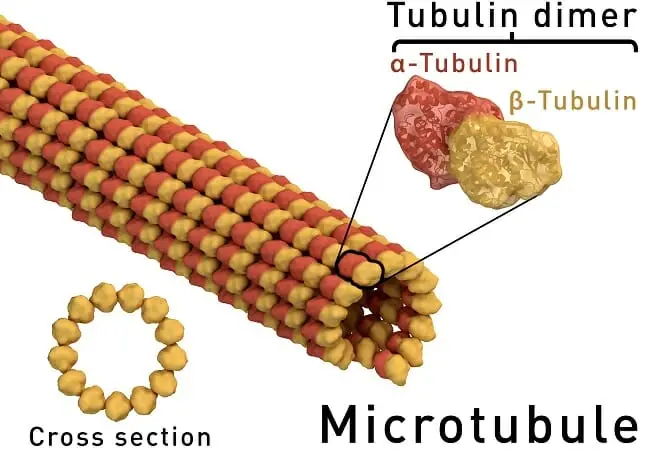
- Microtubules and Their Composition: Microtubules are cylindrical entities constituted by the protein tubulin. These structures are foundational in the formation of the spindle apparatus during cell division.
- Spindle Fiber Formation in Mitosis:
- Origin from Centrosomes: During mitosis, Spindle Fibers originate from centrosomes situated at the cell’s opposing ends.
- Centrioles and Their Role: Centrosomes house centrioles, which are intricate structures composed of microtubules. As the cell commences mitosis, centrosomes diverge.
- Microtubule Extension: Subsequent to the divergence of centrosomes, microtubules emanate from the centrioles, culminating in the formation of Spindle Fibers. These fibers subsequently connect to chromosomes aligned centrally within the cell, facilitating their segregation during cell division.
- Spindle Fiber Formation in Meiosis:
- Origin from Centrosomes and Meiotic Spindle Pole Body: In meiosis, Spindle Fibers are derived not only from centrosomes but also from an entity termed the meiotic spindle pole body.
- Location of the Meiotic Spindle Pole Body: Unlike centrosomes, which are positioned at the cell’s poles, the meiotic spindle pole body resides within the cell’s cytoplasm.
- Microtubule Extension in Meiosis: Microtubules project from both the centrosomes and the meiotic spindle pole body, leading to the formation of Spindle Fibers. These fibers then associate with chromosomes, ensuring their accurate segregation during meiosis.
Structure of Spindle Fibers – What are Spindle Fibres made up of?
Spindle Fibers, integral to the process of cell division, are sophisticated structures with a multifaceted composition. Their formation and function are underpinned by a myriad of proteins and molecular components.

- Microtubules: The Core Component:
- Composition: Microtubules are elongated, cylindrical entities predominantly composed of protein tubulin units. These units are the foundational elements of Spindle Fibers.
- Tubulin Subunits: Microtubules are constituted by two distinct tubulin subunits: Alpha-tubulin and Beta-tubulin. These subunits are organized in a specific configuration, with the Beta-tubulin subunit situated externally and the Alpha-tubulin subunit positioned internally.
- Dynamics of Microtubules: Characterized by their dynamic nature, microtubules undergo continual growth and contraction via the addition and removal of tubulin subunits. This dynamicity is pivotal for the optimal operation of Spindle Fibers during cell division.
- Centrosomes: Microtubule Organizing Centers:
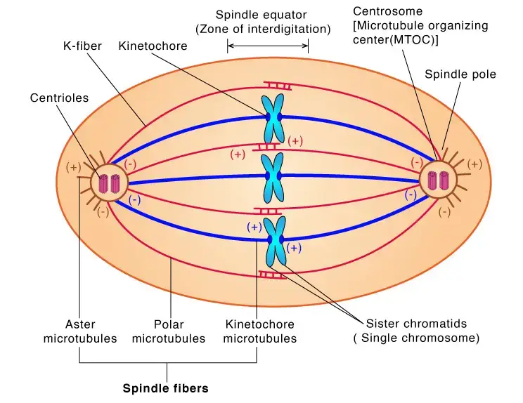
- Centrosomes serve as the nucleating centers for the microtubules constituting the Spindle Fibers. Often referred to as the Microtubule Organizing Center (MTOC), centrosomes harbor proteins that modulate the elongation and contraction of microtubules in response to cellular signals.
- Kinetochores: Chromosome Connectors:
- Kinetochores are intricate protein complexes that affix to chromosomes. Their primary function is to align chromosomes during cell division, ensuring their accurate segregation.
- Motor Proteins: Movers and Organizers:
- Motor proteins, including kinesin and dynein, play a cardinal role in the movement and organization of chromosomes during cell division. These proteins facilitate the transport of cellular components along the microtubules.
Types of Spindle Fibres
Spindle fibers, essential structures in cell division, can be categorized based on their function and location within the cell. These fibers play distinct roles in ensuring the accurate segregation of chromosomes during cell division.

- Kinetochore Fibers:
- Definition: Kinetochore fibers are spindle fibers that connect directly to the kinetochore, a specialized protein structure situated at the centromere of a chromosome.
- Function: Their primary role is to segregate chromosomes during cell division. By attaching to the kinetochore, these fibers exert force to pull chromosomes apart, ensuring that each daughter cell inherits a complete set of chromosomes.
- Composition: These fibers are essentially kinetochore microtubules that form a link between the DNA and the microtubules, facilitating the separation of sister chromatids during cell division.
- Astral Fibers:
- Definition: Astral fibers are spindle fibers that radiate outward from the spindle poles.
- Function: They play a pivotal role in aligning chromosomes prior to cell division. By interacting with the cell’s microtubules and actin filaments, astral fibers ensure that chromosomes are optimally positioned before segregation. Additionally, in animal cells, they anchor the spindle poles to the cell membrane, determining the cleavage furrow’s location during cytokinesis.
- Composition: Termed as aster microtubules, these fibers are radial microtubular assemblies that orient the mitotic spindle apparatus based on cellular polarity.
- Polar Fibers:
- Definition: Polar fibers are spindle fibers that span between the spindle poles.
- Function: Their primary role is to maintain the spindle’s structural integrity and stability during cell division. By anchoring the spindle poles and preventing spindle collapse, they ensure the chromosomes’ proper alignment before segregation. Additionally, they contribute to cell elongation by pushing the spindle poles apart via motor proteins.
- Composition: These are the spindle microtubules that do not connect to chromosomes. At the spindle midzone, they overlap and interact with motor proteins.
Spindle Fibres in Mitosis
Spindle fibers, intricate structures within the cell, are paramount in the process of mitosis. Their primary function is to ensure the accurate segregation of chromosomes, facilitating the formation of two genetically identical daughter cells. Let’s elucidates the role and dynamics of spindle fibers during the various phases of mitosis.
- General Function of Spindle Fibers:
- Spindle fibers are responsible for orchestrating the movement and alignment of chromosomes during mitosis. Their dynamic nature allows them to remodel continuously throughout the process.
- Their primary role is to pull chromosomes to the cell’s opposite ends, ensuring that each daughter cell inherits an identical set of chromosomes.
- Role in Specific Phases of Mitosis:
- Prophase: During this initial phase, spindle fibers commence their formation at the cell’s poles. They then project towards the cell’s center, aiming to connect with chromosomes. The attachment occurs at kinetochores, specialized protein structures situated at each chromosome’s centromere. Subsequently, spindle fibers initiate the process of aligning chromosomes at the metaphase plate, the cell’s central region.
- Metaphase: In this phase, the spindle fibers persistently pull the chromosomes, ensuring their alignment at the cell’s center. Once alignment is achieved, the spindle fibers start to contract, initiating the separation of chromosomes, preparing them for distribution to the daughter cells.
- Anaphase: The contraction of spindle fibers continues in anaphase, further segregating the chromosomes. This phase witnesses the culmination of the spindle fibers’ primary function, ensuring the chromosomes’ proper distribution to the emerging daughter cells. This pivotal process is termed the “Anaphase spindle pull.”
- Telophase: As mitosis nears its conclusion, spindle fibers undergo disintegration during telophase. Concurrently, chromosomes decondense, marking the genesis of the nuclei in the nascent daughter cells.
Spindle Fibres in Meiosis
Meiosis, a specialized form of cell division, is instrumental in producing haploid cells, which are genetically diverse. Spindle fibers, intricate cellular structures, are pivotal in ensuring the successful progression of meiosis. Their primary function is to segregate chromosomes, ensuring that each resultant cell receives an appropriate number and combination of chromosomes.
- Significance of Spindle Fibers in Meiosis:
- Spindle fibers are indispensable for the meiotic process. Their absence or malfunction can impede meiosis, leading to the non-formation of haploid cells. This disruption can consequently hinder sexual reproduction and the generation of genetic diversity.
- Role in Specific Phases of Meiosis:
- Meiosis I:
- In this initial phase of meiosis, spindle fibers are tasked with segregating homologous chromosomes. These are chromosome pairs that, while having identical genes, might possess different gene variants or alleles.
- The spindle fibers anchor themselves to the centromeres, regions where chromosomes conjoin. Subsequently, they exert force to separate the homologous chromosomes, ensuring their distribution into distinct daughter cells.
- Meiosis II:
- The second phase of meiosis witnesses spindle fibers once again anchoring to the centromeres of chromosomes. However, their role here diverges from Meiosis I.
- In Meiosis II, spindle fibers segregate sister chromatids, which are identical chromosome copies formed during Meiosis I.
- The action of the spindle fibers culminates in the formation of four haploid cells, each possessing half the chromosome number of the original cell.
- Meiosis I:
Functions of Spindle Fibers
Spindle fibers are essential cellular structures that play a pivotal role during cell division, both in mitosis (somatic cell division) and meiosis (germ cell division). Here are the primary functions of spindle fibers:
- Chromosome Segregation:
- Spindle fibers are responsible for ensuring the accurate segregation of chromosomes into the daughter cells. They attach to chromosomes and pull them apart, ensuring that each daughter cell receives the correct number of chromosomes.
- Chromosome Alignment:
- Before the actual segregation, spindle fibers help align the chromosomes at the metaphase plate, a central region in the cell. This alignment ensures that the chromosomes are correctly positioned for the subsequent separation.
- Determination of Cell’s Division Plane:
- In animal cells, astral microtubules (a type of spindle fiber) extend from the spindle poles to the cell membrane. These fibers help determine the position of the cleavage furrow, which is where the cell will eventually split during cytokinesis.
- Stabilization of the Spindle Apparatus:
- Polar fibers, another type of spindle fiber, overlap at the cell’s center and help stabilize the spindle apparatus, maintaining its structure and function during cell division.
- Dynamic Remodeling:
- Spindle fibers are dynamic structures that can rapidly assemble and disassemble. This dynamic behavior allows them to quickly respond and adapt during the different stages of cell division.
- Ensuring Genetic Fidelity:
- By ensuring that each daughter cell receives an accurate set of chromosomes, spindle fibers play a crucial role in maintaining the genetic fidelity of cells. Any malfunction in this process can lead to genetic abnormalities.
- Regulation of Cell Cycle Progression:
- The interactions between spindle fibers and chromosomes can send signals that regulate the progression of the cell cycle. For instance, if chromosomes are not properly attached to spindle fibers, the cell can halt progression to allow for corrections, ensuring the accuracy of cell division.
Quiz
What is the primary function of spindle fibers during cell division?
a) DNA replication
b) Protein synthesis
c) Segregation of chromosomes
d) Synthesis of ATP
[expand title=”Show answer” swaptitle=”Hide answer”] c) Segregation of chromosomes [/expand]
Which protein is the main component of spindle fibers?
a) Actin
b) Myosin
c) Tubulin
d) Keratin
[expand title=”Show answer” swaptitle=”Hide answer”] c) Tubulin [/expand]
During which phase of mitosis do spindle fibers begin to form?
a) Telophase
b) Anaphase
c) Metaphase
d) Prophase
[expand title=”Show answer” swaptitle=”Hide answer”] d) Prophase [/expand]
Where do spindle fibers attach on the chromosomes?
a) Telomere
b) Centrosome
c) Kinetochore
d) Centriole
[expand title=”Show answer” swaptitle=”Hide answer”] c) Kinetochore [/expand]
Which type of spindle fiber extends from the spindle poles and plays a role in maintaining the shape and stability of the spindle during cell division?
a) Astral fibers
b) Kinetochore fibers
c) Polar fibers
d) Centriolar fibers
[expand title=”Show answer” swaptitle=”Hide answer”] c) Polar fibers [/expand]
In which cell division process do spindle fibers help separate homologous chromosomes?
a) Mitosis
b) Meiosis I
c) Meiosis II
d) Cytokinesis
[expand title=”Show answer” swaptitle=”Hide answer”] b) Meiosis I [/expand]
Which cellular structure is responsible for organizing and nucleating spindle fibers?
a) Nucleus
b) Golgi apparatus
c) Centrosome
d) Endoplasmic reticulum
[expand title=”Show answer” swaptitle=”Hide answer”] c) Centrosome [/expand]
Which motor proteins are associated with the movement and organization of chromosomes during cell division via spindle fibers?
a) Actin and Myosin
b) Kinesin and Dynein
c) Collagen and Elastin
d) Hemoglobin and Myoglobin
[expand title=”Show answer” swaptitle=”Hide answer”] b) Kinesin and Dynein [/expand]
Spindle fibers are dynamic structures that can:
a) Replicate DNA
b) Synthesize proteins
c) Assemble and disassemble rapidly
d) Produce ATP
[expand title=”Show answer” swaptitle=”Hide answer”] c) Assemble and disassemble rapidly [/expand]
In which phase of mitosis do spindle fibers ensure that chromosomes are aligned at the metaphase plate?
a) Telophase
b) Anaphase
c) Metaphase
d) Prophase
[expand title=”Show answer” swaptitle=”Hide answer”] c) Metaphase [/expand]
FAQ
What are spindle fibers?
Spindle fibers are microtubule-based structures that form during cell division to segregate chromosomes into the daughter cells.
Why are spindle fibers important?
They ensure the proper segregation of chromosomes during cell division, guaranteeing that each daughter cell receives an accurate copy of the genetic material.
What are spindle fibers made of?
Spindle fibers are primarily composed of protein tubulin subunits, which assemble to form microtubules.
How do spindle fibers function in mitosis?
During mitosis, spindle fibers attach to chromosomes at the kinetochores and pull them apart, ensuring each daughter cell receives an identical set of chromosomes.
How do spindle fibers differ in their role during meiosis?
In meiosis, spindle fibers first separate homologous chromosomes during Meiosis I and then separate sister chromatids during Meiosis II, resulting in four genetically diverse haploid cells.
Where do spindle fibers originate in the cell?
They originate from centrosomes, which are the microtubule organizing centers of the cell.
What happens if spindle fibers do not function correctly?
Improper functioning of spindle fibers can lead to errors in chromosome segregation, potentially resulting in genetic abnormalities or cell death.
How do spindle fibers ensure chromosomes align properly during cell division?
Spindle fibers exert forces on chromosomes, aligning them at the cell’s equatorial plane, known as the metaphase plate, before segregation.
What is the relationship between kinetochores and spindle fibers?
Kinetochores are protein complexes on chromosomes where spindle fibers attach, ensuring proper chromosome alignment and segregation during cell division.
Do all cells have spindle fibers?
All eukaryotic cells, both plant and animal, form spindle fibers during cell division. However, the presence and structure of centrosomes, from which spindle fibers emanate, can vary between cell types.
References
- https://classnotes123.com/spindle-fibers-definition-importance-composition-types-and-role-in-mitosis-and-meiosis/
- https://biologydictionary.net/spindle-fibers/
- https://www.sciencefacts.net/spindle-fibers.html
- https://www.biologyonline.com/dictionary/spindle-fiber
- https://unacademy.com/content/neet-ug/study-material/biology/spindle-fibres/
- https://www.thoughtco.com/spindle-fibers-373548