Simple staining is a basic staining procedure in microbiology where a single dye solution is used to color the microbial cells so that these cells becomes visible on a clear background. It is the process that helps in observing the morphology (shape), size and the arrangement of the bacterial cells because most microorganisms are transparent in nature and cannot be seen clearly without introducing color. The organisms appear in one color as only one stain is applied, and this image is referred to as monochromatic.
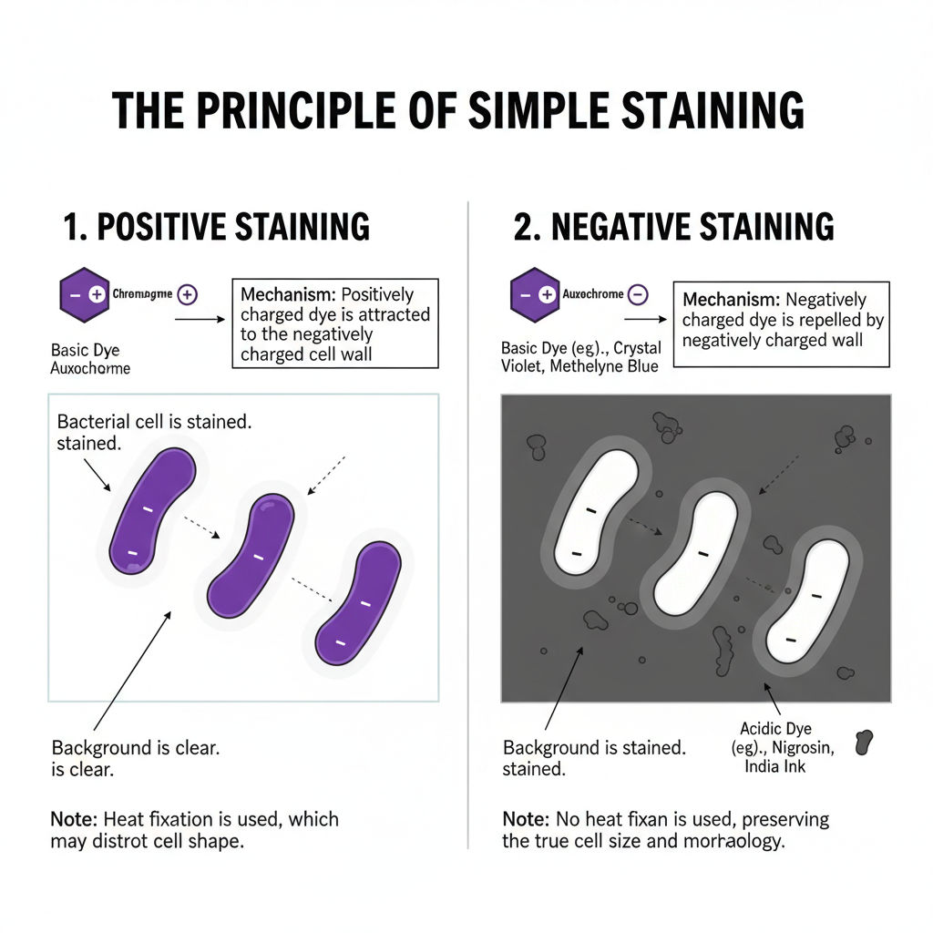
It is the interaction between the dye and the cell surface that allows the stain to bind. The bacterial cell surface generally carries a negative charge, so basic dyes having positively charged chromophores bind easily with the cell wall. Examples of basic dyes are methylene blue, crystal violet and safranin. In negative staining, acidic dyes like nigrosin or India ink is used which carries negative charge, so the stain is repelled by the cell surface. As a result, the background becomes dark and the cells appear clear without any color. These are used when the true size and shape is required as heat fixation is not used in negative staining.
In positive simple staining, a smear is made on a glass slide and it is heat fixed. Heat fixation kills the microorganisms and helps them to attach to the slide so that the cells do not get washed away during staining. However, heat can cause slight shrinkage or distortion of cells. It is important to note that simple staining cannot differentiate between types of bacteria as it does not provide information about internal structures or chemical composition like differential staining methods do.
Objective of Simple Staining Technique
- The primary goal of simple staining is to reveal the morphology (shape) of bacterial cells. Common bacterial shapes include cocci (spherical), bacilli (rod-shaped), and spirilla (spiral-shaped).
- To the arrangement of bacterial cells, such as single cells, pairs, chains, or clusters.
Simple Staining Principle
The principle of simple staining is based on the use of a single dye solution that binds to the microbial cell so the cell becomes visible on a clear background. It is the process that helps in showing the morphology, size, and arrangement of the bacterial cells because the organisms are naturally transparent. The image is monochromatic as only one stain is applied, so all cells appear in the same color.
It is the electrostatic attraction between the dye and the cell surface that explains how the stain is absorbed. The stains contain a colored compound called chromogen, and the charged part of this chromogen is referred to as the auxochrome. In positive staining, basic dyes like methylene blue, crystal violet or safranin is used, and these dyes carry a positive charge. The bacterial cell surface is generally negatively charged, so the positively charged dye ions is attracted towards the cell wall where they bind and the cells becomes colored.
In negative staining, the principle is different because acidic dyes like India ink or nigrosin have a negative charge. The dye is repelled by the negatively charged cell surface, so the background is stained while the cells remain clear. Heat fixation is usually used in positive staining to attach the cells to the slide, but negative staining often avoids heat so that the true size and shape of the cells is maintained.
Requirement
Reagents
- Basic dyes (positive stains) – these dyes carry positive charge and bind with the negatively charged bacterial cell wall.
– Methylene blue
– Crystal violet
– Safranin
– Malachite green
– Basic fuchsin
– Carbol fuchsin - Acidic dyes (negative stains) – these dyes is negatively charged and stains the background only.
– India ink
– Nigrosin
– Congo red
– Eosin
– Rose bengal
– Acid fuchsin - Solvents and mounting media – distilled water is used in preparing smear and rinsing, and ethanol or water is used for dissolving the chromogen.
– Distilled water
– Ethanol or water
– Immersion oil - Fixatives (optional) – sometimes chemical fixatives is used when heat fixation is not preferred.
– Methanol
– Ethanol
Equipment
- For smear preparation and fixation – these are used in making smear and attaching the cells.
– Clean glass slides
– Inoculating loop
– Dropper
– Bunsen burner
– Microincinerator
– Slide warmer - For staining process – slides are placed and rinsed during staining.
– Staining tray
– Wash bottle
– Bibulous paper - For visualization – these are required for observing the stained smear.
– Light microscope (bright-field type)
– Lens paper
– Oil immersion objective lens (100x)
Simple Staining Procedure
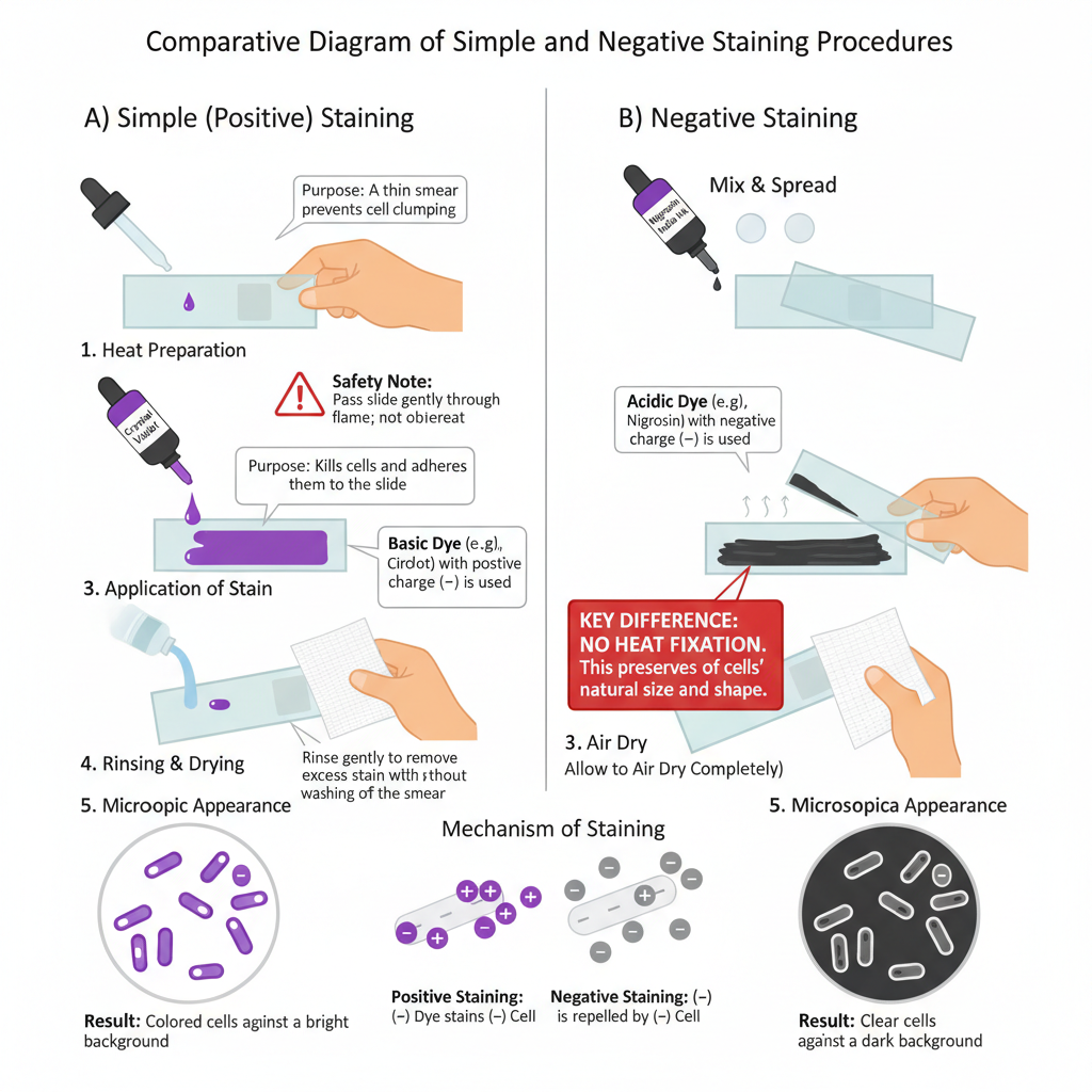
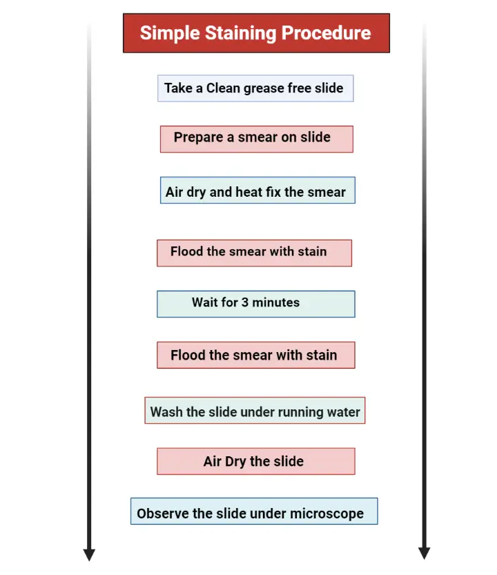
- Preparation of smear. a clean grease-free slide is taken and a small amount of inoculum is placed. From liquid culture the drop is placed directly, and from solid culture a drop of distilled water is first added and then the colony is mixed to make a thin smear. The smear is spread uniformly so that the cells is not clumped and then allowed to air dry completely.
- Fixation of smear. the air-dried smear is fixed so that the cells attach to the slide. The slide is passed gently over the flame 3–4 times, or it is placed on a slide warmer. Chemical fixatives like methanol can also be used when heat is not preferred. Fixation kills the cells and prevent them from washing away during staining.
- Application of stain. a basic dye such as methylene blue, crystal violet or safranin is selected. The smear is flooded with the dye solution and left for the required time to allow proper staining of the cells.
- Rinsing of slide. the slide is washed carefully with distilled water so that the excess stain is removed but the fixed cells is not washed off.
- Drying the stained smear. the slide is blotted dry with bibulous paper. The slide is not rubbed because the smear can be removed.
- Observation under microscope. the stained smear is viewed under the bright-field microscope. The observation usually starts with low power and then the oil immersion objective (100×) is used to visualise the morphology, size and arrangement of the bacterial cells.
- Negative staining alternative. in negative staining, an acidic dye like nigrosin or India ink is used. The dye and organism is mixed on the slide and spread into a thin film. Heat fixation is not done. After air drying, the cells appear clear against a dark background because the negatively charged dye is repelled by the cell surface.
Simple Staining Diagram
Result Interpretation of Simple Staining
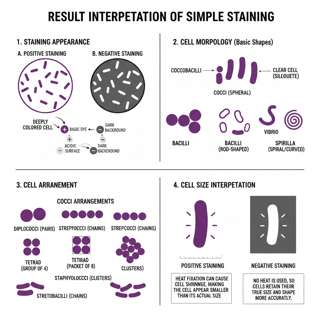
- Appearance of stained field – in positive staining the cells appears deeply colored while the background remains clear because the basic dyes attach with the negatively charged cell surface. In negative staining the background becomes dark and the cells appears as clear silhouettes since the acidic dye is repelled by the cell.
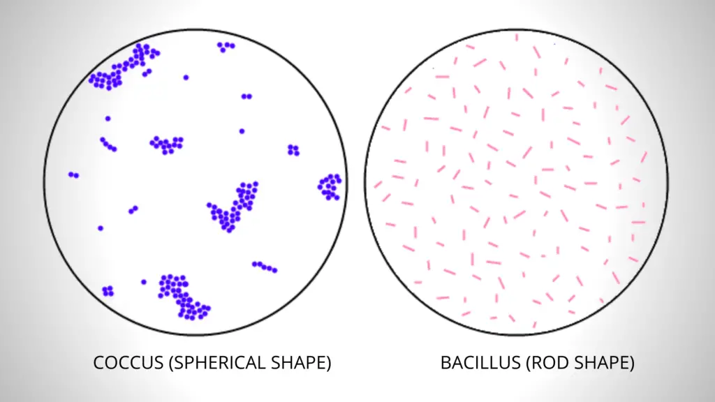
- Interpretation of cell morphology – the simple stain shows the basic shapes of bacteria.
– Cocci are round or slightly flattened spherical cells.
– Bacilli are rod-shaped cells and may sometimes appear short and oval (coccobacilli).
– Spirilla represent spiral or curved forms such as vibrio (comma-shaped) and longer spirals. - Interpretation of cell arrangement – the way cells remain attached after division is observed.
– Diplococci are cocci in pairs.
– Streptococci are cocci arranged in chains.
– Tetrads are groups of four cocci.
– Sarcina are packets of eight cocci.
– Staphylococci are irregular grape-like clusters.
– Bacilli may occur as single rods, diplobacilli or streptobacilli because they divide only across the short axis. - Interpretation of cell size – the size is estimated from the stained smear but some distortion can occur. Heat fixation in positive staining may cause slight shrinkage of the cells, so the size appears smaller. In negative staining heat is not used, so the cells retain their actual size and shape more accurately.
Uses Simple Staining
- It is used to observe the basic morphology of bacteria, as this technique makes the cells visible by providing contrast between cell and background.
- It is the process that helps in identifying common shapes like cocci, bacilli, and spirilla, which is the first step in studying an unknown culture.
- These staining is used for observing the arrangement of cells, such as chains, clusters, or pairs, which is important in primary classification.
- It is used to check the purity of a culture since stained cells can be examined to see if unwanted cells is present.
- The technique is helpful in estimating the quantity of cells because stained cells can be counted easily under the microscope.
- It is used when rapid examination is required, as a single dye can show the general features of cells without complex procedures.
- Positive simple staining is used to study the shape of the cell, but the heat fixation in this step kills the bacteria.
- Negative staining is used for viewing the actual size of cells since it does not require heat fixation and the cells are not distorted.
- It is the process used to visualize delicate structures like capsules because the background is stained while the cell remains clear, showing external features more accurately.
Advantages of Simple Staining
- It is a very simple technique because only a single dye is used and the procedure is easy to perform.
- It is the process that provides rapid results, so it is useful in quick examination of microbial samples.
- The method is cost–effective since very few reagents and equipment is required, making it suitable for routine laboratory work.
- It is used to obtain a preliminary idea about the specimen before doing more complex staining procedures.
- The staining provides clear contrast between the cells and the background, which makes transparent microorganisms visible for observation.
- It helps in identifying the general cell morphology like cocci, bacilli, and spirilla in a very short time.
- These staining is also used to study the arrangement of cells such as pairs, chains, or clusters, which is an important diagnostic feature.
- It is used to estimate the approximate size of the bacterial cell because stained cells can be observed clearly under the microscope.
- Negative simple staining has an advantage since it does not require heat fixation and thus preserves the natural size and shape of the cells.
- It is the process used to visualize delicate external structures like capsules, as the background is stained and the cell remains unstained, avoiding distortion.
Disadvantages of Simple Staining
- It is the process that uses only a single dye, so all the cells appear in the same colour and no differentiation between bacterial groups is possible.
- These staining cannot indicate differences in cell wall composition, hence Gram-positive and Gram-negative cells cannot be separated.
- It does not identify acid-fast organisms because the technique lacks the chemical specificity required for such diagnostic tests.
- Simple staining cannot show delicate external structures like capsules since basic dyes do not bind properly to these structures.
- It is not suitable for observing flagella because these structures are extremely thin and require special staining methods to be visualized.
- Internal structures like nucleoid region or other organelles is not visible due to limited contrast provided by a single dye.
- Heat fixation in positive simple staining causes shrinkage and distortion of cells, so the actual cell size cannot be measured accurately.
- It is the process where staining errors such as over-staining may cause dye crystals which can be confused with bacterial cells.
- Incomplete washing of the dye may leave artefacts on the slide, making the microscopic observation unclear.
Difference between Simple Staining and Gram Staining
- Number of dyes used – simple staining uses only one basic dye, while Gram staining uses multiple reagents including crystal violet, iodine, alcohol and safranin.
- Type of staining – simple staining is a monochromatic staining because all the cells take the same color. Gram staining is a differential staining as it divides bacteria into Gram-positive and Gram-negative groups.
- Complexity of procedure – simple staining is a very simple process where a single stain is applied and rinsed. Gram staining involves primary stain, mordant, decolorizer and counterstain in a stepwise process.
- Basis of staining reaction – in simple staining the positively charged dye attaches with the negatively charged bacterial surface without distinguishing cell wall differences. In Gram staining the result depends on the thickness of peptidoglycan; thick peptidoglycan retains the crystal violet-iodine complex, while thin peptidoglycan loses it during decolorization.
- Appearance of stained cells – in simple staining all cells show the same color of the dye used. In Gram staining Gram-positive cells appear purple and Gram-negative cells appear pink.
- Information obtained – simple staining provides morphology, arrangement and approximate size of the cells. Gram staining provides cell wall characteristics which are important in identifying the organism.
- Diagnostic value – simple staining is used as an initial screening tool to confirm bacterial presence and basic structure. Gram staining is more important in diagnosis because it helps in selecting further tests and guides antibiotic choice.
| Feature | Simple Staining | Gram Staining |
|---|---|---|
| Type of staining | Monochromatic staining using one dye | Differential staining using multiple dyes |
| Reagents used | A single basic dye (methylene blue, crystal violet, safranin etc.) | Crystal violet, iodine, alcohol (decolorizer), safranin |
| Complexity of procedure | Very simple process with one staining step | Multi-step process involving primary stain, mordant, decolorizer and counterstain |
| Basis of staining reaction | Dye binds with negatively charged bacterial surface | Based on peptidoglycan thickness of the cell wall |
| Appearance of cells | All cells appear in the same color | Gram-positive cells appear purple, Gram-negative cells appear pink |
| Information obtained | Morphology, arrangement, approximate size | Cell wall characteristics and Gram reaction |
| Diagnostic value | Used as a quick preliminary screening method | Important diagnostic tool guiding identification and antibiotic selection |
FAQ
Q1. What is simple staining?
A. It is the process where a single dye is used to color the microbial cells so the morphology, size and arrangement of the bacteria becomes visible on a clear background.
Q2. What is the purpose of simple staining?
A. The purpose is to observe the basic shape, arrangement and approximate size of the bacterial cells because the organisms are transparent in their natural state.
Q3. What are the steps in simple staining?
A. A thin smear is prepared on a clean slide, the smear is air dried and then heat fixed. A basic dye is applied for the required time, the slide is rinsed gently with water and finally it is blotted dry and observed under the microscope.
Q4. What bacterial features can be observed with simple staining?
A. The cell morphology (cocci, bacilli, spirilla), the arrangement (single, pairs, chains, clusters) and the approximate cell size is observed.
Q5. What are common basic dyes used in simple staining?
A. Methylene blue, crystal violet and safranin are commonly used basic dyes.
Q6. Why are basic dyes preferred for simple staining?
A. Basic dyes carry positive charge and the bacterial cell surface is negatively charged, so the dye binds easily with the cell giving clear staining.
Q7. What is the difference between simple and differential staining?
A. Simple staining uses one dye and gives a single color to all cells, while differential staining uses multiple reagents and separates bacteria into different groups based on their staining reaction.
Q8. Why is heat fixation important in simple staining?
A. It attaches the cells to the slide, kills the microorganisms and prevents the smear from washing away during staining.
Q9. What features cannot be determined with simple staining?
A. It cannot distinguish between species or between Gram-positive and Gram-negative bacteria, and internal structures like nucleus or spores are not clearly seen.
Q10. How do basic and acidic dyes differ in simple staining?
A. Basic dyes are positively charged and stain the cells. Acidic dyes are negatively charged and stain the background while the cells remain clear.
Q11. What is the most common mistake in simple staining?
A. Making the smear too thick or overheating during fixation which causes cell distortion and poor visualization.
Q12. How long should the dye typically be left on the sample in a simple staining procedure?
A. Most basic dyes are kept for about 30 seconds to 2 minutes depending on the dye used.
Q13. What is the charge of bacterial cells?
A. Bacterial cells generally carry a negative charge.
Q14. How is a bacterial smear prepared for simple staining?
A. A drop of culture (or water for solid culture) is placed on the slide, the organism is mixed, spread into a thin film and then air dried.
Q15. Can simple staining differentiate between different types of bacteria?
A. No, simple staining cannot differentiate types of bacteria because all organisms take the same color.
- AAT Bioquest. (2023, March 9). What are the advantages of differential staining over simple staining? https://www.aatbio.com/resources/faq-frequently-asked-questions/what-are-the-advantages-of-differential-staining-over-simple-staining
- AAT Bioquest. (2023, March 9). What are the differences between simple and differential staining? https://www.aatbio.com/resources/faq-frequently-asked-questions/what-are-the-differences-between-simple-and-differential-staining
- Al-Kadmy, I. (n.d.). Lab 6: Simple staining- Principle, procedure and result interpretation. Mustansiriyah University. https://uomustansiriyah.edu.iq/media/lectures/6/6_2025_12_04!12_09_21_AM.pdf
- Comprehensive monograph on simple staining: Principles, procedure, and microscopic analysis. (n.d.).
- Kosker, F. B., Aydin, O., & Icoz, K. (2022). Simple staining of cells on a chip. Biosensors, 12(11), 1013. https://doi.org/10.3390/bios12111013
- Microbiology Learning. (n.d.). The simple stains. Weebly. https://microbiologylearning.weebly.com/the-simple-stains.html
- Parker, N., Schneegurt, M., Tu, A. T., Lister, P., & Forster, B. M. (2016). Microbiology. OpenStax. https://openstax.org/books/microbiology/pages/2-4-staining-microscopic-specimens
- Pearson. (n.d.). Bacterial cell morphology & arrangements: Videos & practice problems. https://www.pearson.com/channels/microbiology/learn/jason/ch-7-prokaryotic-cell-structures-functions/bacterial-morphology
- Rao, B. S. S., Rao, N. M., & Shanthi, V. (n.d.). Fixation. Histopathology.guru. https://www.histopathology.guru/academics/post-graduate-academics/fixation/
- Salton, M. R. J., & Kim, K. S. (1996). Structure. In S. Baron (Ed.), Medical microbiology (4th ed.). University of Texas Medical Branch at Galveston. https://www.ncbi.nlm.nih.gov/books/NBK8477/
- Staining. (n.d.). In Wikipedia. Retrieved from https://en.wikipedia.org/w/index.php?title=Staining&oldid=1310157740