What is Paramecium?
- Paramecium is a type of single-celled organism belonging to the group of eukaryotes. These organisms are called ciliates and are commonly studied to understand certain biological processes. Paramecia are found in various aquatic environments, such as freshwater, brackish, and marine habitats. They are particularly abundant in still water bodies like ponds and basins. Due to their ease of cultivation and ability to reproduce through both sexual and asexual methods, they have become valuable subjects for scientific research in classrooms and laboratories.
- These organisms have a distinct appearance, with a footprint-like shape that measures between 50 to 330 micrometers in length. They are covered in hair-like structures known as cilia, which they use for movement and capturing food. Paramecia move by beating their cilia, and they feed by using these structures to sweep food particles into their mouth-like opening. While some larger paramecia can be seen without a microscope, most species require magnification to observe their details.
- Paramecium falls into a specific category known as “ciliates” due to the presence of cilia on their cells. These cilia serve vital functions in their movement and feeding processes. The classification of Paramecium includes its placement within the Kingdom Protista, Subkingdom Protozoa, Phylum Ciliophora, Class Oligohymenophorea, Subclass Hymenostomatia, Order Hymenostomatida, Suborder Peniculina, Family Parameciiade, and finally, Genus Paramecium.
- This overview provides insight into the anatomy, behavior, and classification of Paramecium, making it a helpful resource for those interested in learning more about this fascinating microorganism. It encompasses topics such as how Paramecium moves, feeds, and reproduces, as well as its historical significance in scientific research.
Definition of Paramecium
Paramecium is a microscopic, single-celled organism with hair-like structures called cilia, found in various aquatic environments, often studied for its role as a model organism in biological research.
Paramecium Scientific classification
Paramecium is divided into this following phylum and subphylum;
| Domain: | Eukaryota |
| Clade: | SAR |
| Infrakingdom: | Alveolata |
| Phylum: | Ciliophora |
| Class: | Oligohymenophorea |
| Order: | Peniculida |
| Family: | Parameciidae |
| Genus: | Paramecium |
Habitat of Paramecium
Paramecium is a type of organism that lives on its own without being attached to anything. It is commonly found in places where water is not moving much, like still pools, lakes, ditches, and ponds. You can also find Paramecium in fresh water and in places where the water is flowing slowly. These environments often have old, decaying material in the water.
Types of Paramecium
Paramecium species exhibit a variety of types, primarily distinguished by their body shapes and genetic characteristics. Let’s explore the main types:
- Aurelia Group: The Aurelia group comprises 15 distinct species of Paramecium. These organisms share common characteristics, such as relatively long bodies with a pointed end. Among the species in this group are:
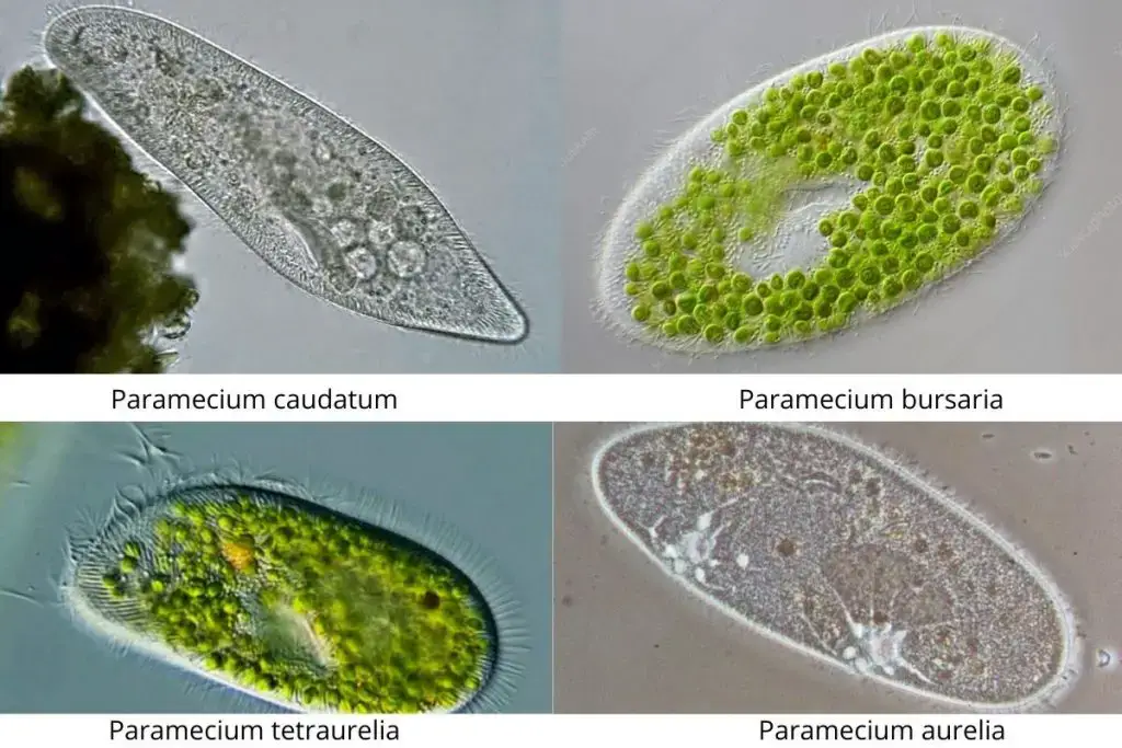
- Paramecium Aurelia
- Paramecium Caudatum
- Paramecium Multimicronucleatum
- Bursaria Group: The Bursaria group features a different body shape, characterized by shorter and broader dimensions. These Paramecia are flatter in the dorsoventral position and have a more rounded rear. Notable species in this group include:
- Paramecium Bursaria
- Paramecium Calkinsi
- Paramecium Woodruffi
- Paramecium Polycaryum
- Paramecium Trichium
It’s important to note that the classification of certain Paramecium species has been subject to dispute due to various factors. The differentiation between these groups is not only based on physical characteristics but also involves genetic and biochemical distinctions. Overall, the diversity within the Paramecium genus showcases a range of body shapes and features, contributing to their intriguing biology and classification.
Paramecium Size
The size of paramecium varies depending on the species:
- Paramecium aurelia: This type can be about 120 to 180 micrometers long.
- Paramecium bursaria: These are larger, measuring around 180 to 300 micrometers in length.
- Paramecium calkinsi: This species is about 100 to 150 micrometers long and 50 micrometers wide.
- Paramecium caudatum: Similar to Paramecium bursaria, these are also in the range of 180 to 300 micrometers long.
- Paramecium jenningsi: These measure about 115 to 218 micrometers in length.
- Paramecium multimicronucleatum: Among the largest species, these can reach sizes of 200 to 330 micrometers.
- Paramecium polycaryum: This type is smaller, with lengths of about 70 to 110 micrometers.
- Paramecium putrinum: These are around 80 to 150 micrometers long.
- Paramecium trichium: Smaller still, these measure about 50 to 105 micrometers, with a range of 80 to 90 micrometers.
- Paramecium woodruffi: These fall within the range of 150 to 210 micrometers in length.
Paramecium Species
| Paramecium biaurelia | Paramecium biaurelia |
| Paramecium primaurelia | Paramecium tetraurelia |
| Paramecium sonneborni | Paramecium quadecaurelia |
| Paramecium tredecaurelia | Paramecium dodecaurelia |
| Paramecium undecaurelia | Paramecium decaurelia |
| Paramecium novaurelia | Paramecium pentaurelia |
| Paramecium sexaurelia | Paramecium septaurelia |
| Paramecium octaurelia | Paramecium triaurelia |
| Paramecium bursaria | Paramecium buetschlii |
| Paramecium multimicronucleatum | Paramecium woodruffi |
| Paramecium schewiakoffi | Paramecium putrinum |
| Paramecium polycaryum | Paramecium nephridiatum |
| Paramecium jenningsi | Paramecium grohmannae |
| Paramecium duboscqui | Paramecium chlorelligerum |
| Paramecium caudatum | Paramecium calkinsi |
Characteristics of Paramecium
Paramecium, a single-celled organism, exhibits several distinctive features:
- Cell Size and Shape: Paramecium cells vary in size, ranging from 50 to 300 micrometers. They can have an ovoid, slipper-like, or cigar-like shape.
- Pellicle: The cellular cytoplasm is enclosed by a pellicle, which comprises an outer plasma membrane, inner epiplasm, and a layer of alveoli in between. This pellicle is elastic, giving the cell a defined yet changeable shape.
- Cilia: Tiny hair-like structures called cilia project from depressions in the pellicle, covering the entire cell surface. Cilia serve both for movement and for capturing nutrient-rich water, which is taken inside the cell through a gullet.
- Protoplasm: The cell’s protoplasm is divided into outer ectoplasm and inner endoplasm, which is granular in nature.
- Trichocysts: Defensive trichocysts are embedded in the ectoplasm, serving as protective organs.
- Endoplasmic Granules: Granules within the endoplasm serve as reserves for food. Some granules may also serve secretory or excretory functions.
- Nuclei: Paramecia contain at least two nuclei – micronuclei (one or more) and one macronucleus. Micronuclei participate in reproduction, containing diploid chromosomes. The macronucleus oversees vital metabolic activities and growth, housing multiple copies of the genome.
- Contractile Vacuoles: Varying in number among species, these vacuoles help with osmoregulation and expelling excess water.
- Oral Groove: An oral groove on the ventral side, known as the vestibule, draws food into the cell due to coordinated cilia movement. This leads to the mouth (cytostome) and further into the gullet or pharynx.
- Food Vacuoles: Numerous food vacuoles aid in digesting food particles.
- Anal Pore: An anal pore, situated on the ventral surface’s posterior half, known as cytoproct or cytopyge, helps expel undigested food.
Paramecium caudatum is the most familiar and extensively studied species within the genus.
Paramecium Structure With Labeled Diagram
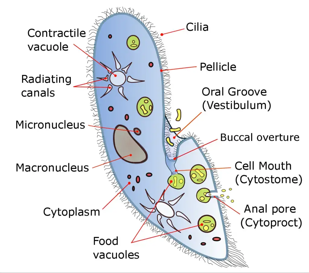
The structure of a Paramecium, a single-celled organism, can be understood through various key features:
- Cilia: Cilia are hair-like projections on the cell’s surface, used for both movement and ingesting food. The cilia responsible for food intake are situated in a funnel-shaped depression called the gullet. Caudal cilia, longer in length, are involved in mating during conjugation.
- Contractile Vacuole: Paramecium usually possesses two contractile vacuoles, located at opposite ends of the cell. These vacuoles help expel waste liquids from the cell. Alternating contractions empty the vacuoles through pores, crucial for maintaining water balance and preventing cell rupture.
- Pellicle: The pellicle, consisting of the plasma membrane, alveolar system, and epiplasm, surrounds the cell, providing elasticity and shape. The pellicle’s ridges mold into shapes like hexagons and parallelograms on the cell surface.
- Radiating Canals: Radiating canals absorb waste and materials from the surrounding cytoplasm, eventually expelled from the cell by the contractile vacuoles.
- Vestibulum (Oral Groove): A funnel-shaped indentation known as the vestibulum or oral groove leads to the mouth region (cytostome) of the Paramecium. This groove connects to the buccal overture.
- Micronucleus and Macronucleus: Paramecia have at least two nuclei – the micronucleus, responsible for reproduction and carrying genetic information, and the macronucleus, controlling cellular metabolism.
- Cytostome: The cytostome functions as the “mouth” of the Paramecium, transferring prey into the food vacuole.
- Cytoplasm: This jelly-like substance holds the cell’s organelles, including vesicles, ribosomes, and food storage reserves. It also provides the necessary components for protein synthesis.
- Food Vacuoles: Food vacuoles accumulate gathered food particles through the cytostome. Once they reach a certain size, these vacuoles travel through the cell, digesting the food with enzymes. Useful materials remain in the cytoplasm, while undigested waste is expelled through the cytoproct.
- Cytoproct: Also known as the “anal pore,” the cytoproct expels waste from the cell’s rear end.
- Trichocyst: These are involved in the defense of the Paramecium. Trichocysts, shaped like spindles with a wider end resembling an upside-down golf tee, are used to deter attackers.
In essence, the Paramecium’s structure is multifaceted, encompassing cilia for movement and feeding, contractile vacuoles for water balance, nuclei for reproduction and metabolism control, and various other components supporting vital functions.
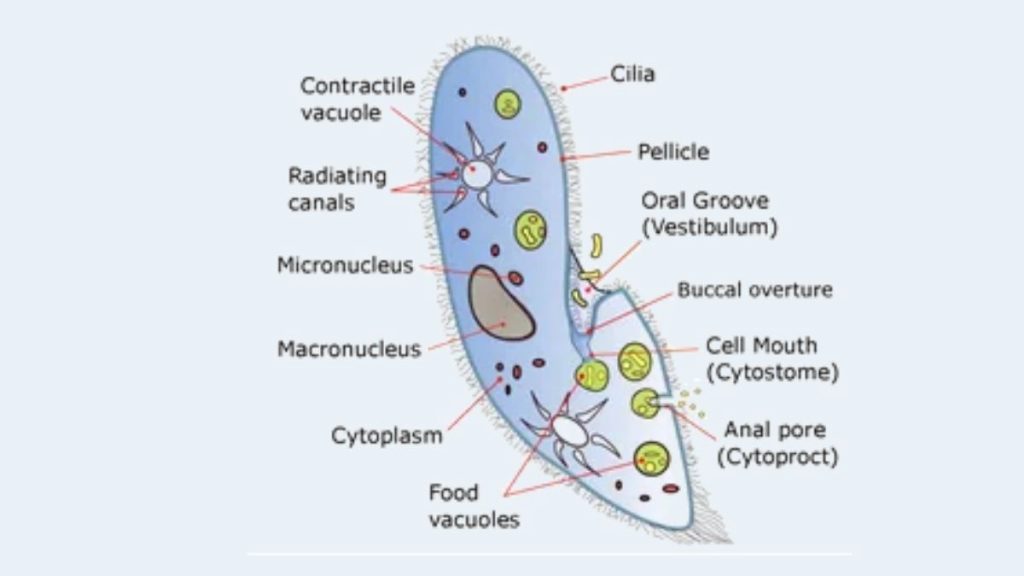
Paramecium Labeled Diagram
Paramecium Movement
- Paramecium, while not known for graceful movement, navigates its environment using a unique method. The organism uses hair-like structures called cilia, which are arranged in closely spaced rows around its body, to propel itself. This movement involves two phases: a swift “effective stroke,” during which the cilia become rigid and move forward, followed by a slower “recovery stroke,” where the cilia curl loosely and sweep in a counter-clockwise direction.
- The cilia work together in a synchronized manner, creating a wave-like pattern across the organism’s surface, akin to wind moving through a field of grain. This coordinated action allows the Paramecium to spiral through the water. When it encounters an obstacle, it swiftly reverses the “effective stroke,” causing it to momentarily swim backward. This reaction is known as avoidance, and the Paramecium repeats this process until it finds a way around the obstacle.
- Although the Paramecium’s movements appear rapid and jerky under a microscope, they are driven by the coordinated whipping motion of the cilia. These movements are energetically costly, with over half of the organism’s energy expended in propelling itself through the water. Despite its seemingly inefficient locomotion, the ciliary method employed by Paramecium is close to the maximum theoretical efficiency achievable for an organism with such short cilia.
- The Paramecium’s lack of visual perception means it relies on this repeated avoidance reaction to navigate its surroundings effectively. Through these distinctive movements driven by its cilia, the Paramecium ensures its continued progress and survival in its aquatic habitat.
Feeding of Paramecium / Paramecium Nutrition
- Paramecium primarily follows a heterotrophic mode of nutrition. They obtain their food by consuming various microorganisms such as bacteria, algae, and yeast. This process is known as holozoic nutrition. Paramecia use their cilia, which are hair-like structures on their surface, to create movements that draw in water containing food particles. This water is directed towards the cytostome, a funnel-shaped opening, and then into the gullet (cytopharynx).
- In the cytopharynx, the food accumulates near the posterior end. Vacuoles form around the gathered food, eventually pinching off and moving through the endoplasm, the inner granular part of the cell. Within the food vacuoles, digestive enzymes break down the ingested food, aiding in digestion.
- Any undigested remnants are expelled from the cell through a temporary anal pore known as the cytopyge.
- Interestingly, some Paramecium species, like Paramecium bursaria, establish a symbiotic relationship with green algae. In this mutualistic partnership, algae live inside the Paramecium and provide sustenance through photosynthesis. In return, the algae benefit from a protected environment.
- It’s worth noting that certain Paramecium may harbor intracellular bacteria called kappa particles. When present, these particles grant Paramecium the ability to eliminate other strains of Paramecium, showcasing a unique aspect of their complex nutritional interactions.
Feeding Mechanism of Paramecium
Paramecium has a specific way of getting its food, mainly bacteria, algae, and yeasts. Here’s how they go about it:
- Trapping the Prey: Paramecium uses its cilia, which are tiny hair-like structures, to catch the organisms it wants to eat. The cilia create movements that help trap the prey.
- Oral Groove and Gullet: Once trapped, the prey is moved along an opening called the oral groove. This groove guides the food particles into the cell.
- Buccal Cavity and Cytostome: From the oral groove, the food particles are directed into the buccal cavity or gullet. This is like a passage that leads the food deeper into the cell.
- Interior of the Cell: The food particles then travel through the cytostome, which is like the cell’s mouth. This takes them from the outer part of the cell to the inside.
- Food Vacuoles: Inside the cell, the food particles are stored in structures called food vacuoles. These vacuoles are like storage compartments where the food is kept.
- Circulation and Digestion: The food vacuoles move around within the cell with the help of a process called cyclosis or cytoplasmic streaming. This circulation helps mix the food and digestive enzymes.
- Digestive Enzymes: Digestive enzymes and hydrochloric acids are released into the food vacuoles. These enzymes break down the food and make the vacuoles more acidic.
- Absorption and Nutrients: As the food particles are broken down, the nutrients are absorbed into the cell’s cytoplasm, which is the jelly-like substance inside the cell.
- Waste Release: When the food vacuoles reach the anal pore, they rupture, releasing waste materials outside the cell. This is how Paramecium gets rid of the parts of the food that it can’t use.
So, Paramecium uses its cilia to catch and guide food into its cell, where it’s processed, digested, and absorbed, with waste being expelled through the anal pore.
Reproduction of Paramecium
Paramecium reproduces in two main ways: asexually through binary fission and sexually through conjugation.
- Asexual Reproduction (Binary Fission): In binary fission, a mature Paramecium cell divides into two new cells. Each of these cells grows rapidly and develops into a new individual. This process allows Paramecium to multiply quickly, especially under favorable conditions, occurring up to three times a day. During binary fission, the cell divides transversely, and the micronucleus undergoes mitotic division, while the macronucleus divides amitotically. The gullet, where food is ingested, also divides into two halves.
- Sexual Reproduction (Conjugation): Paramecium can also reproduce sexually through a process called conjugation. This occurs when there is a scarcity of food. During conjugation, two compatible Paramecia come together, forming a connection known as the conjugation bridge. The united Paramecia are called conjugants. In this process, the macronuclei of both cells disappear, and the micronucleus of each conjugant undergoes meiosis to form four haploid nuclei. Three of these nuclei degenerate, and the remaining haploid nuclei fuse to form diploid micronuclei, resulting in cross-fertilization. The conjugants then separate, forming exconjugants. Each exconjugate further divides to produce four daughter Paramecia. A new macronucleus is formed from the micronuclei.
- Other Reproductive Modes: Paramecium also exhibits autogamy, which is self-fertilization. In this process, a new macronucleus is produced, enhancing vitality and rejuvenation.
Cytogamy, a less frequent mode of reproduction, involves two Paramecia coming into contact without exchanging nuclei. Despite lacking nuclear exchange, cytogamy rejuvenates the Paramecium, leading to the formation of a new macronucleus.
It’s important to note that Paramecia have a finite lifespan, aging and eventually dying after around 100 to 200 cycles of fission if they do not undergo conjugation. The macronucleus plays a role in clonal aging due to DNA damage.
In summary, Paramecium can reproduce asexually through binary fission, and when conditions demand, they engage in sexual reproduction through conjugation or other forms like autogamy and cytogamy.
Transverse Binary Fission of Paramecium
Transverse binary fission is a process of cell division that Paramecium uses to reproduce. Here’s how it works:
- Starting Division: The process begins with the division of the nuclei, which are like the control centers of the cell. Alongside this, the structures that help with feeding, such as oral grooves and buccal structures, disappear.
- Macronucleus Division: The macronucleus, one of the nuclei, divides amitotically. This means it elongates and then gets constricted in the middle, like a squeezed balloon.
- Micronucleus Division: The other nucleus, called the micronucleus, divides mitotically. This involves a series of phases—prophase, metaphase, anaphase, and telophase. During telophase, the micronuclei elongate, and new oral grooves and contractile vacuoles form.
- Constriction and Splitting: As the nucleus division wraps up, a constriction appears in the middle of the cell. This constriction deepens until the cell splits into two distinct cells. These two new cells are essentially copies of the “parent” cell since they contain the same DNA.
- Outcome: The result is the creation of two daughter cells that are identical to each other and the original cell. This process happens approximately two to three times a day and usually takes around 30 minutes to complete.
In summary, transverse binary fission is a way Paramecium reproduces by dividing its nuclei and gradually splitting into two identical daughter cells. This process ensures the continuation of Paramecium’s population.
Conjugation of Paramecium
In the process of conjugation in Paramecium caudatum, the stages can be outlined as follows (see diagram for reference):
- Meeting and Partial Fusion: Two compatible mating strains of Paramecium come into contact and partially fuse together.
- Meiosis of Micronuclei: The micronuclei within each Paramecium undergo meiosis, resulting in the formation of four haploid micronuclei in each cell.
- Micronuclei Disintegration: Out of the four micronuclei produced, three of them disintegrate, leaving one micronucleus intact.
- Micronucleus Mitosis: The remaining micronucleus undergoes mitosis, leading to its division into two identical nuclei.
- Micronucleus Exchange: The two Paramecia exchange one of their micronuclei, contributing to the mixing of genetic material.
- Cell Separation: After the micronucleus exchange, the two Paramecia separate from each other.
- Micronucleus Fusion: Within each Paramecium, the micronucleus that was exchanged fuses with the original micronucleus, resulting in the formation of a diploid micronucleus.
- Multiple Micronuclei Formation: Mitosis occurs three times, generating a total of eight micronuclei within each cell.
- Macronucleus Transformation: Four of the new micronuclei transform into macronuclei, while the old macronucleus disintegrates.
- Daughter Cell Formation: Binary fission takes place twice, producing four identical daughter cells with a mix of genetic material from both parent cells.
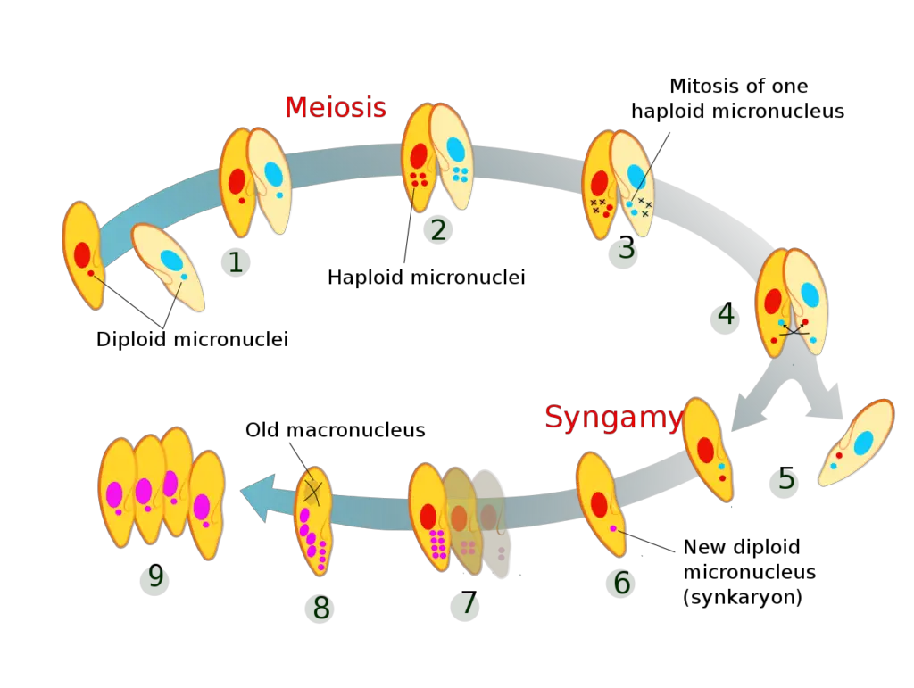
In summary, the stages of conjugation in Paramecium caudatum involve a series of intricate processes, including meiosis, micronucleus exchange, fusion, and multiple divisions, ultimately resulting in the formation of genetically diverse daughter cells through binary fission.
Paramecium binary fission
Video Author: Deuterostome
Paramecium in conjugation
Video Author: Antonio Guillén
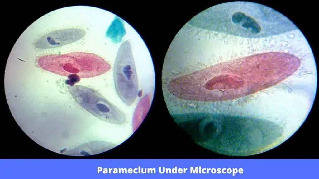
Paramecium Under Microscope
To observe Paramecium Under Microscope, take a jar with mud, grass and pond water. Then seal the lid and keep where it can get a lot of sunlight.
After a few days, place a drop of water from the jar on a slide and cover it with a cover slip. Then observe it under the microscope, starting at 40x.
Paramecium Aging
- In the phase of asexual fission growth, when cells divide through mitosis, a phenomenon known as “clonal aging” takes place in Paramecium. This gradual process leads to a decline in vitality over time. One example of this is observed in Paramecium tetraurelia, a well-studied species. In the absence of autogamy or conjugation, the clonally aging Paramecia in the asexual lineage experience a decrease in vitality and eventually perish after around 200 cell divisions.
- The understanding of clonal aging was advanced through transplantation experiments conducted by Aufderheide in 1986. These experiments involved injecting macronuclei from clonally young Paramecia into Paramecia of standard clonal age. The results showed that the recipient’s lifespan, measured in terms of clonal divisions, was extended. However, the transfer of cytoplasm from young Paramecia did not have the same effect on lifespan. This highlighted that the macronucleus, rather than the cytoplasm, is the key factor responsible for clonal aging.
- Further studies by researchers like Smith-Sonneborn, Holmes and Holmes, and Gilley and Blackburn provided insights into the mechanisms behind clonal aging. These experiments demonstrated a significant increase in DNA damage during clonal aging. It was revealed that DNA damage in the macronucleus is a crucial factor contributing to the aging process in Paramecium tetraurelia.
- Interestingly, the aging process in this single-celled protist appears to share similarities with the aging processes observed in multicellular eukaryotes. The theory of DNA damage as a cause of aging, commonly known as the “DNA damage theory of aging,” provides a framework to understand the aging phenomenon in Paramecium and suggests parallels between single-celled organisms and more complex life forms.
Paramecium Genome
The sequencing of P. tetraurelia provides strong evidence for the three whole-genome duplications.
The UAA and UAG codon in Stylonychia and Paramecium are reassigned as sense codons whereas UGA as a stop codon.
Paramecium Example
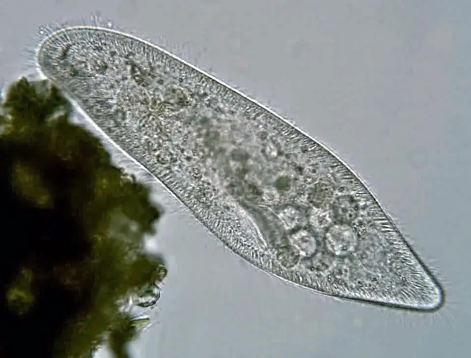
(i) Paramecium Caudatum
- It is an unicellular protist of phylum Ciliophora.
- Their size can be 0.33 mm in length.
- They contain small hair-like structures all over the surface area known as cilia which help them in locomotion and feeding.
- They are spindle-shaped, the front portion is rounded and tapering at the posterior to a blunt point.
- They contain 2 contractile vacuoles.
Paramecium Caudatum Reproduction
Paramecium Caudatum reproduces through the binary fission which is an asexual method. Under unfavorable conditions they reproduce by self-fertilization (autogamy) or conjugation.
Paramecium Caudatum Under Microscope
Here is the picture of Paramecium Caudatum Under Microscope
Paramecium Caudatum Under Microscope video:
Author: Alexander Klepnev
(ii) Paramecium aurelia
- They can be found in freshwater environments, and are especially in scums.
- Acidic conditions attract them.
- They feed on bacteria and dead organic matter.
- They contain hair-like cilia all over the surface.
- They can reproduce by sexually, asexually, or by the process of endomixis.
Other examples of Paramecium are Paramecium tetraurelia, Paramecium bursaria.
Video: Paramecium bursaria | Author: Deuterostome
Paramecium putrinum | Author: Deuterostome
FAQ on Paramecium
What is Paramecium?
Paramecium is a single-celled, microscopic organism belonging to the ciliate group. It is commonly found in freshwater environments and is known for its unique shape and cilia-covered surface.
How does Paramecium move?
Paramecium moves using hair-like structures called cilia that cover its body. The coordinated beating of cilia propels it through the water, giving it a distinct jerky movement.
How does Paramecium feed?
Paramecium is heterotrophic and feeds on bacteria, algae, and other microorganisms. It uses its cilia to create currents that draw in food particles through its oral groove.
How does Paramecium reproduce?
Paramecium reproduces asexually through binary fission, where a cell divides into two identical daughter cells. It can also reproduce sexually through a process called conjugation, where genetic material is exchanged between two compatible cells.
What is clonal aging in Paramecium?
Clonal aging is a gradual loss of vitality observed in asexual Paramecium populations during cell divisions. It leads to decreased vitality and eventual death if conjugation or autogamy does not occur.
What are the different species of Paramecium?
Paramecium species can be categorized into groups based on body shape, such as the “Aurelia” group with pointed ends and the “Bursaria” group with a broader shape. Species include Paramecium Aurelia, Paramecium Caudatum, and Paramecium Bursaria, among others.
What is the role of the macronucleus and micronucleus in Paramecium?
Paramecium has two nuclei: the macronucleus controls vital metabolic activities, while the micronucleus is involved in reproduction. The micronucleus undergoes meiosis and mitosis during conjugation.
What is the significance of conjugation in Paramecium?
Conjugation is a sexual reproduction process in Paramecium where genetic material is exchanged between two compatible cells. It promotes genetic diversity and rejuvenation in the population.
How does Paramecium respond to obstacles in its path?
When Paramecium encounters an obstacle, it uses its cilia to reverse its direction and swim backward, a behavior known as an avoidance reaction. This helps it navigate around obstacles.
How is Paramecium used in scientific research?
Paramecium has been widely studied as a model organism to understand biological processes. Its simple structure, rapid reproduction, and ease of cultivation make it a valuable tool in classrooms and laboratories for studying cell biology and genetics.
References
- https://microscopeclarity.com/paramecium-everything-you-need-to-know/
- https://microbiologynotes.com/general-description-of-paramecium/
- https://www.livescience.com/55178-paramecium.html
- https://doi.org/10.1002/jez.1402600111
- https://www.uniprot.org/locations/SL-0268
- https://doi.org/10.1080/00039896.1967.10664819
- https://www.ncbi.nlm.nih.gov/pubmed/1244399
- https://en.wikipedia.org/wiki/Paramecium
- https://link.springer.com/chapter/10.1007%2F978-3-319-32211-7_16
- https://doi.org/10.1016/0273-1177(81)90249-0
- Text Highlighting: Select any text in the post content to highlight it
- Text Annotation: Select text and add comments with annotations
- Comment Management: Edit or delete your own comments
- Highlight Management: Remove your own highlights
How to use: Simply select any text in the post content above, and you'll see annotation options. Login here or create an account to get started.