What is Muscle Cell?
- Muscle cells, scientifically termed myocytes, are specialized cells in animals designed for contraction. These cells possess a unique arrangement of motor proteins that enable them to reduce their length. Central to this contraction mechanism are the proteins actin and myosin, which form the thick and thin filaments, respectively.
- These filaments interact and slide past one another, leading to the contraction of structures known as sarcomeres. Multiple sarcomeres are aligned end-to-end within a larger structure termed a myofibril.
- Distinctively, a muscle cell is elongated compared to other cellular types and houses multiple nuclei that are situated adjacent to the cell membrane. These cells often link end-to-end, culminating in the formation of extended fibers characteristic of muscle tissue.
- The term “myocyte” is not restricted to a single type of muscle cell. It encompasses both cardiac muscle cells (cardiomyocytes) and smooth muscle cells. While both are relatively smaller cells, the skeletal muscle cell stands out due to its elongated, thread-like structure, housing multiple nuclei, and is thus termed a muscle fiber.
- The genesis of muscle cells, including myocytes and muscle fibers, can be traced back to embryonic precursor cells labeled myoblasts. These myoblasts undergo fusion to produce multinucleated skeletal muscle cells, a phenomenon termed syncytia, during the process of myogenesis.
- Both skeletal and cardiac muscle cells are equipped with myofibrils and sarcomeres, giving rise to a striated appearance in the muscle tissue. Cardiomyocytes, specifically, constitute the cardiac muscle found lining the heart chambers. Unlike their skeletal counterparts, these cells typically contain a singular, centrally located nucleus. A distinguishing feature of cardiac muscle cells is their connection to neighboring cells via structures called intercalated discs. Collectively, they form what is referred to as a cardiac muscle fiber.
- In contrast, smooth muscle cells regulate involuntary actions, such as the rhythmic contractions observed in the esophagus and stomach, known as peristalsis. Unlike the striated counterparts, smooth muscle lacks myofibrils and sarcomeres, resulting in a non-striated appearance. Each smooth muscle cell contains a solitary nucleus.
Definition of Muscle Cell
A muscle cell, or myocyte, is a specialized animal cell designed for contraction, facilitated by organized motor proteins, primarily actin and myosin. These cells can be found in various forms, including skeletal, cardiac, and smooth muscle tissues, each serving distinct functions within the body.
Structure of Muscle Cell
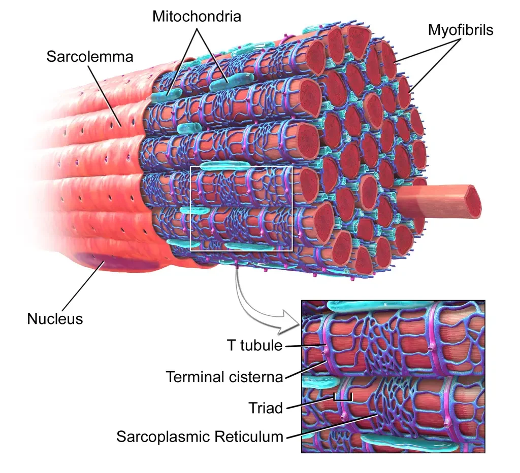
The intricate microanatomy of a muscle cell has led to its unique nomenclature. In these cells, the cytoplasm is referred to as the sarcoplasm, the smooth endoplasmic reticulum is termed the sarcoplasmic reticulum, and the cell membrane is labeled the sarcolemma. The sarcolemma is crucial as it receives and conducts stimuli.
Skeletal Muscle Cells
- Skeletal muscle cells, often termed muscle fibers due to their elongated appearance, are the primary contractile units within a muscle. A single muscle, such as the biceps brachii, can house approximately 253,000 of these fibers. Unique to skeletal muscle fibers is their multinucleated nature, a result of the fusion of myoblasts during myogenesis. This fusion process relies on specific fusogens, namely myomaker and myomerger.
- Within a striated muscle fiber, myofibrils are present, composed of extended protein chains called myofilaments. These myofilaments can be categorized into three types: thin, thick, and elastic. The thin filaments primarily consist of actin, while the thick ones are predominantly made of myosin. These filaments interact, leading to muscle contraction. The elastic filament, on the other hand, is primarily composed of the protein titin.
- Striations in muscle bands are formed due to the presence of myosin (dark filaments) in the A band and actin (light filaments) in the I band. The smallest contractile segment in the fiber is the sarcomere, situated between two Z bands. The sarcoplasm is rich in glycogen, providing energy during intense physical activity, and myoglobin, which stores oxygen.
- The sarcoplasmic reticulum, a specialized smooth endoplasmic reticulum, envelopes each myofibril. This network comprises terminal cisternae and T-tubules, forming triads. The primary role of the sarcoplasmic reticulum is to act as a calcium ion reservoir. Upon receiving an action potential via the T-tubule, it releases calcium ions to initiate muscle contraction.

Cardiac Muscle Cells
- Cardiac muscle cells, or cardiomyocytes, possess a cell membrane with specialized regions, including intercalated discs and transverse tubules. This membrane is enveloped by a lamina coat, roughly 50 nm in width. Like skeletal muscle, cardiac muscle is striated, containing myofibrils, myofilaments, and sarcomeres. The cell’s cytoskeleton provides structural stability and defines the cell’s size and shape, influencing its electrical properties.
Smooth Muscle Cells
- Distinct from their striated counterparts, smooth muscle cells lack myofibrils and sarcomeres, resulting in a non-striated appearance. These cells are prevalent in hollow organ walls, blood vessels, and various body tracts. Smooth muscle cells are spindle-shaped, with a single nucleus. Despite the absence of sarcomeres, they are rich in contractile proteins, actin, and myosin. Actin filaments in these cells are anchored to the sarcolemma by dense bodies.
Development of Muscle Cell
- Origin of Myoblasts:
- Muscle cell development begins with embryonic precursor cells known as myoblasts.
- Regulation of Differentiation:
- Myoblast differentiation is governed by myogenic regulatory factors.
- Key factors include MyoD, Myf5, myogenin, and MRF4.
- Additionally, GATA4 and GATA6 are involved in the differentiation process.
- Formation of Skeletal Muscle Fibers:
- Myoblasts fuse together to form skeletal muscle fibers.
- As a result of this fusion, muscle fibers become multinucleated.
- Each nucleus within the muscle fiber, termed a myonucleus, originates from a distinct myoblast.
- Exclusivity to Skeletal Muscle:
- The fusion process of myoblasts is specific to skeletal muscle development and does not occur in cardiac or smooth muscle.
- Dedifferentiation of Myoblasts:
- Some myoblasts that don’t form muscle fibers revert to a state known as myosatellite cells.
- These cells position themselves adjacent to skeletal muscle fibers.
- Location of Myosatellite Cells:
- Satellite cells are found between the sarcolemma and the basement membrane of the endomysium.
- Reactivation for Muscle Growth:
- To contribute to muscle growth or repair, satellite cells can be stimulated to differentiate and form new muscle fibers.
- In Vitro Generation:
- Modern scientific techniques allow for the directed differentiation of pluripotent stem cells to produce myoblasts and related cells in a laboratory setting.
- Role of Kindlin-2:
- During muscle development, the protein Kindlin-2 is crucial for the elongation phase of myogenesis.
Functions of Muscle Cell
Muscle cells, also known as myocytes, play a pivotal role in various bodily functions. Here are the primary functions of muscle cells:
- Movement:
- Muscle cells contract and relax, enabling movement of body parts. This includes voluntary movements, such as walking or lifting, and involuntary movements, like the beating of the heart.
- Posture and Body Support:
- Skeletal muscle cells provide the necessary tension and support to maintain body posture, preventing us from collapsing.
- Heat Production:
- Through muscle contraction, muscle cells generate heat, which helps maintain the body’s core temperature, a process essential for metabolic reactions.
- Protection:
- Muscle cells, especially those in the abdominal region, protect internal organs from potential damage due to external impacts.
- Facilitation of Bodily Functions:
- Smooth muscle cells in organs and structures like the intestines, uterus, and blood vessels contract and relax to facilitate functions such as digestion, circulation, and childbirth.
- Stabilization of Joints:
- Muscle cells, in coordination with tendons and ligaments, help stabilize joints, ensuring coordinated and safe movement.
- Regulation of Organ Volume:
- Muscle cells in the walls of hollow organs (like the bladder) can contract or relax to regulate the volume of the organ.
- Generation of Reflexes:
- Muscle cells are involved in reflex actions, which are rapid, involuntary responses to stimuli. For example, the quick withdrawal of a hand from a hot surface.
- Conduction of Electrical Impulses:
- Cardiac muscle cells are specialized to conduct electrical impulses that regulate the heart’s rhythmic contractions.
- Storage and Movement of Materials:
- Smooth muscle cells in organs like the stomach and intestines help in the storage of materials (like food) and move them along the digestive tract through peristaltic movements.
In essence, muscle cells are indispensable for a myriad of physiological processes, ranging from gross motor functions to intricate cellular activities.
Examples of Muscle Cell
Muscle cells, also known as myocytes, come in various types, each with specific functions and characteristics. Here are examples of muscle cells:
- Skeletal Muscle Cells (Muscle Fibers):
- Location: Attached to bones by tendons.
- Function: Facilitate voluntary movements such as walking, jumping, and lifting.
- Characteristics: Multinucleated, long, cylindrical, and striated appearance.
- Cardiac Muscle Cells (Cardiomyocytes):
- Location: Heart.
- Function: Pump blood throughout the body.
- Characteristics: Single nucleus, branched structure, striated, and connected by intercalated discs which allow synchronized contractions.
- Smooth Muscle Cells:
- Location: Walls of hollow organs (e.g., stomach, intestines, blood vessels, bladder, uterus).
- Function: Control involuntary movements such as the peristalsis contractions in the digestive tract.
- Characteristics: Single nucleus, spindle-shaped, and lack striations.
Each of these muscle cell types plays a unique role in the body, from facilitating movement to pumping blood and controlling involuntary functions.
MCQ Practice
What is another term for a muscle cell?
a) Myofibril
b) Sarcomere
c) Myocyte
d) Actin
Which protein is primarily found in the thin filaments of muscle cells?
a) Myosin
b) Titin
c) Actin
d) Tropomyosin
Which type of muscle cell is multinucleated?
a) Cardiac muscle cell
b) Smooth muscle cell
c) Skeletal muscle cell
d) None of the above
What is the primary function of smooth muscle cells?
a) Voluntary movement
b) Pumping blood
c) Facilitating bodily functions like digestion
d) Maintaining posture
Which muscle cells are responsible for the heart’s rhythmic contractions?
a) Skeletal muscle cells
b) Smooth muscle cells
c) Cardiac muscle cells
d) Myoblasts
The smallest contractile unit of a muscle fiber is called:
a) Myofibril
b) Sarcomere
c) Sarcoplasm
d) Sarcolemma
Which protein is responsible for the elastic properties of muscle cells?
a) Actin
b) Myosin
c) Titin
d) Tropomyosin
The cell membrane of a muscle cell is termed:
a) Sarcoplasm
b) Sarcomere
c) Sarcolemma
d) Myofibril
Which cells are the precursors to muscle cells?
a) Myocytes
b) Myofibrils
c) Myoblasts
d) Sarcomeres
Which type of muscle cell lacks striations?
a) Skeletal muscle cell
b) Cardiac muscle cell
c) Smooth muscle cell
d) All of the above
FAQ
What is a muscle cell?
A muscle cell, also known as a myocyte, is a specialized cell designed for contraction, facilitating movement and other bodily functions.
How do muscle cells contract?
Muscle cells contract through the sliding filament theory, where actin and myosin filaments slide past each other, shortening the muscle cell.
What are the different types of muscle cells?
The three main types are skeletal muscle cells, cardiac muscle cells, and smooth muscle cells.
Why are skeletal muscle cells multinucleated?
Skeletal muscle cells are multinucleated because they form from the fusion of multiple myoblasts, each contributing a nucleus.
What is the function of smooth muscle cells?
Smooth muscle cells control involuntary movements in various organs and structures, such as the intestines, blood vessels, and bladder.
How do cardiac muscle cells differ from skeletal muscle cells?
Cardiac muscle cells are found in the heart, have a single central nucleus, and are connected by intercalated discs. In contrast, skeletal muscle cells are multinucleated and don’t have intercalated discs.
What is the role of the sarcolemma in muscle cells?
The sarcolemma is the cell membrane of a muscle cell, responsible for receiving and conducting stimuli.
Why do muscle cells have a high number of mitochondria?
Muscle cells require a significant amount of energy for contraction, and mitochondria are the cell’s “powerhouses,” producing ATP, the primary energy molecule.
What are myofibrils, and why are they important?
Myofibrils are long protein chains within muscle cells made up of actin and myosin filaments. They play a crucial role in muscle contraction.
How do muscle cells repair after injury?
Satellite cells, which are a type of stem cell found adjacent to muscle fibers, become activated upon muscle injury. They proliferate and differentiate to repair and replace damaged muscle fibers.
References
- Mukherjee, S., Skrede, S., Haugstøyl, H., Lópéz, L., & Fernø, J. (2023). Peripheral and central macrophages in obesity. Frontiers in Endocrinology, 14, 1232171. doi:10.3389/fendo.2023.1232171
- Lodish, H., Berk, A., Kaiser, C. A., Krieger, M., Scott, M. P., Bretscher, A., . . . Matsudaira, P. (2008). Molecular Cell Biology 6th. ed. New York: W.H. Freeman and Company.
- Reece, J. B., Urry, L. A., Cain, M. L., Wasserman, S. A., Minorsky, P. V., & Jackson, R. B. (2014). Campbell Biology, Tenth Edition (Vol. 1). Boston: Pearson Learning Solutions.
- Blausen.com staff (2014). “Medical gallery of Blausen Medical 2014”. WikiJournal of Medicine 1 (2). DOI:10.15347/wjm/2014.010. ISSN 2002-4436.