What is Mitosis?
- Mitosis, a fundamental process in the cell cycle, facilitates the separation of duplicated DNA into two distinct cells. This cellular division is pivotal for eukaryotic organisms, both single-celled and multi-celled. In single-celled eukaryotes, mitosis serves as the mechanism for asexual reproduction. Conversely, in multi-celled eukaryotes, mitosis enables the transformation of a singular zygote into a full-fledged organism.
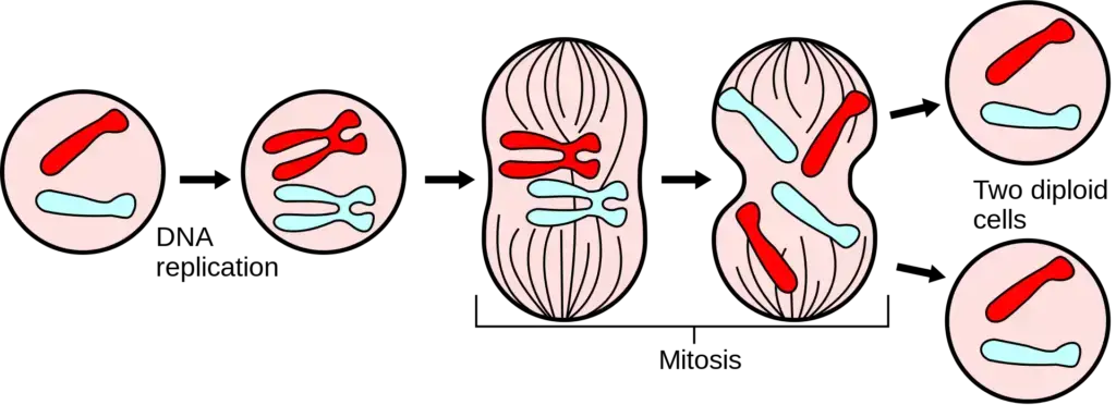
- The essence of mitosis is captured in its definition: a phase in the cell cycle where the newly synthesized DNA is segregated, culminating in the formation of two new cells, each inheriting the same number and type of chromosomes as the original nucleus. This ensures genetic consistency across cells, a feature crucial for maintaining the integrity of an organism.
- Mitosis is characterized by a series of stages, each delineating specific cellular activities. These stages encompass preprophase (exclusive to plant cells), prophase, prometaphase, metaphase, anaphase, and telophase. The chromosomes, post-duplication, undergo condensation and subsequently attach to spindle fibers. These fibers orchestrate the movement of each chromosome copy to opposing ends of the cell. The culmination of this process yields two genetically congruent daughter nuclei. Subsequent cellular division, termed cytokinesis, results in the formation of two daughter cells.
- It’s imperative to note that mitosis is a brief interval of chromosome condensation, segregation, and cytoplasmic division. This process is ubiquitous in somatic cells, facilitating cell multiplication during embryogenesis and blastogenesis in both flora and fauna. Despite the diversity of life, the process of mitosis exhibits remarkable consistency across various animal and plant species.
- In the broader context of cell biology, mitosis is a segment of the cell cycle where replicated chromosomes segregate into fresh nuclei. This division yields genetically identical cells, preserving the total chromosomal count, hence its designation as an equational division. Typically, mitosis is preceded by the S phase of interphase, where DNA replication transpires. Often, mitosis is succeeded by telophase and cytokinesis, distributing the cytoplasm, organelles, and cell membrane into two new cells, each receiving approximately equal cellular components.
- Errors during mitosis can lead to anomalies such as tripolar mitosis or even induce apoptosis, the programmed cell death. Such aberrations can be precursors to mutations, some of which may be implicated in carcinogenesis.
- Historically, the phenomenon of cell division has been documented since the 18th and 19th centuries. The term “mitosis” was introduced by Walther Flemming in 1882, derived from the Greek word μίτος (mitos), meaning “warp thread”. Over time, other terminologies like “karyokinesis” and “equational division” have been proposed, but “mitosis” remains the most widely accepted term in scientific literature.
- In summary, mitosis is a critical cellular process ensuring genetic uniformity across daughter cells, underpinning the growth, development, and repair mechanisms in eukaryotic organisms.
Definition of Mitosis
Mitosis is a cellular process in which a single eukaryotic cell divides to produce two genetically identical daughter cells, each maintaining the same number of chromosomes as the parent cell.
Features of Mitosis
- Daughter Cell Formation: Each cycle of mitotic cell division results in the generation of two daughter cells derived from a single parent cell.
- Equational Division: Termed as an equational cell division, mitosis ensures that both the parent and the daughter cells possess an identical number of chromosomes.
- Role in Plant Growth: In plants, mitosis predominantly occurs in vegetative parts, facilitating growth in regions such as the root and stem tips.
- Absence of Genetic Recombination: Unlike meiosis, mitosis does not involve genetic recombination. Segregation and combination of genetic material are absent in this process.
- Cell Replacement and Repair: Mitosis plays a pivotal role in tissue repair and regeneration. When cells are damaged, neighboring cells undergo mitosis to replace the lost or damaged cells, ensuring tissue integrity.
- Growth and Development: Mitosis is instrumental in the overall growth of an organism. By producing genetically identical cells, it ensures consistent growth and development.
- Consistency in Genetic Information: One of the defining features of mitosis is the preservation of genetic information. There is no exchange or recombination of genetic material, ensuring that daughter cells are genetically identical to the parent cell.
In essence, mitosis is a meticulously regulated process that ensures genetic consistency, facilitates growth, and aids in repair and regeneration across eukaryotic organisms.
Purpose of Mitosis
- Growth and Development: One of the primary purposes of mitosis is to facilitate the growth of an organism. Through continuous mitotic divisions, an organism evolves from a singular cell to a multifaceted, multicellular entity.
- Cell Renewal: The human body, as well as other organisms, undergoes constant cellular turnover. For instance, human skin cells and red blood cells are perpetually replaced through mitotic divisions. It is estimated that approximately 5×10^9 cells are generated daily in humans as a result of mitosis.
- Repair and Regeneration: Mitosis plays a crucial role in the body’s innate ability to repair and regenerate. For example, certain marine organisms like starfish can regenerate lost body parts, a process underpinned by mitotic cell divisions.
- Asexual Reproduction: In many organisms, particularly unicellular ones, mitosis serves as the primary mechanism for asexual reproduction. This mode of reproduction ensures the generation of offspring that are genetically identical to the parent.
In summation, mitosis is an indispensable cellular process, ensuring growth, continuous cell renewal, repair, and asexual reproduction in various organisms. Its role is paramount in maintaining the structural and functional integrity of living entities.
Cell Cycle Overview
The cell cycle is a meticulously orchestrated sequence of events that eukaryotic somatic cells undergo, ensuring growth, DNA replication, and cell division. This cycle can be delineated into several distinct phases:
- Resting Phase (Gap 0): This is a phase where the cell is not actively preparing for division but might re-enter the cycle under certain conditions.
- Interphase: Comprising approximately 22 hours of a typical 24-hour mammalian cell cycle, interphase is further divided into:
- Gap 1 (G1) Phase: Occurring post-mitosis, this phase witnesses cellular growth. Cells prepare for DNA replication, and in the absence of essential nutrients or growth signals, progression can be halted or decelerated.
- Synthetic (S) Phase: This is the phase where DNA replication transpires. Each DNA molecule is duplicated, resulting in two identical daughter DNA molecules.
- Gap 2 (G2) Phase: This phase succeeds DNA replication. Here, cells evaluate the integrity of the replicated DNA. If anomalies or unreplicated segments are detected, the progression to mitosis is delayed. Otherwise, cells transition to the mitotic phase.
- Mitotic Phase: This phase, which spans roughly 2 hours, encompasses the division of the cell nucleus (karyokinesis) and is succeeded by cytokinesis, the division of the cell’s cytoplasm. Occasionally, cytokinesis might not follow karyokinesis, leading to multinucleate cells.
- Cytokinesis: This is the cytoplasmic division, ensuring that the cell’s contents are evenly distributed between the two daughter cells.
- Centrosome Cycle: Integral to the cell cycle, the centrosome cycle ensures the accurate inheritance and duplication of the centrosome, which establishes the poles of the mitotic spindle.
- Control Mechanisms: The cell cycle is punctuated with checkpoints, such as the G2-M DNA damage checkpoint, which ascertain the cell’s readiness for the subsequent phase. These checkpoints are pivotal in maintaining the fidelity of cell division.
In essence, the cell cycle is a harmonized succession of events, alternating between DNA synthesis, cell growth, and division. It encompasses the chromosome cycle, where DNA synthesis and mitosis alternate; the cytoplasmic cycle, alternating between cell growth and division; and the centrosome cycle, vital for mitotic spindle formation. This intricate process ensures the continuity and integrity of genetic information across generations of cells.
Spindle Structure
The mitotic spindle, a pivotal structure during metaphase, is characterized by its bilateral symmetry and is primarily composed of microtubules. This structure plays a crucial role in ensuring the accurate segregation of chromosomes during cell division. Here are the salient features of the spindle structure:
- Bipolarity: The spindle is inherently bipolar, with two spindle poles delineating its axis. These poles serve as the sites to which chromatids are transported during anaphase.
- Centrosome Composition: In many animal cells, the spindle poles house a centrosome, which comprises a pair of centrioles accompanied by pericentriolar material that nucleates microtubules.
- Microtubule Composition: The spindle’s structural integrity is primarily maintained by microtubules, dynamic cytoskeletal filaments that project from the spindle poles. These microtubules are instrumental in attaching chromosomes to the spindle and facilitating their movement.
- Types of Spindle Microtubules:
- Interpolar Microtubules: These originate from each spindle pole and intersect at the spindle’s center.
- Kinetochore Microtubules: One end of these microtubules is embedded in the kinetochore, a specialized region on the chromosome.
- Astral Microtubules: Radiating from the centrosomes toward the cell’s periphery, these microtubules play roles in spindle positioning and determining the contractile ring’s location during cytokinesis.
- Variability in Spindle Structure: The architecture of the spindle exhibits diversity across different cell types. For instance, higher plant cells possess a spindle pole that lacks focus and is devoid of centrioles, resulting in anastral spindles without astral microtubules. Yeasts, on the other hand, have a spindle pole body at each spindle end, which nucleates microtubules extending into both the nucleus and cytoplasm.
- Microtubule Quantity: The number of spindle microtubules varies among cell types. For example, in yeast, a single microtubule connects each kinetochore to the spindle pole body. In contrast, a typical mammalian cell’s kinetochore is associated with approximately 20 microtubules, with each half-spindle comprising hundreds of microtubules.
In conclusion, while the spindle structure may exhibit variations across different organisms and cell types, its fundamental role remains consistent: ensuring the precise and equal distribution of genetic material to the daughter cells during mitosis.
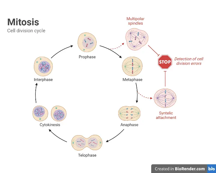
Phases of mitosis

1. Interphase
Interphase, a critical segment of the cell cycle, is characterized by the cell’s preparation for the subsequent mitotic phase (M phase) through DNA replication. Contrary to its seemingly quiescent nature, interphase is a hive of cellular activity, often regarded as the most dynamic phase of the cell cycle. This phase encompasses a series of metabolic transformations, systematically categorized into three distinct sub-phases:
- G1-Phase (Pre-DNA Synthesis Phase):
- Duration: This phase, following the M phase of the preceding cell cycle, is the lengthiest segment of the cell cycle.
- Characterization: Often dubbed the “resting phase,” the G1 phase is somewhat misleadingly named, as DNA synthesis is absent, but the cell is far from inactive.
- Cellular Activities: During this phase, cellular organelles undergo enlargement, and there’s a brisk synthesis of diverse RNA types and proteins. Noteworthy processes include the transcription of various RNAs, production of regulatory proteins, enzymes vital for DNA synthesis, and the assembly of tubulin proteins and other components essential for mitosis.
- S-Phase (DNA Synthesis Phase):
- DNA Replication: The hallmark of the S-phase is the replication of nuclear DNA, accompanied by the synthesis of histone proteins. It’s worth noting that cytoplasmic DNA replication can transpire at any juncture within the cell cycle.
- Chromosomal Duplication: Post the S-phase, every chromosome comprises two DNA molecules, effectively duplicating the gene set.
- Duration: This phase spans approximately 6 to 10 hours, ensuring the cell is equipped with a complete set of genetic information for the ensuing division.
- G2-Phase (Post DNA Synthesis Phase):
- Characterization: Often referred to as the second gap or another “resting phase,” the G2 phase is a preparatory interval post-DNA synthesis and prior to cell division.
- Cellular Activities: This phase witnesses the continued synthesis of RNA and proteins requisite for the cell. Given that cell division demands a significant energy outlay, the cell accumulates ATP reserves during the G2 phase.
- Transition to M-Phase: Upon the culmination of the G2 phase, the cell is primed to transition into the mitotic or M-phase, marking the commencement of cell division.
In essence, interphase is a preparatory phase, ensuring that the cell is genetically and energetically equipped for the intricate process of mitosis. Through systematic DNA replication and metabolic activities, the cell sets the stage for accurate and efficient cell division.
Preprophase (plant cells)
In the realm of plant cell mitosis, preprophase emerges as a distinct phase preceding the conventional prophase. This phase is characterized by specific cellular events that lay the groundwork for subsequent stages of cell division. Here’s a detailed examination of the events and significance of preprophase in plant cells:
- Preprophase Band Formation: A hallmark of preprophase is the emergence of the preprophase band. This band, composed of densely packed microtubules, transiently appears beneath the plasma membrane. Its primary role is to demarcate the future plane of cell division, thereby providing a blueprint for where the new cell wall will be established during cytokinesis.
- Significance of the Preprophase Band: The pivotal role of the preprophase band in plant mitosis was elucidated through studies on the Arabidopsis plant. In particular, variants of this plant that lacked the genetic machinery to form the preprophase band served as a testament to its importance in ensuring accurate cell division (Ref.1).
- Microtubule Nucleation: Concurrently, during preprophase, the initiation of microtubule nucleation at the nuclear envelope transpires. This event is of paramount importance in the context of plant cells, given their lack of centrosomes, which are prominent in animal cells.
- Role of the Nuclear Envelope: In the absence of centrosomes, which in animal cells serve as the epicenter for organizing mitotic spindles, plant cells rely on their nuclear envelope. The nuclear envelope, or nuclear membrane, assumes the role of the microtubule-organizing center (MTOC) in plant cells. This adaptation ensures that plant cells can effectively orchestrate the mitotic spindles, facilitating the precise segregation of chromosomes during mitosis.
In summary, preprophase in plant cells is a preparatory stage that sets the stage for the intricate process of mitosis. Through the formation of the preprophase band and the initiation of microtubule nucleation at the nuclear envelope, plant cells ensure that they are primed for the subsequent phases of cell division, ensuring the accurate and efficient partitioning of genetic material.
2. Prophase
Prophase, the inaugural stage of mitosis, is marked by a series of cellular transformations that prepare the cell for the subsequent phases of cell division. Here’s a detailed exploration of the events that transpire during prophase:
- Chromosomal Condensation: The onset of prophase is characterized by the emergence of chromosomes that begin to condense into thin, thread-like structures. These chromosomes, initially in a relaxed chromatin state during interphase, undergo condensation to prevent tangling and potential breakage during subsequent mitotic stages.
- Chromatid Formation: Each chromosome in prophase comprises two coiled strands known as chromatids. These chromatids are the product of DNA replication that occurred during the S phase of interphase.
- Centromere Role: As prophase advances, the chromatids thicken and shorten. The two sister chromatids of each chromosome remain connected at a specific DNA-rich region termed the centromere, ensuring their coordinated movement during mitosis.
- Nuclear Alterations: The chromosomes migrate toward the nuclear envelope, rendering the nucleus’s central region vacant. Concurrently, the nucleolus, a vital nuclear structure, starts to disintegrate, signaling the culmination of prophase. Notably, in certain primitive plant and animal species, the nuclear envelope remains intact throughout mitosis.
- Centriole Migration: Parallelly, pairs of centrioles, encircled by radiating microtubules, traverse to the cell’s opposite poles. These centrioles play a pivotal role in orchestrating the spindle apparatus, which will subsequently engage with the chromosomes.
- DNA Packaging: To facilitate efficient movement, the cell’s machinery wraps the DNA around specialized proteins known as histones. This wrapping enables the DNA to condense into compact structures, ensuring its safe and organized segregation.
- Microtubule Formation: During prophase, centrioles emerge as microtubule-organizing centers. These microtubules extend and are primed to interact with the chromosomes, ensuring their proper alignment and segregation in subsequent phases.
- Plant Cell Specificity: In plant cells, a unique preprophase stage precedes prophase. This stage repositions the nucleus centrally, countering its typical peripheral location due to expansive vacuoles. This reorganization ensures optimal organelle arrangement for ensuing division.
In essence, prophase is a preparatory phase, setting the stage for the intricate process of mitosis. Through systematic chromosomal condensation, nuclear reorganization, and microtubule formation, the cell ensures that the genetic material is poised for accurate and efficient segregation.
3. Prometaphase
Prometaphase is a critical transitional phase in mitosis, bridging the gap between prophase and metaphase. This stage is marked by a series of cellular events that facilitate the alignment and subsequent segregation of chromosomes. Here’s an in-depth exploration of the events and mechanisms underpinning prometaphase:
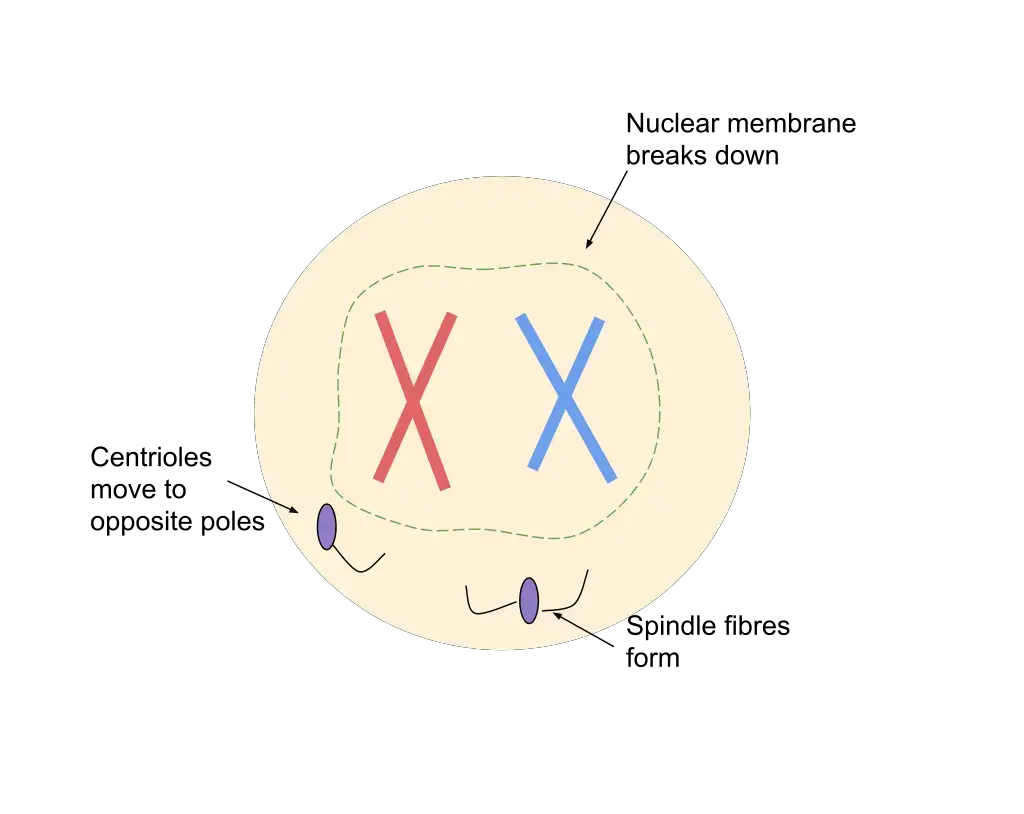
- Nuclear Envelope Breakdown: The onset of prometaphase is characterized by the disintegration of the nuclear envelope. This disassembly allows the spindle fibers, which are microtubule structures, to access and interact with the chromosomes.
- Chromosomal Movement: As the chromosomes become accessible to spindle fibers, they undergo vigorous movements. This dynamic behavior results from the centromeres of the chromosomes capturing the ends of microtubules. Consequently, the chromosomes oscillate and rotate between the spindle poles due to the pulling forces exerted by the captured microtubules.
- Kinetochore Attachment: By the culmination of prometaphase, each sister chromatid becomes anchored to spindle fibers emanating from opposite poles. This attachment occurs at the kinetochore, a specialized protein structure that forms on the centromere of each chromatid. As a result, the sister chromatids are positioned along the metaphase plate, poised for subsequent segregation.
- Open vs. Closed Mitosis: In animal cells, the nuclear envelope’s disintegration, facilitated by the phosphorylation of nuclear lamins, typifies what is termed “open mitosis.” This process allows microtubules to invade the erstwhile nuclear space. In contrast, certain fungi, protists, and algae exhibit “closed mitosis,” where the mitotic spindle either forms within an intact nucleus or the microtubules penetrate the undissolved nuclear envelope.
- Microtubule Dynamics: During the latter stages of prometaphase, kinetochore microtubules actively seek and bind to chromosomal kinetochores. Concurrently, polar microtubules from one centrosome interact with their counterparts from the opposing centrosome, culminating in the formation of the mitotic spindle. The kinetochore, though not entirely understood, houses a molecular motor. Upon microtubule attachment, this motor is activated, utilizing ATP energy to traverse the microtubule toward its originating centrosome. This motor-driven movement, in conjunction with microtubule polymerization and depolymerization, generates the requisite force to eventually segregate the chromatids.
In essence, prometaphase is a dynamic phase that ensures the chromosomes are aptly aligned and anchored, setting the stage for their precise and orderly segregation during the subsequent phases of mitosis.
4. Metaphase
Metaphase is a pivotal stage in mitosis, characterized by the precise alignment of chromosomes along the cell’s equatorial plane. This alignment ensures the accurate and equal segregation of genetic material to the daughter cells. Here’s a detailed exploration of the events and mechanisms underpinning metaphase:
- Chromosomal Characteristics: At the onset of metaphase, chromosomes attain their maximum condensation, becoming the shortest and thickest. This compact structure facilitates their alignment and subsequent segregation.
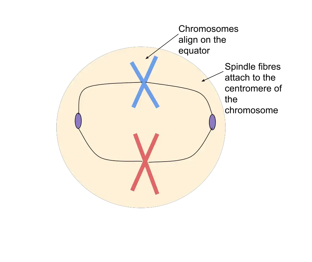
- Formation of the Metaphase Plate: The centromeres of the sister chromatids align along an imaginary plane termed the metaphase plate or equatorial plane. This plane is equidistant from the two centrosomes, positioning itself approximately at the cell’s midpoint. The arms of the chromatids, meanwhile, are oriented toward the cell’s poles.
- Chromatid Dynamics: Within each chromosome, the two sister chromatids exhibit a repulsive interaction. This repulsion, combined with the tension exerted by the stationary microtubules, ensures that the chromatids remain distinct and poised for subsequent separation.
- Microtubule-Driven Alignment: Following the kinetochore-microtubule attachment established during prometaphase, the centrosomes exert forces on the chromosomes, pulling them in opposite directions. This tug-of-war culminates in the chromosomes’ alignment along the metaphase plate. The balanced forces ensure that each chromosome is equidistant from the two poles, optimizing their subsequent segregation.
- The Metaphase Checkpoint: Cellular fidelity is paramount during mitosis. The metaphase checkpoint serves as a surveillance mechanism, ensuring that all chromosomes are correctly attached to the mitotic spindle and are aligned along the metaphase plate. Only upon satisfying these criteria does the cell receive the green light to transition to anaphase, the next stage of mitosis.
In summary, metaphase is a meticulously orchestrated stage that ensures the chromosomes are aptly aligned for their subsequent and equitable distribution to the daughter cells. This alignment is crucial for maintaining genetic stability across cellular generations.
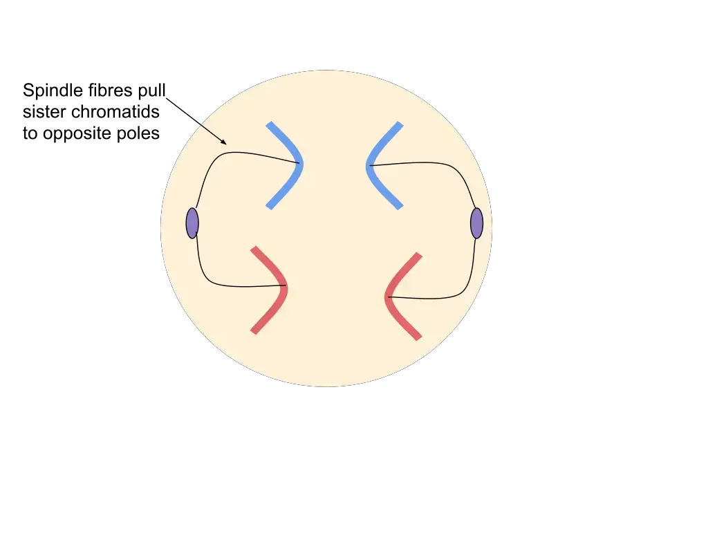
5. Anaphase
Anaphase is a critical juncture in mitosis, marked by the synchronous separation of sister chromatids, now termed daughter chromosomes. This phase ensures the equitable distribution of genetic material to the ensuing daughter cells. Here’s an in-depth exploration of the events and mechanisms that define anaphase:
- Initiation of Anaphase: Anaphase commences with the simultaneous cleavage of each chromosome into its constituent sister chromatids. This cleavage is facilitated by a surge in cytosolic Ca2+ levels, which triggers the splitting of the centromere, the region binding the chromatids.
- Chromatid Movement: Post cleavage, the daughter chromosomes migrate poleward. This movement is driven by the shortening of kinetochore microtubules, which effectively “reel in” the chromosomes towards the poles. As they travel, the centromeres lead the way, resulting in the chromosomes adopting characteristic U, V, or J-shaped configurations.
- Role of Interzonal Fibers: These fibers, which span the region between the two poles, elongate during anaphase. They play a pivotal role in facilitating and supporting the poleward movement of the chromosomes.
- Energy Consumption: The migration of chromosomes is an energy-intensive process. It is estimated that approximately 30 ATP molecules are expended for each chromosome’s journey to its respective pole.
- Anaphase Subdivisions: Anaphase can be further delineated into two sub-phases:
- Anaphase A: During this phase, the cohesins binding the sister chromatids are cleaved, resulting in the formation of two identical daughter chromosomes. The shortening of kinetochore microtubules then pulls these chromosomes towards opposite cell poles.
- Anaphase B: In this phase, polar microtubules, which extend from each spindle pole and overlap in the cell center, push against each other. This action causes the cell to elongate, further aiding in chromosome segregation.
- Chromosomal Condensation: As anaphase progresses, chromosomes reach their peak level of condensation. This heightened condensation facilitates the segregation of chromosomes and the subsequent re-establishment of the nucleus.
In essence, anaphase is a meticulously coordinated stage of mitosis, ensuring the precise and equal distribution of genetic material, setting the stage for the culmination of cell division.
6. Telophase
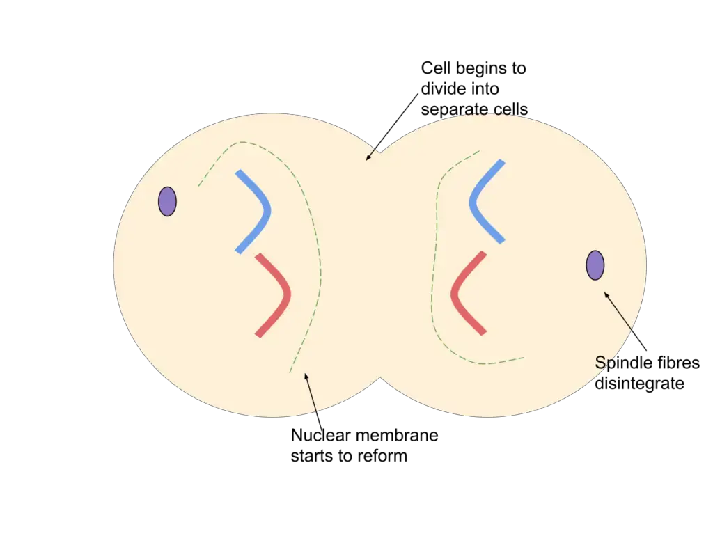
Telophase, derived from the Greek term τελος signifying “end,” represents the concluding stage of mitosis, characterized by the reversal of events witnessed during prophase and prometaphase. This phase ensures the final preparations for cell division, setting the stage for cytokinesis. Here’s a comprehensive examination of the events and intricacies of telophase:
- Commencement of Telophase: Telophase is initiated once the daughter chromosomes complete their migration to the respective poles. This phase signifies the culmination of the mitotic process, preparing the cell for division.
- Reformation of Nuclear Structures: One of the hallmark events of telophase is the reassembly of the nuclear envelope. Utilizing membrane vesicles from the parent cell’s previous nuclear envelope, a new nuclear envelope forms around each cluster of daughter chromosomes, thereby delineating two distinct daughter nuclei within the cell.
- Chromosomal Decondensation: Post their migration, the chromosomes undergo decondensation, reverting to their elongated, slender forms. This relaxed state facilitates the cell’s transition into interphase, where the genetic material is more accessible for transcription.
- Reappearance of the Nucleolus: The nucleolus, a vital cellular structure responsible for ribosomal RNA synthesis, reemerges at the culmination of telophase. Its reappearance indicates the cell’s readiness to resume its regular metabolic activities.
- Dissolution of the Mitotic Apparatus: The intricate machinery that facilitated chromosome movement and segregation, including the spindle fibers, disassembles during telophase. Concurrently, the cytoplasm’s viscosity decreases, and RNA synthesis recommences, indicating the cell’s return to a non-divisive state.
- Cell Elongation: The polar microtubules persist in their elongation, further stretching the cell. This elongation prepares the cell for the subsequent division process, ensuring that each daughter cell receives an equitable share of the cellular contents.
- Completion of Mitosis: With the conclusion of telophase, mitosis is deemed complete. The cell now possesses two genetically identical daughter nuclei, each encompassing an identical set of chromosomes. The subsequent step, cytokinesis, may then proceed, culminating in the division of the cell into two distinct daughter cells.
In essence, telophase is a restorative phase, reinstating the nuclear structures and preparing the cell for its eventual division. It ensures the preservation of genetic fidelity and sets the stage for the cell’s return to its regular metabolic functions.
7. Cytokinesis
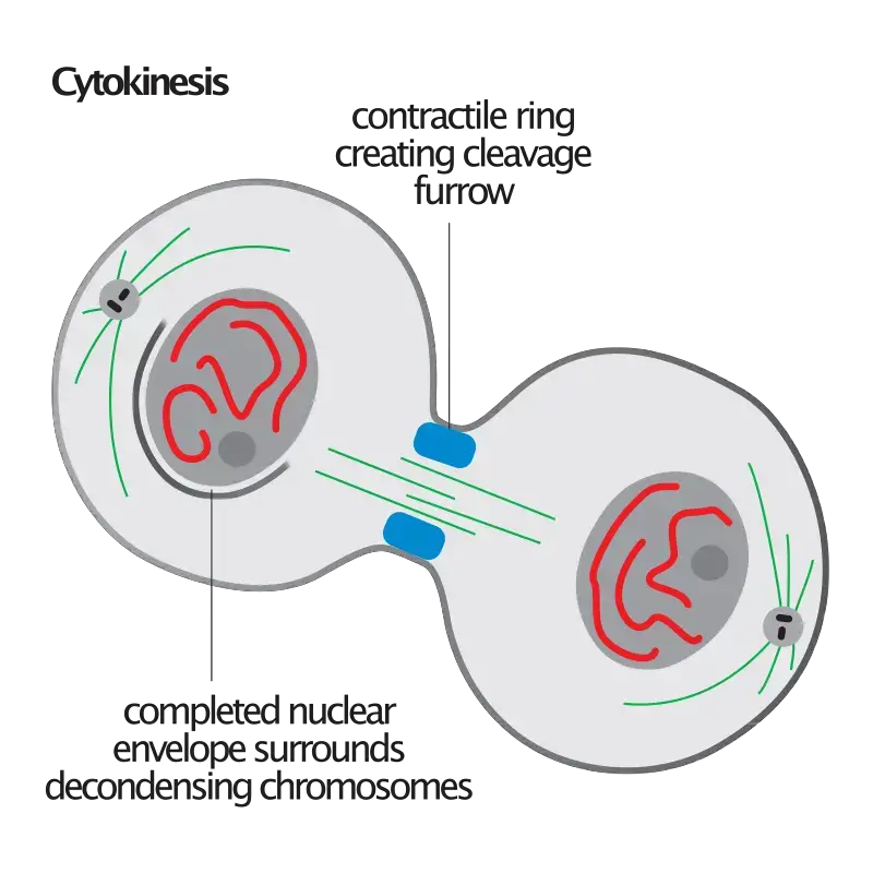
Cytokinesis, distinct from mitosis, is the pivotal process responsible for the division of the cytoplasm, culminating in the formation of two distinct daughter cells. This process ensures that the genetic material segregated during mitosis is enclosed within individual cellular entities, each equipped with its own set of organelles and cytoplasmic components. Here’s an in-depth exploration of cytokinesis:
- Temporal Occurrence: Cytokinesis commences during the anaphase stage of mitosis and extends through telophase, eventually transitioning into interphase. It is the concluding step post-mitotic segregation, ensuring cellular individuality.
- Mechanisms in Animal Cells: In animal cells, cytokinesis is orchestrated through a process known as cleavage. The initial indication of this cleavage is the observable constriction of the plasma membrane during the anaphase stage. This constriction, invariably aligned with the plane of the former metaphase plate, deepens progressively, eventually leading to the separation of the cell into two daughter entities.
- Mechanisms in Plant Cells: Contrary to animal cells, plant cells undergo cytokinesis via the formation of a cell plate. The presence of a rigid cell wall in plant cells precludes the possibility of membrane constriction. Instead, components from the Golgi apparatus align at the equatorial region, forming a structure termed the phragmoplast. This phragmoplast subsequently evolves into the cell plate, which matures into the cell wall, demarcating the two daughter cells.
- Distinctive Features in Various Organisms: While the fundamental principle of cytokinesis remains consistent across eukaryotes, the mechanisms can exhibit variations. For instance, certain green algae employ a phycoplast microtubule array during cytokinesis, a deviation from the typical phragmoplast observed in higher plants.
- Instances of Independent Occurrence: In certain organisms, mitosis and cytokinesis can function independently. This results in the formation of multinucleated cells, where a single cell houses multiple nuclei. Such a phenomenon is prominently observed in fungi, slime molds, and certain algae. Even in the animal kingdom, specific developmental stages, such as in fruit fly embryos, can witness independent occurrences of mitosis and cytokinesis.
- Conclusion of Cytokinesis: The termination of cytokinesis signifies the end of the M-phase, with each daughter cell possessing an identical genomic copy of its progenitor cell.
In essence, cytokinesis is the cellular mechanism that ensures the equitable distribution of cellular components post-mitotic segregation, facilitating the continuity of life through cellular proliferation.
| Phase | Description |
|---|---|
| Prophase | Chromosomes condense and become visible. Nuclear envelope breaks down. Spindle fibers form from the centrosomes, which move to opposite poles. |
| Prometaphase | Nuclear envelope disappears completely. Spindle fibers attach to the kinetochores on the chromosomes. |
| Metaphase | Chromosomes align at the metaphase plate (equatorial plane). Spindle fibers connect the centromeres of the chromosomes to the poles of the cell. |
| Anaphase | Sister chromatids are pulled apart to opposite poles of the cell. Each chromatid now becomes an individual chromosome. |
| Telophase | Chromosomes begin to decondense and return to their thread-like form. Nuclear envelope re-forms around each set of chromosomes, resulting in two separate nuclei within the cell. |
Regulation of Mitosis
Mitosis, the process of cell division, is meticulously regulated to ensure the accurate replication and distribution of genetic material. The primary regulators of this process are cyclins and Cyclin-Dependent Kinases (CDKs), which orchestrate the progression of cells through various phases of the cell cycle.
- G1 Phase (Gap 1): During this preparatory phase, G1 cyclins activate CDKs. These activated CDKs phosphorylate specific proteins, setting the stage for DNA synthesis in the subsequent phase. The G1 checkpoint ensures that conditions are optimal for DNA replication, considering factors such as DNA integrity, cell size, nutrient availability, and appropriate signaling molecule cues.
- S Phase (Synthesis): In this phase, DNA replication occurs. Cyclins and CDKs oversee the completion of DNA synthesis, ensuring that the genetic material is accurately duplicated.
- G2 Phase (Gap 2): G2 cyclins activate CDKs, which then phosphorylate proteins essential for initiating mitosis. The G2 checkpoint verifies that DNA replication is complete and without errors, and that the cell has adequate resources to proceed to mitosis.
- M Phase (Mitosis): M cyclins activate CDKs during this phase, driving the cell into mitosis. These CDKs regulate critical mitotic events such as chromosome condensation, spindle assembly, and the separation of sister chromatids. The metaphase checkpoint ensures that chromosomes are properly attached to spindle fibers and correctly aligned on the metaphase plate before proceeding to anaphase.
- Checkpoints: These are critical regulatory points within the cell cycle that assess whether conditions are favorable for progression to the subsequent phase. If conditions are suboptimal, the cell cycle may halt, allowing the cell to repair any issues or enter a resting state, known as Gap 0 (G0).
For instance:
- At the G1 checkpoint, the cell evaluates DNA integrity, its size, nutrient availability, and the presence of appropriate signaling molecules.
- The G2 checkpoint ensures complete DNA replication, checks for DNA errors, and confirms sufficient resources for mitosis.
- The Metaphase checkpoint during mitosis verifies the correct attachment of chromosomes to spindle fibers and their proper alignment on the metaphase plate.
In essence, the intricate interplay of cyclins, CDKs, and checkpoints ensures that mitosis proceeds accurately, safeguarding the integrity of the cell’s genetic material. This regulatory framework is crucial for maintaining cellular health and preventing anomalies that could lead to diseases like cancer.
Forms of mitosis
Mitosis, the process of cell division in eukaryotic organisms, exhibits variations in its execution, leading to the classification of different forms. These forms are primarily based on the behavior of the nuclear envelope, the symmetry of the spindle apparatus, and the location of the central spindle. Here’s a comprehensive overview of the various forms of mitosis:
- Based on Nuclear Envelope Integrity:
- Closed Mitosis: The nuclear envelope remains intact throughout the process.
- Open Mitosis: The nuclear envelope disintegrates, allowing the mitotic spindle to interact directly with the chromosomes.
- Semiopen Mitosis: An intermediate form where the nuclear envelope partially degrades.
- Based on Spindle Apparatus Symmetry:
- Orthomitosis: The spindle apparatus is axially symmetric and centered during metaphase.
- Pleuromitosis: The spindle apparatus exhibits bilateral symmetry, being eccentric in its positioning.
- Based on Central Spindle Location in Closed Pleuromitosis:
- Intranuclear Pleuromitosis: The central spindle is located within the nucleus.
- Extranuclear Pleuromitosis: The central spindle is positioned in the cytoplasm.

Specific Occurrences:
- Closed Intranuclear Pleuromitosis: Observed in Foraminifera, certain fungi, some Prasinomonadida, Kinetoplastida, Oxymonadida, Haplosporidia, and some Radiolaria. This form is believed to be the most primitive type.
- Closed Extranuclear Pleuromitosis: Characteristic of Trichomonadida and Dinoflagellata.
- Closed Orthomitosis: Found in diatoms, ciliates, certain fungi, and some Microsporidia.
- Semiopen Pleuromitosis: Predominantly seen in most Apicomplexa.
- Semiopen Orthomitosis: Exhibited by some amoebae (Lobosa) and green flagellates like Raphidophyta and Volvox.
- Open Orthomitosis: Common in mammals, other Metazoa, and land plants. Some protists also exhibit this form.
It’s noteworthy that nuclear division, as described above, is exclusive to eukaryotic organisms. Prokaryotic entities, such as bacteria and archaea, lack a nucleus and undergo a distinct type of division. Within the eukaryotic domain, both open and closed forms of mitosis can be found across different supergroups, with the exception of Excavata, which exclusively exhibits closed mitosis.
In summary, the intricate variations in mitotic processes across different organisms highlight the evolutionary adaptability and complexity of cell division mechanisms in the eukaryotic domain.
Errors and other variations of mitosis
Mitosis, a fundamental process of cell division, is not exempt from errors. While checkpoints typically regulate each phase of mitosis to ensure its proper progression, anomalies can arise, particularly during early embryonic development in humans.
- Aneuploidy: Errors during mitosis can lead to the formation of aneuploid cells, which possess an abnormal number of chromosomes. Such cells may have either an excess or a deficiency of one or more chromosomes. Aneuploidy is notably linked to cancerous growths. In certain conditions, cells, especially those that are cancerous, infected, or intoxicated, might undergo pathological division, resulting in three or more daughter cells. This tripolar or multipolar mitosis can lead to significant chromosomal aberrations.
- Nondisjunction: This error occurs when sister chromatids do not separate appropriately during anaphase. Consequently, one daughter cell inherits both sister chromatids, leading to trisomy (three copies of a chromosome), while the other cell receives none, resulting in monosomy (a single copy). In some instances, cells experiencing nondisjunction might not complete cytokinesis, producing binucleated cells.
- Anaphase Lag: This phenomenon arises when a chromatid’s movement is hindered during anaphase, often due to improper attachment of the mitotic spindle to the chromosome. The lagging chromatid is excluded from both daughter cells and is subsequently lost, causing one daughter cell to be monosomic for that specific chromosome.
- Endoreduplication and Endomitosis: Endoreduplication involves chromosome duplication without subsequent cell division, leading to polyploid cells or, in cases of repeated duplication, polytene chromosomes. This process is a standard aspect of development in many species. Endomitosis, a variant, sees cells replicate chromosomes and initiate but prematurely terminate mitosis. Instead of dividing into two nuclei, the replicated chromosomes remain within the original nucleus. This cycle can repeat, increasing the chromosome count with each iteration. Notably, platelet-producing megakaryocytes undergo endomitosis during differentiation.
- Amitosis: Observed in ciliates and placental tissues of animals, amitosis results in a random distribution of parental alleles.
- Karyokinesis without Cytokinesis: This process leads to the formation of multinucleated cells, termed coenocytes.
In summary, while mitosis is a tightly regulated process, deviations can occur, leading to various cellular anomalies. Understanding these errors and variations is crucial, especially given their implications in conditions like cancer and developmental abnormalities.
Mitosis vs. Meiosis
Mitosis and meiosis are two distinct processes of cell division, each serving a unique purpose and resulting in different outcomes. While both are fundamental to the continuity of life, they differ in their mechanisms, functions, and outcomes.
- Purpose of Division:
- Mitosis: Occurs in somatic cells (non-sex cells) and is responsible for growth, repair, and general maintenance of an organism.
- Meiosis: Takes place in sex cells or gametes and is essential for sexual reproduction, leading to the formation of offspring with genetic variation.
- Genetic Composition:
- Mitosis: Produces daughter cells that are genetically identical to the parent cell. There is no exchange or recombination of genetic material.
- Meiosis: Results in four non-identical daughter cells, each with half the number of chromosomes of the parent cell. This process introduces genetic diversity through recombination and independent assortment.
- Number of Divisions:
- Mitosis: Involves a single cell division, resulting in two daughter cells.
- Meiosis: Consists of two successive cell divisions – meiosis I and meiosis II. This leads to the formation of four daughter cells from a single parent cell.
- Chromosome Number:
- Mitosis: The chromosome number remains unchanged. If a parent cell is diploid (2n), the two daughter cells will also be diploid.
- Meiosis: The chromosome number is halved. A diploid parent cell (2n) produces four haploid (n) daughter cells.
- Genetic Recombination:
- Mitosis: There is no genetic recombination or crossing over during mitosis.
- Meiosis: Genetic recombination occurs during prophase I of meiosis I, leading to increased genetic diversity in the offspring.
- Role in Evolution:
- Mitosis: Since it produces genetically identical cells, mitosis does not contribute directly to genetic variation in populations.
- Meiosis: By generating genetic diversity, meiosis plays a pivotal role in evolution, allowing for adaptation and survival in changing environments.
In summary, while mitosis ensures the maintenance and repair of tissues by producing identical cells, meiosis is crucial for sexual reproduction and genetic diversity, which are essential for the survival and evolution of species. Understanding the differences between these two processes is fundamental to the study of biology and genetics.
| Criteria | Mitosis | Meiosis |
|---|---|---|
| Purpose of Division | Growth, repair, and maintenance of an organism | Sexual reproduction |
| Type of Cells | Somatic cells | Sex cells |
| Number of Divisions | One | Two |
| Resulting Cells | Two genetically identical daughter cells | Four genetically different daughter cells |
| Genetic Recombination | Does not occur | Occurs during crossover |
| Chromosome Number | Remains the same | Reduced to half |
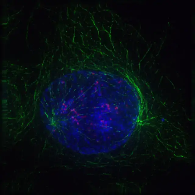
Microscopically visualization of Mitosis
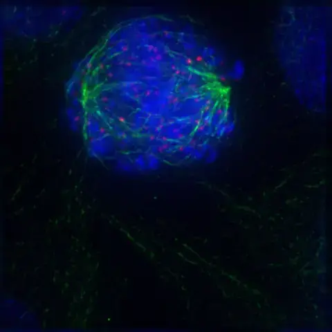
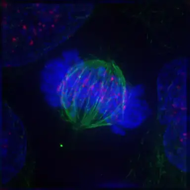

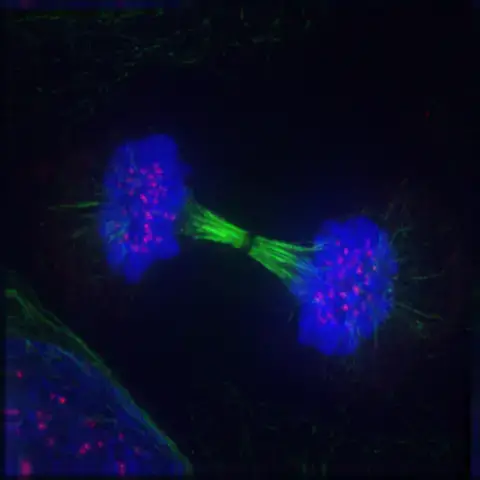
Functions of Mitosis
Mitosis, a fundamental cellular process, ensures the precise and equal distribution of genetic material from a parent cell to its daughter cells. This process is pivotal for maintaining the integrity of the chromosomal set, guaranteeing that each resultant cell inherits a chromosomal composition identical to that of the progenitor cell. Here’s a detailed exploration of the multifaceted functions of mitosis:
- Development and Growth:
- Description: Mitosis serves as the cellular mechanism underpinning the expansion of an organism’s cell count.
- Significance: It facilitates the transformation of a unicellular zygote into a multicellular organism. Moreover, as organisms mature, mitosis drives their growth by increasing the total number of cells.
- Cell Replacement:
- Description: Certain regions of organisms, such as the skin and the digestive tract, experience continuous cell loss. Mitosis compensates for this loss by generating new cells.
- Significance: This process ensures that cells lost to natural wear and tear, injury, or aging are promptly replaced. For instance, erythrocytes (red blood cells) have a limited lifespan, necessitating their periodic replacement through mitotic cell production.
- Regeneration:
- Description: Regeneration refers to the ability of organisms to restore lost or damaged body parts.
- Significance: Mitosis plays a central role in regeneration by producing the requisite cells for tissue repair and growth. A classic example is the starfish, which can regenerate amputated arms via mitotic cell divisions.
- Asexual Reproduction:
- Description: Asexual reproduction allows organisms to produce offspring without the fusion of gametes. Offspring produced in this manner are genetically identical to the parent.
- Significance: Mitosis is the cellular foundation of asexual reproduction, ensuring the offspring inherit an exact genetic copy of the parent. Organisms like the hydra utilize mitosis for budding, where surface cells undergo mitotic divisions to form a genetically identical bud. Similarly, in plants, mitotic divisions facilitate vegetative propagation, a form of asexual reproduction.
In essence, mitosis is indispensable for an array of biological processes, from individual cell function to the broader aspects of organismal growth, repair, and reproduction. Its role in ensuring genetic consistency across cell generations underscores its importance in the continuity and vitality of life.
Significance of Mitosis
Mitosis, a fundamental cellular division process, holds paramount significance in the life cycle of organisms, ensuring genetic continuity and cellular functionality. Here’s an elucidation of the multifaceted significance of mitosis:
- Developmental Role:
- Description: Mitosis is instrumental in the transformation of a zygote, a single cell, into a fully developed multicellular organism.
- Significance: This process ensures that as an organism develops from a fertilized egg to maturity, cells proliferate in a controlled and organized manner, preserving genetic integrity.
- Genetic Consistency:
- Description: One of the primary roles of mitosis is the equal distribution of chromosomes to the resultant daughter cells.
- Significance: This ensures that every cell in an organism possesses an identical set of chromosomes, maintaining the organism’s genetic homogeneity.
- Growth and Repair:
- Description: Mitosis underpins the growth of an organism by producing new cells. Additionally, it plays a crucial role in the repair and regeneration of tissues.
- Significance: Through mitosis, organisms can replace old, damaged, or dead cells, ensuring tissue integrity and functionality. For instance, the continuous replacement of gut epithelium and blood cells in animals is facilitated by mitotic divisions.
- Genomic Stability:
- Description: Mitosis ensures the preservation of the genome’s purity across generations of cells.
- Significance: Since mitosis does not involve genetic recombination or crossing over, the genetic content remains unaltered, ensuring stability and preventing anomalies.
- Asexual Reproduction and Vegetative Propagation:
- Description: Mitosis is the cellular mechanism underlying asexual reproduction in many organisms and vegetative propagation in plants.
- Significance: Through mitosis, organisms can reproduce without the need for gamete fusion, leading to offspring that are genetically identical to the parent. This mode of reproduction is especially advantageous in stable environments where genetic consistency is beneficial.
In summary, mitosis is a cornerstone of cellular biology, ensuring genetic continuity, facilitating growth, repair, and reproduction, and maintaining the stability and purity of the genome. Its role is indispensable in the life cycle of organisms, underscoring its evolutionary and biological importance.
Applications of Mitosis
Mitosis, the fundamental process of eukaryotic cell division, has been harnessed for a plethora of applications in molecular biology, biotechnology, and medicine. This process ensures the accurate replication and segregation of the genetic material, enabling the generation of two genetically identical daughter cells. Here’s an exploration of the pivotal applications of mitosis:
- Cloning:
- Definition: Cloning refers to the biotechnological technique aimed at producing genetically identical copies of cells or specific DNA sequences.
- Mitotic Role: The foundational principle of cloning is the amplification of organisms through mitosis, ensuring genetic uniformity.
- Applications: Cloning has been instrumental in various biological assays, including DNA fingerprinting, which aids in genetic mapping and forensic investigations.
- Tissue Culture:
- Definition: Tissue culture encompasses the in vitro cultivation of cells or tissues in a controlled environment, typically using specialized growth media.
- Mitotic Role: The exponential expansion of cells in tissue culture is underpinned by mitotic divisions, facilitating the generation of a substantial biomass from a limited initial sample.
- Extended Applications: Beyond mere tissue cultivation, this technique can be extrapolated to organ culture, enabling the growth of entire organs ex vivo, which holds promise for transplantation and regenerative medicine.
- Stem Cell Regeneration:
- Definition: Stem cells are undifferentiated cells characterized by their potential to differentiate into various specialized cell types and their capacity for self-renewal.
- Mitotic Role: Stem cells leverage mitosis for their proliferation, ensuring a consistent reservoir of these cells for therapeutic applications.
- Therapeutic Implications: The regenerative potential of stem cells, driven by mitotic divisions, offers a promising avenue for the treatment of degenerative diseases, tissue injuries, and organ failures. By directing stem cells towards specific lineages, it is possible to replace damaged or diseased tissues, heralding a new era in regenerative medicine.
In conclusion, the intricate process of mitosis, beyond its natural biological significance, has been ingeniously employed in various scientific and medical domains. Its applications, ranging from cloning to tissue regeneration, underscore the centrality of mitosis in contemporary biotechnological advancements and medical therapies.
Quiz
Which phase of mitosis is characterized by the condensation of chromosomes and the breakdown of the nuclear envelope?
a) Anaphase
b) Telophase
c) Prophase
d) Metaphase
[expand title=”Show answer” swaptitle=”Hide answer”] c) Prophase [/expand]
During which phase of mitosis do sister chromatids separate and move to opposite poles of the cell?
a) Prophase
b) Metaphase
c) Anaphase
d) Telophase
[expand title=”Show answer” swaptitle=”Hide answer”] c) Anaphase [/expand]
The alignment of chromosomes at the equatorial plane of the cell occurs during which phase?
a) Prophase
b) Metaphase
c) Anaphase
d) Telophase
[expand title=”Show answer” swaptitle=”Hide answer”] b) Metaphase [/expand]
Which structure is responsible for pulling the chromosomes apart during mitosis?
a) Centrioles
b) Lysosomes
c) Spindle fibers
d) Golgi apparatus
[expand title=”Show answer” swaptitle=”Hide answer”] c) Spindle fibers [/expand]
The reformation of the nuclear envelope around the chromosomes occurs during which phase?
a) Prophase
b) Metaphase
c) Anaphase
d) Telophase
[expand title=”Show answer” swaptitle=”Hide answer”] d) Telophase [/expand]
Which of the following is NOT a phase of mitosis?
a) Prophase
b) Cytokinesis
c) Metaphase
d) Anaphase
[expand title=”Show answer” swaptitle=”Hide answer”] b) Cytokinesis [/expand]
How many daughter cells are produced at the end of mitosis?
a) One
b) Two
c) Three
d) Four
[expand title=”Show answer” swaptitle=”Hide answer”] b) Two [/expand]
Which phase of mitosis is characterized by the decondensation of chromosomes and the reformation of the nuclear envelope?
a) Prophase
b) Metaphase
c) Anaphase
d) Telophase
[expand title=”Show answer” swaptitle=”Hide answer”] d) Telophase [/expand]
During which phase of mitosis do chromosomes become visible under a light microscope?
a) Prophase
b) Metaphase
c) Anaphase
d) Telophase
[expand title=”Show answer” swaptitle=”Hide answer”] a) Prophase [/expand]
Which organelle plays a crucial role in organizing the spindle fibers during mitosis in animal cells?
a) Mitochondria
b) Centrosome
c) Lysosome
d) Endoplasmic reticulum
[expand title=”Show answer” swaptitle=”Hide answer”] b) Centrosome [/expand]
FAQ
What is mitosis?
Mitosis is a process of cell division that results in two genetically identical daughter cells, each having the same number of chromosomes as the parent nucleus.
Why is mitosis important?
Mitosis is essential for growth, tissue repair, and regeneration in multicellular organisms. It also allows for the replacement of old or damaged cells.
How is mitosis different from meiosis?
While mitosis produces two genetically identical daughter cells, meiosis results in four genetically diverse daughter cells with half the number of chromosomes of the parent cell. Meiosis is responsible for producing gametes for sexual reproduction.
What are the main phases of mitosis?
The primary phases of mitosis are Prophase, Metaphase, Anaphase, and Telophase.
What happens during Prophase?
During Prophase, chromosomes condense and become visible, the nuclear envelope breaks down, and spindle fibers begin to form.
How do chromosomes align during Metaphase?
During Metaphase, chromosomes align at the cell’s equatorial plane, often referred to as the metaphase plate.
What is the significance of the Anaphase stage?
In Anaphase, sister chromatids separate and are pulled to opposite poles of the cell by the spindle fibers.
When does the cell start to divide into two during mitosis?
The cell begins to divide into two distinct cells during Telophase, the final phase of mitosis.
What role do spindle fibers play in mitosis?
Spindle fibers are essential for separating sister chromatids and ensuring that each daughter cell receives an equal number of chromosomes.
Can errors occur during mitosis?
Yes, errors can occur during mitosis, leading to conditions like aneuploidy, where cells have an abnormal number of chromosomes. Such errors are often associated with diseases like cancer.
References
- Sullivan, K. F. (2001). Mitosis. Encyclopedia of Genetics, 1224–1227. doi:10.1006/rwgn.2001.0839
- Teusel, F., Henschke, L., & Mayer, T. U. (2018). Small molecule tools in mitosis research. Methods in Cell Biology, 137–155. doi:10.1016/bs.mcb.2018.03.005
- Wadsworth, P., & Rusan, N. M. (2004). Mitosis. Encyclopedia of Biological Chemistry, 743–747. doi:10.1016/b0-12-443710-9/00094-6
- Schatten, H. (2013). Mitosis. Brenner’s Encyclopedia of Genetics, 448–451. doi:10.1016/b978-0-12-374984-0.00962-1
- Schaefer, E., Belcram, K., Uyttewaal, M., Duroc, Y., Goussot, M., Legland, D., Laruelle, E., de Tauzia-Moreau, M.-L., Pastuglia, M., & Bouchez, D. (2017). The preprophase band of microtubules controls the robustness of division orientation in plants. Science, 356(6334), 186–189. https://doi.org/10.1126/science.aal3016
- prometaphase | Learn Science at Scitable. (2014). Nature.Com. https://www.nature.com/scitable/definition/prometaphase-281/
- De Souza, C. P. C., & Osmani, S. A. (2007). Mitosis, Not Just Open or Closed. Eukaryotic Cell, 6(9), 1521–1527. https://doi.org/10.1128/ec.00178-07