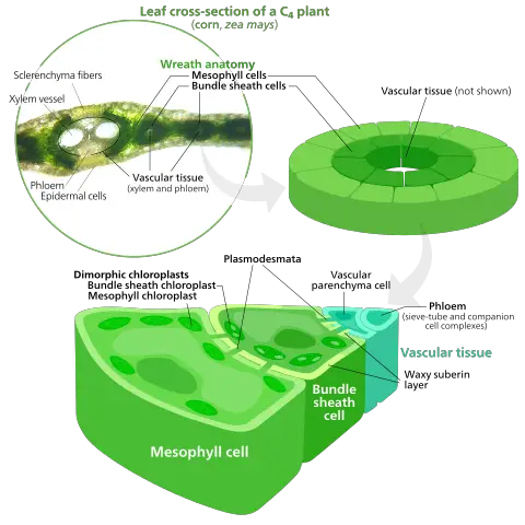
Definition of Mesophyll Cells
The mesophyll cell is a group of highly differentiated cells which make up the mesophyll layer in plant leaves. The mesophyll layer in the leaves of dicotyledonous plant species is made up two types of cells: the spongy, and the palisade. This makes the mesophyll a site of photosynthesis.
These are some of the most important characteristics.
- Found between the upper and below epidermis
- Leaves’ inner tissues are made up of this
- Variation in shape
- Form a type of ground tissue
The mesophyll is a combination of two Greek words: mesos (which means middle) and phyllo (which means leaf).
While mesophyll tissue consists of two layers (spongy, palisade) of cells, monocots’ mesophyll is mostly composed of isodiametric (cells that are spherical and polyhedral).

Origin of Mesophyll Cells
- Mesophyll cells are the inner mesophyll tissue in a leaf’s mesophyll. These cells remake up the cortex which is made up of parenchyma cells.
- In vascular plants, the mesophyll is a product of a group of cells called ground meristematic, which are themselves made by cells from the apical, or lateral meristem.
- One group of stem cells is found in plants. It is located in the meristem (meristematic tissues). As such, they divide to create cells that can be differentiated to perform different functions in plants.
- Ground meristem cells divide and differentiate to form a variety of tissues such as the cortex, pith, and pith. They produce the parenchyma mesophyll cells (palisade, spongy mesophyll) which are involved in photosynthesis. This can be represented as:
Location
- It’s important that you look at the overall structure of a leaf in order to understand the mesophyll-cell arrangement and location.
- A leaf consists of many tissues. These include the epidermis, mesophyll layer, and vascular tissue.
- The epidermis is made up of epidermal cells and covers the leaf’s upper (adaxial), as well as lower (abaxial), surfaces. Although the epidermis can be considered a separate tissue, it protects the cells from material that may enter or leave them.
- The mesophyll (ground tissues) is located between upper and lower epidermis. This is where, particularly in dicots and dicots, mesophyll is composed of two types of cells. They are the palisade parenchyma cells just below the epidermis as well as the spongy Parenchyma s cells, which are located below the palisade cell and above the lower epidermis.
- The mesophyll layers contain the vascular tissue. This is where they help to move material to the cells. The mesophyll layers, which are mesophyll-rich cells, are sandwiched between the lower and upper epidermis, with the vascular bundles xylem (and phloem), running between their cells.

The two types of cells that make up the mesophyll layer are They differ in morphology, and serve different purposes.
Structure of Mesophyll cells
As mentioned earlier, there are two types of cells in the mesophyll layers. These are:
1. Palisade Cells
- Palisade cells form part of mesophyll tissues in plant leaves. This layer (palisade) is located under the upper epidermis. It consists of cells that are columnar/cylindrical.
- Apart from the nucleus, palisade cells also have a cell membrane, large vacuole and chloroplasts. The cells are arranged vertically below the epidermis. There are also small separations that allow different materials to flow between them.
- The structure and arrangement in which palisade cell are placed in mesophyll tissues plays an important role in photosynthesis. Palisade-shaped cells have a lot of chloroplasts.
- Palisade cells can absorb more light needed for photosynthesis, in addition to all these characteristics. As mentioned above, palisade is located under the epidermis. This thin layer of cells allows palisade to absorb light needed for photosynthesis.
Also, the structure/morphology and photosynthesis of palisade cell cells are beneficial to chloroplasts. These include:
Chloroplast movement
- It has been demonstrated that light conditions can cause the movement of chloroplasts inside a cell. Low light conditions (low light levels) are conditions in which the amount of sunlight available is low. In these conditions, chloroplasts can move along the cell wall and accumulate so that they are parallel to incident rays.
- The ability to move within the cell to avoid being too exposed to too much light has been demonstrated. The palisade cell’s long shape allows for sufficient space for chloroplasts to move within the cell (moved via specific structural proteins).
Large vacuole
- Palisade cells are able to move due to their shape, however, the large vacuole in the middle of the cell prevents chloroplasts from reaching the membrane. This allows for light to reach the chloroplasts easily and photosynthesis can take place.
2. Spongy Mesophyll Cells
The cells of the spongy layer are located above the palisade tissues and the lower epidermis. The cells of spongy mesophyll tissue are more spherical than those of the palisade layers.
The microscope shows between 4 to 6 layers of spongy, mesophyll cells beneath the palisade cells. Spongy mesophyll cells contain organelles similar to palisade cell nucleus, a vacuum, and a cell membrane along with chloroplasts.
However, the number of these chloroplasts is lower than that found in palisade cell cells. Some of the leaves’ spongy cell bodies have crystal inclusions. Crystal inclusion (e.g. druse Crystals are shorter/smaller than other cells in the same region of the leaf.
The thickness and density of the spongy parenchyma ranges between 1.5 to 2 times that of palisade. There are three versions of the spongy Parenchyma, depending on the plant:
- A typical parenchyma cell
- Palisade-like spongy cells
- Aerenchymatous spongy cells
Although spongy cells of mesophyll do not have as many chloroplasts than palisade cells they still play an important role in photosynthesis. Because they are loosely packed, photosynthesis can be enhanced by gas exchange.
Mechanism in Photosynthesis
Palisade Cells
- Palisade cell is a parenchyma type that contains the majority of the chloroplasts present in plant leaves. Palisade cells, which are located below the epidermis’s upper layer, are well placed to absorb the light necessary for photosynthesis.
- Additionally, the location of these chloroplasts ensures that carbon dioxide needed for photosynthesis does NOT have to travel long distances to reach the chloroplasts.
- Also, because the palisade cells are below the epidermis, which allows light and water to reach the cells easily and there are small spaces between them, these spaces ensure that the entire cell is in direct contact with the air. Palisade cells’ thin cell walls allow gases to easily diffuse.
- Due to the conditions created by palisade cells, the chloroplasts can easily access the vital material needed for photosynthesis to occur
- Here, the photosynthesis pigment called chlorophyll is absorbed by wavelengths of sunlight. This provides the energy needed for the photosynthetic reaction in which carbon dioxide and water produce a sugarmolecule and oxygen.
Spongy Cells
- Spongy cells have some chloroplasts just like palisade cells. Spongy cells contain some photosynthesis, just like palisade cells. However, they are found deeper within the leaf than the palisade tissue and the upper epidermis.
- This is a disadvantage in terms of photosynthesis because light can’t penetrate into this region. Spongy cells don’t get enough sunlight to enable photosynthesis to take place optimally.
- Spongy cells aren’t well suited to photosynthesis, but they are great for gaseous exchange. Spongy cells have a looser packing above the lower epidermis, which makes them ideal for gaseous exchanging.
- These gases, such as carbon dioxide, can enter the leaf through small holes in the epidermis. The mesophyll also produces oxygen through photosynthetic processes.
- This region of the cell contains loosely packed cells (spongy) that allow for exchange of gases. In this region, oxygen can be released while carbon dioxide can be used for photosynthesis.
- Photosynthesis occurs in spongy cells with high light intensities.
Microscopic observation of Mesophyll Cells
It is possible to observe the architecture of the Thylakoid membrane and mesophyll cell structures using an electron microscope. For the purpose of viewing mesophyll cells, however, a light microscope suffices.
Requirements
- Cassava cork
- Microscope – compound microscope
- Alcohol -30 percent, 50 percent, 70 percent, and 96 percent
- Safranin-O
- Clamp-on hand sliding microtome
- Young leaf
- Microscope glass slide and cover slips
- Preservation liquid (consisting of 70 percent alcohol and glycerin)
Procedure
To cut thin sections from various samples, a vibratome may be used. Some samples, such very thin leaves may require alternative cutting methods.
- Cassava corks can be used to hold and cut thin leaves. The cassava stem, which is young and thin, is cleaned and dried.
- Insert the cassava Cork (with the sample in between) into the Microtome hole. Using the blade, cut the cork to create several transversal pieces – Try to get thin slices (almost transparent).
- Carefully collect the section using a sharp needle from a paintbrush’s tip.
- Use a graded series to dehydrate the sections you have obtained. Each section should be between 30 percent, 50 percent and 70 percent.
- Discard the alcohol from the glass and check that the alcohol has been drained completely.
- Combine 70 percent alcohol and 70% glycerin. This is a preservation solution.
- Slide onto a clear glass slide. Cover the slide with a cover slip.
Observation
Well prepared slices will show preserved mesophyll cells when viewed under the microscope. This will make the epidermis appear thinner and darker than usual, while the scattered cells of spongy cells beneath well-organized palisade cell bodies will be visible.