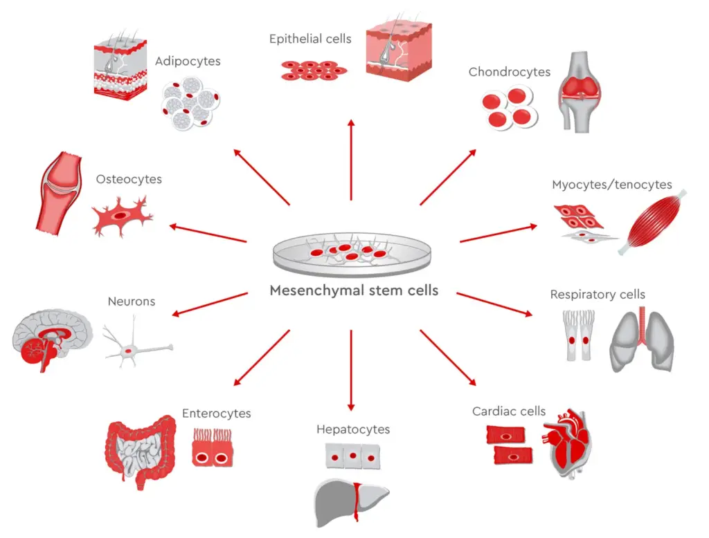
Mesenchymal stem cell (MSCs) are also referred to as mesenchymal stromal cell (also known as medicinal signaling cells) are stromal cells with multi-potency which can transform into a variety of cells, including osteoblasts (bone cells) as well as the chondrocytes (cartilage cells) and myocytes (muscle cells) and Adipocytes (fat cells that contribute to the adipose tissue of the marrow).
Definition of Mesenchymal stromal cells
The terms mesenchymal stromal cell (MSC) and Marrow stromal cells have been employed interchangeably for several years, neither of them is adequate in its description:
- Mesenchyme is an embryonic connective tissue that originates from the mesoderm. It differentiates into connective and hematopoietic tissue, while MSCs don’t differentiate into the hematopoietic cells.
- Stromal cell are the connective tissue-like cells that create the supporting structure within which the functional cells of the tissue are located. While this is a valid description for the function of MSCs, the word is not sufficient to describe the recent findings of their roles in the process of repairing tissue.
- The term covers multipotent cells that originate from non-marrow tissueslike the placenta, blood from the umbilical cord and adipose tissue. They also include corneal stroma and the tooth pulp from teeth that are deciduous (baby) teeth.The cells are not able to regenerate whole organs.

Structure of Mesenchymal stromal cells
Mesenchymal stem cells can be described in morphology by a tiny cell body that has some cell processes that are lengthy and thin. The cell body is comprised of an enormous, round nucleus with a prominent nucleolus which is covered with finely distributed chromatin molecules that give the nucleus an attractive appearance. The rest of the cell’s body has a small quantity of Golgi apparatus and rough endoplasmic-reticulum polyribosomes and mitochondria. Cells, that are large and thin, can be well dispersed. The surrounding extracellular matrix is populated with some reticular fibrils, but is not populated by the other collagen fibrils. These distinct morphological characteristics of mesenchymal stem cell can be observed without labeling using Live cell imaging.
Location of Mesenchymal stromal cells
1. Bone marrow
Bone marrow is the primary source of MSCs and is the most commonly used. The bone stem cells from the bone marrow do not aid in the development of blood cells, and therefore don’t express the markers for hematopoietic stem cells, CD34. They are often called bone marrow stem cells.
2. Cord cells
The earliest and most primitive MSCs are derived from umbilical cord tissue including Wharton’s Jelly and cord blood. But MSCs are at a higher level in Wharton’s jelly as when compared with cord blood which is a significant source of the hematopoietic stem cells. The umbilical cord can be found following the birth. It is typically removed and is not a risk for the collection. MSCs are a good source for collection. MSCs could prove to be an effective resource for MSCs for clinical use because of their basic properties and rapid growth rate.
They also have a number of advantages over MSCs derived from bone marrow. Adipose tissues-derived MSCs (AdMSCs) as well as to being more dependable and easier to extract than bone marrow-derived MSCs are available in greater quantities.
3. Molar cells
The tooth bud that is developing in the mandibular 3rd molar is an excellent resource of MSCs. Although they are described as multipotent the possibility is that they could be also pluripotent. They will eventually develop enamel dentin, blood vessels as well as dental pulp and nervous tissues. The stem cells are capable of transforming into cardiomyocytes, chondrocytes melanocytes, and hepatocytes-like cells in the laboratory.
4. Amniotic fluid
Stem cells can be found in the amniotic fluid. Up to 1 out of 100 cells collected in amniocentesis contain pluripotent mesenchymal stem cells.
Function of Mesenchymal stromal cells
1. Differentiation capacity
MSCs possess a remarkable capacity for self-renewal, while maintaining their multipotency. Recent research suggests that b-catenin through its the regulation of EZH2 is a key protein that helps maintain their “stemness” of MSC’s. The most reliable test to prove multipotency is the differentiation of cells into osteoblasts, adipocytes , and myocytes, as well as chondrocytes.
MSCs are known to differentiate into neuron-like cells, however it is unclear if the MSC-derived neuron are actually functional. The extent to which the culture differentiates varies between individuals, as does the method by which differentiation is initiated, e.g., chemical as opposed to. mechanical. It is unclear if this is due to a distinct amount of “true” progenitor cells in the culture or due to the different capacity of differentiation in the individual’s progenitors. The ability of cells to multiply and differentiate is believed to decline with time of life for the donors and also the length of duration of the culture. It is also unclear if this is due to a decline in the amount of MSCs or an alteration to existing MSCs isn’t known.
2. Immunomodulatory effects
MSCs exert a positive effect on the innate immune system as well as specific cells. MSCs make a number of immunomodulatory substances such as prostaglandin E2 (PGE2), Nitric oxide, indoleamine 2,3 dioxygenase (IDO) and interleukin 6 (IL-6) and other surface markers like FasL and PD-L1.
MSCs can have an impact on macrophages, neutrophils mast cells, NK cells, and dendritic cells within the innate immune system. MSCs can move towards the site of injury, and change their polarization to PGE2 macrophages that exhibit the M2 phenotype , which is distinguished with an anti-inflammatory property. Additionally, PGE2 inhibits the ability of mast cells to degranulate, and to produce TNF-a. The proliferation and cytotoxic activities of NK cells are affected through PGE2 along with IDO. MSCs also inhibit the activity of NK cell receptors, namely NKG2D, NKp44 and NKp30. MSCs block the apoptosis and respiratory flare of neutrophils through the production of cytokines IL-6 as well as the IL-8. Expression and differentiation of the dendritic cell surface markers is blocked by PGE2 and IL-6 of MSCs. The immunosuppressive properties of MSC additionally depend on the IL-10 receptor, however it’s unclear if they generate it by themselves or if they only induce other cells to make it.
MSC produces adhesion molecules ICAM-1 and VCAM-1 that allow T-lymphocytes attach to their surface. In turn, MSC may affect them through molecules with a short duration and their effects are located in the area that of the cells. They include nitric oxide PGE2, HGF, and activation of PD-1’s receptor. MSCs inhibit the proliferation of T cells during G0 to G1 cycle stages and also decrease levels of IFNg from Th1 cells as well as increasing the expression of IL-4 in Th2 cells. MSCs also reduce the proliferation of B-lymphocytes in between the G0 and G1 cell cycle phases.
3. Antimicrobial properties
MSCs produce antibiotic peptides (AMPs) like human cathelicidin, b-defensins such as lipocalin 2, hepcidin and b-defen. These peptides, along and the indoleamine enzyme 2,3 dioxygenase (IDO) are responsible for their broad-spectrum antibacterial function of MSCs.

- Text Highlighting: Select any text in the post content to highlight it
- Text Annotation: Select text and add comments with annotations
- Comment Management: Edit or delete your own comments
- Highlight Management: Remove your own highlights
How to use: Simply select any text in the post content above, and you'll see annotation options. Login here or create an account to get started.