What is Lymph Nodes?
- Lymph nodes are small, solid structures located at various points along the lymphatic system, including the groin, armpit, and mesentery. They play a crucial role in the immune response against foreign antigens that enter the tissues. Lymph nodes contain different types of immune cells, including T and B lymphocytes, as well as accessory cells. These cells work together to mount immune responses and defend the body against harmful invaders.
- The lymphatic system consists of a network of vessels that transport lymph, a clear fluid that carries nutrients, waste products, and immune cells throughout the body. Lymph nodes are strategically positioned along these vessels. They act as filters, removing foreign particles, such as bacteria, viruses, and even cancer cells, from the lymph before it returns to the bloodstream. This filtration process helps prevent the spread of infections and aids in the recognition and elimination of abnormal cells.
- Each lymph node is kidney-shaped and enclosed within a fibrous capsule. It is composed of an outer cortex and an inner medulla. The cortex contains densely packed lymphocytes, primarily B cells, which produce antibodies that target specific antigens. The medulla contains fewer lymphocytes but houses other types of immune cells that support the immune response.
- Lymph nodes can become inflamed or enlarged in response to various diseases. This can range from minor infections, like throat infections, to more serious conditions, including certain types of cancers. The condition of lymph nodes is crucial in cancer staging, which helps determine the appropriate treatment and predicts the prognosis. When lymph nodes are inflamed or enlarged, they may feel firm or tender to the touch.
- In summary, lymph nodes are essential components of the immune system and the lymphatic system. They act as filters, removing foreign particles and abnormal cells from the lymph. Lymph nodes contain different types of immune cells and play a vital role in mounting immune responses against invading antigens. Monitoring the condition of lymph nodes is important for diagnosing and managing diseases, including cancer.
Definition of Lymph Nodes
Lymph nodes are small organs in the lymphatic system that filter lymph and play a critical role in the body’s immune response.
Location of Lymph Nodes
- Lymph nodes are distributed throughout the body, with higher concentrations near and within the trunk, and they are organized into groups. In adults, there are approximately 450 lymph nodes. Some lymph nodes can be felt when they become enlarged, such as the axillary lymph nodes under the arm, the cervical lymph nodes in the head and neck region, and the inguinal lymph nodes near the groin crease.
- While many lymph nodes are located within the trunk, adjacent to other major structures in the body, such as the paraaortic lymph nodes and tracheobronchial lymph nodes, the specific lymphatic drainage patterns can vary between individuals. Additionally, the distribution of lymph nodes can be asymmetrical on each side of the body.
- It’s important to note that there are no lymph nodes within the central nervous system (CNS), which is separated from the rest of the body by the blood-brain barrier. However, lymph from the meningeal lymphatic vessels in the CNS does drain to the deep cervical lymph nodes in the neck region.
Size of Lymph Nodes
The size of lymph nodes can vary depending on their location within the body. Here are some general guidelines for the upper limits of lymph node sizes in different regions of adults:
- Generally: 10 mm
- Inguinal: 10-20 mm
- Pelvis: 10 mm for ovoid lymph nodes, 8 mm for rounded lymph nodes
- Neck (non-retropharyngeal): 10 mm
- Jugulodigastric lymph nodes: 11 mm or 15 mm
- Retropharyngeal: 8 mm
- Lateral retropharyngeal: 5 mm
- Mediastinum (generally): 10 mm
- Superior mediastinum and high paratracheal: 7 mm
- Low paratracheal and subcarinal: 11 mm
- Upper abdominal:
- Retrocrural space: 6 mm
- Paracardiac: 8 mm
- Gastrohepatic ligament: 8 mm
- Upper paraaortic region: 9 mm
- Portacaval space: 10 mm
- Porta hepatis: 7 mm
- Lower paraaortic region: 11 mm
These measurements represent the upper limits of normal lymph node sizes in adults and can be used as a reference for assessing lymph node enlargement or abnormalities. It’s important to note that these values are approximate and can vary depending on individual factors.
Structure of Lymph Nodes
Lymph nodes have a distinct structure that plays a crucial role in their function. Here is an overview of the structure of lymph nodes:
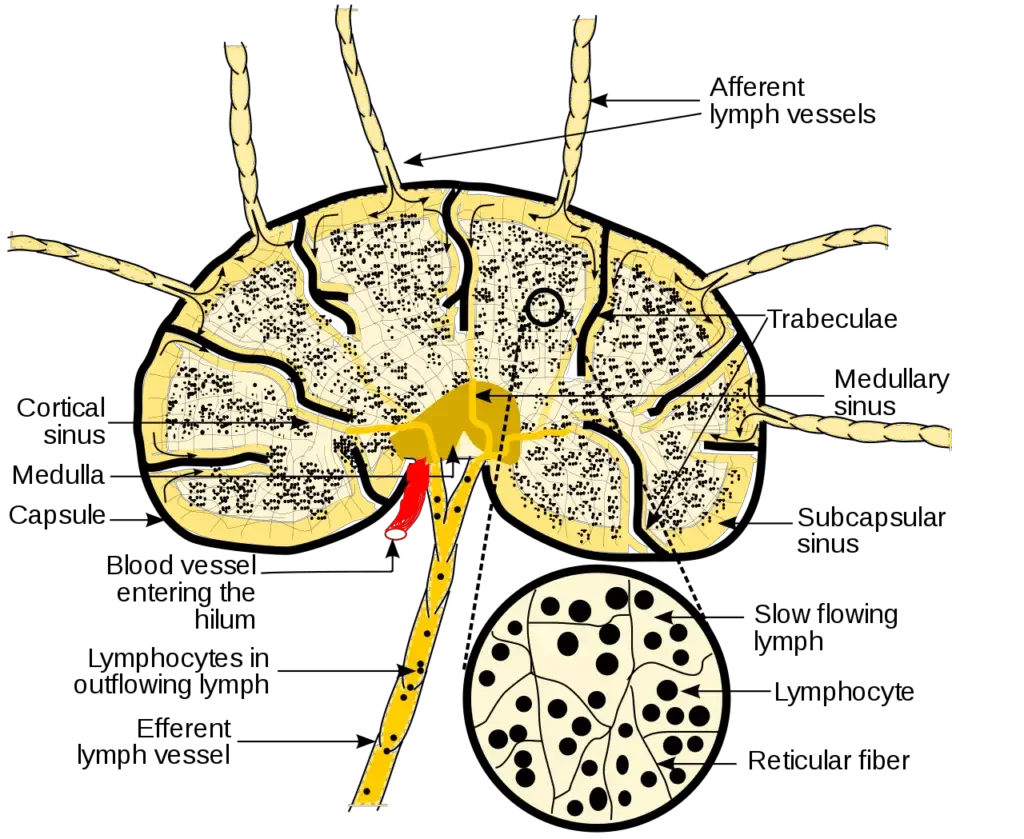
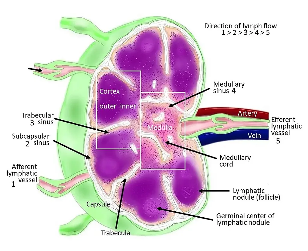
- Capsule: The lymph node is surrounded by a dense connective tissue capsule that sends trabeculae (thin partitions) into the node, radiating towards the center.
- Subscapular Sinus: The subcapsular sinus is the space between the capsule and the cortex of the lymph node. It serves as a pathway for lymphatic fluid and is traversed by reticular fibers and cells. The subcapsular sinus receives the afferent lymphatic vessels, connects with the trabecular sinuses, and ultimately joins the medullary sinus in the medulla of the lymph node.
- Cortex: The cortex is the layer beneath the subcapsular sinus and is composed of two parts: the outer cortex and the paracortex. The outer cortex, also known as the B-cell layer, contains B-cells organized into follicles. Upon antigenic stimulation, these follicles can develop germinal centers. Surrounding the germinal centers is a mantle zone consisting of resting B-cells and dendritic cells. The paracortex, or T-cell layer, is rich in T-cells that interact with dendritic cells and is enriched with CCR7 chemokines.
- Medulla: The medulla is the innermost layer of the lymph node. It contains large blood vessels, sinuses, and medullary cords. Medullary cords contain plasma cells that secrete antibodies, B-cells, and macrophages. Medullary sinuses, also known as sinusoids, are vessel-like spaces that separate the medullary cords. They receive lymph from the trabecular sinuses and cortical sinuses and contain reticular cells and histocytes. The medullary sinuses drain lymph into the efferent lymphatic vessels.
Lymph nodes are encapsulated structures that range in size from 2 to 10 mm. They are spherical in shape and consist of reticular and lymphatic tissues containing mainly lymphocytes and macrophages. Each lymph node has a concave surface called the hilum, where an artery enters, a vein and the efferent lymph vessel leave. Lymph nodes are found in various locations throughout the body, including the neck, collarbone, armpit, and groin. They can be superficial or deep, and groups of lymph nodes are present in these regions.

Lymph Circulation in Lymph Nodes
Lymph circulation within lymph nodes follows a specific pathway. Here is an overview of the lymph circulation in lymph nodes:
- Afferent Lymphatics: Lymph, which contains fluid, waste products, and immune cells, arrives at the lymph node through a network of afferent lymphatic vessels. These vessels bring lymph from the surrounding tissues or from a preceding lymph node in the chain.
- Subcapsular Sinus: Upon entering the lymph node, the lymph flows into the subcapsular sinus, which is the space between the capsule and the cortex of the lymph node. The subcapsular sinus allows the lymphatic fluid to pass through and facilitates its transportation within the lymph node.
- Cortex: From the subcapsular sinus, the lymph moves into the cortex of the lymph node. It flows around the follicles, which are areas rich in B-cells, and continues into the paracortical area. The paracortex is enriched with T-cells and is where interactions between T-cells and dendritic cells occur.
- Medulla: After passing through the cortex, the lymph progresses into the medulla of the lymph node. Within the medulla, the lymph circulates within the medullary sinuses or sinusoids. These are vessel-like spaces that separate the medullary cords, which contain antibody-secreting plasma cells, B-cells, and macrophages.
- Efferent Lymphatics: Lymph in the medullary sinuses drains into efferent lymphatic vessels. These vessels carry the lymph away from the lymph node and join larger lymphatic vessels. Eventually, the lymph flows through the lymphatic system and returns to the bloodstream.
The circulation of lymph through lymph nodes allows for the filtration and processing of lymphatic fluid. As lymph passes through the lymph node, it comes into contact with immune cells, such as B-cells, T-cells, and dendritic cells, which help mount an immune response against any pathogens or foreign substances present in the lymph. The lymph node serves as a critical site for immune surveillance and activation before the lymph is returned to the bloodstream.

Clinical Significance
Lymph nodes have significant clinical importance in various conditions. Here are some key clinical significances related to lymph nodes:
- Swelling (Lymphadenopathy): Swelling or enlargement of lymph nodes, known as lymphadenopathy, can indicate various underlying conditions. It may be caused by drug reactions, autoimmune diseases, infections, tumors, lymphoma, or leukemia. Localized enlargement of lymph nodes can be suggestive of tumors, which may be painful. Generalized swelling of lymph nodes often indicates the risk of infection or autoimmune disease.
- Lymphedema: Lymphedema refers to the swelling of lymph tissues due to inadequate drainage by the lymphatic system. Primary lymphedema is a congenital condition where lymph nodes are either underdeveloped or absent. Secondary lymphedema occurs when lymph nodes are surgically removed, such as in breast cancer treatment, or damaged by radiation therapy. Lymphedema can also occur due to parasitic infections.
- Cancer: Lymph nodes can be affected by primary cancers of the lymphatic tissue or secondary cancers originating from other body parts. Primary cancers of the lymphatic tissue are called lymphomas, which include both Hodgkin and non-Hodgkin lymphomas. Lymphomas primarily involve B-cells and can manifest as painless and slow-growing masses or painful nodes that undergo sudden enlargement.
Secondary cancers from other body parts often metastasize to the lymph nodes, and the presence of cancer cells in the lymph nodes is important for diagnosis, staging, and treatment planning. Lymph nodes are often examined through imaging and biopsy to determine the extent of cancer spread.
Functions of Lymph Nodes
Lymph nodes play important functions in the immune system and are involved in the following processes:
- Filtration: The primary role of lymph nodes is to filter the lymph fluid that circulates through them. Lymph nodes act as checkpoints, removing foreign particles, microbes, damaged cells, and tumor cells from the lymph. This helps in maintaining the quality and integrity of the lymphatic system and preventing the spread of infection or cancer.
- Immune Response: Lymph nodes are crucial for initiating an immune response against trapped microbes and antigens. They contain specialized white blood cells called lymphocytes, including B cells and T cells. B cells produce antibodies, while T cells play a role in cell-mediated immunity. Within the lymph nodes, these lymphocytes interact with antigen-presenting cells, such as dendritic cells, to recognize and respond to specific antigens. This immune response helps in combating infections and mounting a defense against foreign substances.
- Antibody Production: Lymph nodes are sites where B cells undergo a process known as antibody production. B cells, when stimulated by antigens, multiply and differentiate within the lymph nodes. They develop into plasma cells, which are antibody-secreting cells. Antibodies, also called immunoglobulins, are proteins that specifically bind to antigens and help in neutralizing or eliminating them from the body. Lymph nodes play a crucial role in supporting the production of these antibodies, which are essential for immune defense.
- Lymphocyte Development and Activation: Lymph nodes provide an environment for the development and activation of lymphocytes. Naive lymphocytes, which are immature and have not encountered antigens, enter the lymph nodes through specialized high endothelial venules. Within the lymph nodes, B cells and T cells interact with antigen-presenting cells and receive signals that activate them. B cells can differentiate into plasma cells or memory B cells, while T cells undergo activation and differentiation to perform their specific immune functions.
- Lymphatic Drainage: Lymph nodes are part of the lymphatic drainage system. They receive lymph fluid from afferent lymphatic vessels, which carry lymph from various tissues and organs. Within the lymph nodes, the lymph fluid is filtered and processed before it exits through efferent lymphatic vessels. This drainage system helps in maintaining fluid balance, removing waste products, and transporting immune cells throughout the body.
Overall, lymph nodes play a crucial role in filtering lymph, initiating immune responses, producing antibodies, and supporting the development and activation of lymphocytes. Their strategic locations and functions make them vital components of the immune system and important targets for clinical evaluation and diagnosis.
FAQ
What are lymph nodes?
Lymph nodes are small, bean-shaped structures located throughout the body that are part of the lymphatic system, a network of vessels and organs involved in immune function and fluid balance.
What is the function of lymph nodes?
The primary function of lymph nodes is to filter lymph fluid and help in the immune response. They trap and destroy foreign particles, microbes, damaged cells, and cancer cells, and they also produce immune cells and antibodies.
Where are lymph nodes located?
Lymph nodes are present throughout the body, concentrated near the trunk, and arranged in groups. Some commonly known locations include the neck, armpits, groin, and abdomen. However, lymph nodes can be found in various other regions as well.
How can I feel my lymph nodes?
In some cases, lymph nodes can be felt when they are enlarged. Common areas where lymph nodes are palpable include the neck, underarms, and groin. Gently pressing these areas with your fingers may allow you to feel small, round, or bean-shaped structures beneath the skin.
What causes lymph nodes to swell?
Lymph nodes can swell or become enlarged in response to various factors such as infections (e.g., cold, flu, strep throat), immune reactions, autoimmune diseases, drug reactions, tumors, lymphoma, or leukemia. The specific cause of swelling depends on individual circumstances and requires medical evaluation.
Should I be concerned if my lymph nodes are swollen?
Swollen lymph nodes can be a sign that your body is fighting off an infection or responding to another condition. In many cases, the swelling subsides as the underlying cause resolves. However, persistent or rapidly growing swollen lymph nodes, especially in the absence of an apparent cause, should be evaluated by a healthcare professional.
Can lymph nodes be cancerous?
Yes, lymph nodes can be affected by cancer. Primary cancers originating in the lymph nodes are called lymphomas, which include Hodgkin lymphoma and non-Hodgkin lymphoma. Lymph nodes can also be involved in secondary cancers, where cancer from other body parts spreads to the lymph nodes through metastasis.
What is lymphedema?
Lymphedema is a condition characterized by the swelling of lymphatic tissues due to impaired drainage by the lymphatic system. It can occur when lymph nodes are underdeveloped, absent, or surgically removed, such as in breast cancer treatment or due to radiation therapy. Parasitic infections can also cause lymphedema.
How are lymph nodes evaluated clinically?
Clinically, lymph nodes are evaluated through physical examination, where a healthcare professional assesses their size, consistency, tenderness, and mobility. Depending on the findings and clinical suspicion, additional tests such as imaging studies, biopsies, or blood tests may be conducted to determine the underlying cause of lymph node abnormalities.
When should I seek medical attention for lymph node concerns?
It is recommended to seek medical attention if you notice persistent, rapidly growing, or painful lymph node swelling, especially if there are other concerning symptoms present. A healthcare professional can assess your condition, perform necessary evaluations, and provide appropriate guidance or treatment based on the underlying cause.
References
- Bujoreanu I, Gupta V. Anatomy, Lymph Nodes. [Updated 2022 Jul 25]. In: StatPearls [Internet]. Treasure Island (FL): StatPearls Publishing; 2023 Jan-. Available from: https://www.ncbi.nlm.nih.gov/books/NBK557717/
- Tuitui, R., & Suwal, D. S. (2010). Human Anatomy and Physiology. Kathmandu: Vidyarthi Prakashan.
- Lydyard, P.M., Whelan,A.,& Fanger,M.W. (2005).Immunology (2 ed.).London: BIOS Scientific Publishers.
- Playfair, J., & Chain, B. (2001). Immunology at a Glance. London: Blackwell Publishing.
- https://www.verywellhealth.com/understanding-the-purpose-of-lymph-nodes-2249122
- https://training.seer.cancer.gov/anatomy/lymphatic/components/nodes.html
- Text Highlighting: Select any text in the post content to highlight it
- Text Annotation: Select text and add comments with annotations
- Comment Management: Edit or delete your own comments
- Highlight Management: Remove your own highlights
How to use: Simply select any text in the post content above, and you'll see annotation options. Login here or create an account to get started.