Inverted Microscope – Principle, Procedure, Parts, Uses

What is an Inverted Microscope?

  • An inverted microscope, as its name suggests, is a specialized light microscope with its components arranged in an inverted manner. This unique design is characterized by the placement of the light source and the condenser lens above the specimen stage, pointing downwards.
  • Conversely, the objectives and the turret are situated below the stage, directing their focus upwards. This arrangement is in stark contrast to the conventional microscope design where the objective lenses are positioned above the stage and the condenser, along with the light source, are located below.
  • The inception of the inverted microscope can be traced back to 1850. It was the brainchild of J. Lawrence Smith, a distinguished faculty member of the Medical College of Louisiana.
  • The primary motivation behind this innovative design was to facilitate the viewing of specimens from a bottom-up perspective, as opposed to the top-down viewpoint offered by traditional microscopes. Therefore, the term “inverted” aptly describes this reversal in design and functionality.
  • Besides its unique construction, the inverted microscope is renowned for its advanced capabilities, especially in the realm of digital microscopy. The Digital Inverted Microscope, a modern iteration of this instrument, is fortified with cutting-edge video and image processing technologies.
  • This makes it an invaluable tool for meticulous laboratory cell analyses, as well as for capturing live-cell images and recordings. The microscope offers a magnification range between 100X and 400X, providing researchers with detailed insights into their specimens.
  • Furthermore, the Digital Inverted Microscope is equipped with a display screen or can be connected to a microscope camera, enabling users to view the specimen’s image with clarity and precision. It boasts a double-layer mechanical stage, complete with adjustable knobs both horizontally and vertically.
  • This feature, coupled with its three-sized petri dish trays, allows for the observation of a diverse array of samples. Data transfer is also streamlined with the inclusion of a 2.0 USB interface, ensuring rapid and efficient data exchange.
  • In terms of its core components, the inverted microscope’s light source and condenser lens play pivotal roles. The condenser lens, positioned above the specimen, is tasked with concentrating the light onto the sample. Then, the objective, located beneath the stage, focuses this light to generate a clear and detailed image of the specimen.
  • In conclusion, the inverted microscope stands as a testament to the evolution of microscopy. Its design, which emphasizes function and precision, has paved the way for groundbreaking discoveries in the field of biology and beyond.

Inverted Microscope Definition

An inverted microscope is a specialized light microscope where the light source and condenser are positioned above the specimen stage, while the objectives and turret are below the stage, allowing for the viewing of specimens from a bottom-up perspective. This design is opposite to that of conventional microscopes.

Principle of Inverted Microscope

The inverted microscope operates on foundational principles akin to those of the traditional upright light microscope. Essentially, both types of microscopes utilize light rays to magnify and project an image of the specimen for observation. However, the inverted microscope boasts a distinct configuration that sets it apart from its upright counterpart.

In the design of the inverted microscope, the light source and the condenser are strategically positioned above the specimen stage, directing light rays downwards onto the specimen. The primary role of the condenser lens, located above the specimen, is to concentrate and focus the light onto the specimen, ensuring optimal illumination. Following this, the specimen, which is placed on a spacious stage, interacts with the light.

Subsequently, the objectives, which are situated below the stage and oriented upwards, capture the light that has passed through the specimen. These objectives magnify the image, channeling it towards the ocular lens. Through a series of optical components, including mirrors, the image is further refined and directed towards the viewer’s eye.

A pivotal aspect of the inverted microscope’s functionality is the observation of cells or specimens through the base of the cell culture apparatus. For successful observation using an inverted microscope, the bottom of the culture vessel must possess superior optical characteristics. This ensures clarity and precision in viewing. Therefore, components like the ibidi Polymer Coverslip and the ibidi Glass Coverslip are integral. These coverslips, with their exceptional optical properties, enhance the viewing experience, ensuring that the user obtains a clear and detailed image of the specimen.

In summary, while the inverted microscope shares foundational principles with the upright light microscope, its unique design and configuration cater to specific observational needs, especially in cell culture studies. The strategic placement of its components, combined with high-quality optical tools like the ibidi coverslips, ensures that it delivers clear and detailed images for researchers and professionals.

Parts of Inverted Microscope

Parts of Inverted Microscope

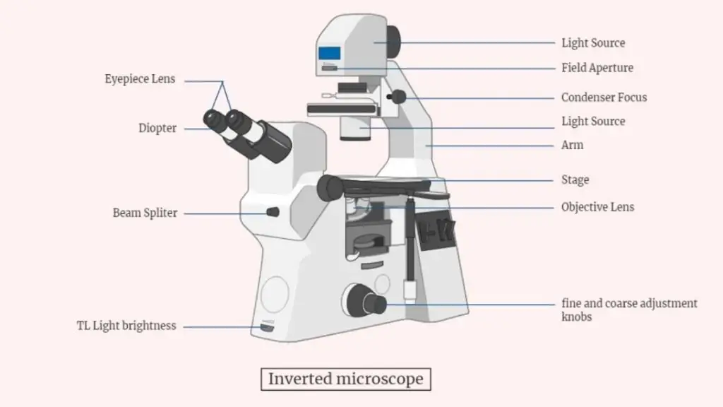
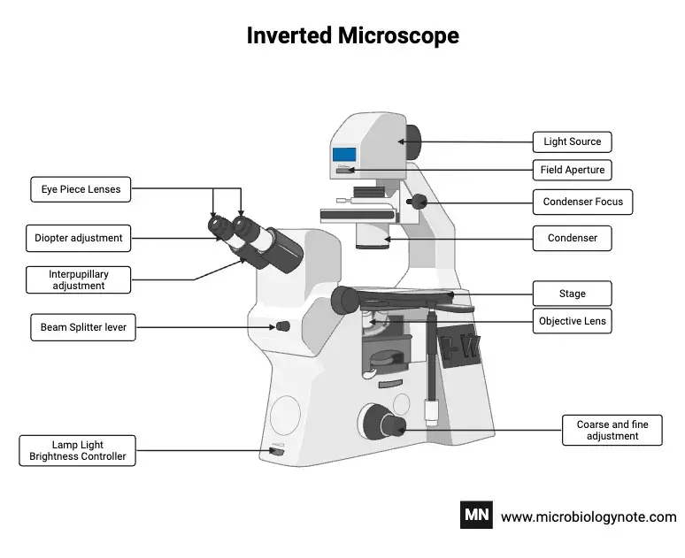
An inverted microscope, while distinct in its design, comprises components that are analogous to those found in traditional microscopes. The primary distinction lies in the arrangement of these components, which are positioned inversely in relation to their placement in standard microscopes. Here’s a detailed breakdown of the parts of an inverted microscope:

  1. Stage: Positioned at the top of the microscope, the stage is a broad, fixed platform designed to accommodate sizable vessels, such as Petri plates. It serves as the surface where specimens or culture dishes are placed for observation.
  2. Objective Lenses: Located at the microscope’s bottom, these lenses play a pivotal role in collecting light from the specimen. Typically, an inverted microscope is equipped with multiple objective lenses, each offering different magnification powers. This array allows users to select the magnification best suited for their specimen.
  3. Nosepiece: This component, also known as a rotating turret, securely holds the objective lenses. It facilitates the swift and seamless switching between different objective lenses.
  4. Dual Concentric Knobs: Comprising both fine and coarse adjustment knobs, these are instrumental in refining the focus on the specimen. They enable users to make both broad and minute adjustments to achieve optimal clarity.
  5. Condenser Lens: Positioned below the stage, the condenser lens concentrates light onto the specimen. Some condenser lenses are equipped with a diaphragm, allowing users to regulate the light intensity directed at the specimen.
  6. Illumination System: This system, essential for illuminating the specimen, might consist of various light sources, such as LEDs or fluorescent lamps. Additionally, it includes a series of lenses and filters that channel the light onto the specimen.
  7. Eyepiece: Situated at the microscope’s top, the eyepiece is the component through which users observe the specimen. Depending on the microscope model, there might be one or multiple eyepieces.
  8. Control Panel: Modern inverted microscopes often feature a control panel. This interface allows users to adjust various microscope settings, potentially including stage positioning, illumination adjustments, and advanced features like fluorescence imaging or time-lapse capabilities.
  9. Digital Camera: An optional yet valuable addition, the digital camera captures or records the specimen’s images, facilitating documentation and analysis.
  10. Stage Clips: These clips ensure that the specimen remains securely in place during observation.
  11. Arm: Serving as the backbone of the microscope, the arm supports both the optical and mechanical components.
Parts of Inverted Microscope

Operating Procedure of Inverted Microscope

Operating an inverted microscope requires a systematic approach to ensure that the specimen is viewed with clarity and precision. The following steps elucidate the procedure for effectively using an inverted microscope:

  1. Setup: Begin by positioning the inverted microscope on a firm and stable surface, such as a laboratory table or a designated microscope platform. It is imperative to ensure that the microscope is stable to prevent any disturbances during the observation process.
  2. Specimen Placement: Once the microscope is securely positioned, place the specimen, which could be on a slide or within a glass container, onto the specimen stage. Given the unique design of the inverted microscope, it’s crucial to ensure that the specimen is aligned correctly for optimal viewing.
  3. Stage Considerations: A notable feature of the inverted microscope is its fixed specimen stage. Unlike the movable stages found in traditional microscopes, the stage in an inverted microscope remains stationary. Therefore, users need not make adjustments to the stage’s position.
  4. Focusing: To bring the specimen into clear view, adjustments to the objective and condenser lenses are necessary. Using the designated knobs or controls, users can fine-tune the focus. The dual concentric knobs, comprising both fine and coarse adjustments, facilitate this process, ensuring that the specimen is viewed with utmost clarity.
  5. Observation: With the specimen in focus, users can proceed to observe the magnified image through the ocular lens or eyepiece. Some advanced models of inverted microscopes are equipped with digital screens, offering users the convenience of viewing the specimen on a display, which can be particularly beneficial for group observations or educational demonstrations.
  6. Advanced Imaging: Inverted microscopes, given their versatility, can be paired with confocal scanners and fluorescent illuminators. These tools augment the microscope’s imaging capabilities, enabling users to engage in advanced imaging techniques, such as fluorescent labeling. When integrating these components, it’s essential to adhere to the manufacturer’s guidelines to ensure proper setup and optimal results.

In conclusion, the operation of an inverted microscope, while distinct from traditional microscopes, is a straightforward process. By following the aforementioned steps and understanding the unique features of the microscope, users can achieve detailed and precise observations of their specimens.

Types of Inverted microscope

There are several different types of inverted microscopes, each designed for specific applications:

  1. Basic inverted microscopes: These are the most basic type of inverted microscopes and are typically used for routine observations of cultures and slides. They may have simple illumination systems and fixed stages, and may not have advanced features such as motorized stages or fluorescence imaging capabilities.
  2. Advanced inverted microscopes: These microscopes are designed for more specialized applications and may have a wider range of features and capabilities. They may have motorized stages, advanced illumination systems, and fluorescence imaging capabilities, as well as other specialized features such as phase contrast or DIC optics.
  3. Live-cell imaging microscopes: These microscopes are designed specifically for studying living cells and tissues and are equipped with features such as temperature-controlled stages, specialized illumination systems, and fluorescence imaging capabilities. They may also have specialized features such as oxygen control or CO2 gas control to maintain a stable environment for the cells.
  4. Industrial inverted microscopes: These microscopes are designed for use in industrial and manufacturing settings and are used to inspect parts and components. They may have specialized features such as high-resolution cameras, motorized stages, and specialized illumination systems to facilitate the inspection process.

The capability of Inverted Microscopes

Inverted microscopes possess a distinct set of capabilities that set them apart from traditional microscopes. Primarily designed for metallurgical sample analysis, they are also indispensable for observing living specimens or tissues. One of the most notable advantages of inverted microscopes is their ability to facilitate the observation of the cell division process, a feat unattainable with conventional compound microscopes. This unique feature allows researchers to monitor living cells over extended periods, providing invaluable insights into various life processes.

Furthermore, the design of inverted microscopes is tailored to accommodate larger specimens, which can be placed in expansive petri dishes rather than the restrictive slides used in standard microscopes. To ensure the longevity and vitality of the specimens, it is imperative to use covered containers. This not only minimizes evaporation but also promotes efficient gas exchange, crucial for sustaining life. In essence, the inverted microscope’s capability to observe and study vital life processes over prolonged durations underscores its superiority over traditional compound light microscopes.

Uses of Inverted Microscope

The inverted microscope, with its unique design and capabilities, has found a myriad of applications in various scientific and diagnostic fields. Here’s a comprehensive overview of its uses:

  1. Microbial Observation: One of the primary uses of the inverted microscope is to observe living microbial cells. These cells often settle at the bottom of laboratory vessels, such as tissue culture flasks and Petri plates. The inverted design allows for direct observation of these cells without the need to prepare a slide, enabling researchers to view them in their natural state.
  2. Fungal Cultures: The microscope plays a pivotal role in diagnosing fungal cultures. For instance, it aids in the detection of species like Phytophthora in cultures. The ability to view cultures from the bottom up ensures that the growth and characteristics of these fungi are observed with precision.
  3. Nematology: In the realm of parasitology, the inverted microscope proves invaluable. It facilitates the diagnosis of nematology extraction specimens, allowing researchers to observe nematodes, such as Vermiform nematodes, in detail.
  4. Microscopic Observation Drug Sensitivity (MODS) Assay: This diagnostic assay is crucial for detecting the early growth of Mycobacterium tuberculosis (MTB). The inverted microscope’s design is especially beneficial for this assay. By viewing the MODS culture from below, it overcomes challenges like condensation on the plate lid and obstruction from the culture medium. This bottom-up observation provides a clearer magnification of MTB colonies, ensuring rapid and accurate detection.
  5. Live Cell Imaging: The digital inverted microscope has revolutionized live-cell imaging. By maintaining a consistent temperature, often at 37°C, it provides focal stability for extended periods. This stability is crucial for observing dynamic cellular processes, such as cell division, growth, and morphogenesis. For instance, studies have been conducted on yeast to understand these processes in detail. Additionally, the microscope has been employed to study fungal hyphae, offering insights into its molecular dynamics at high spatial resolutions.

Advantages of Inverted Microscopy

  1. Wide Stage for Larger Specimens: One of the primary advantages of the inverted microscope is its expansive stage. This design allows researchers to view specimens contained in larger vessels, such as glass tubes and Petri plates. Therefore, it becomes an ideal tool for studying live cells, especially since these cells can be observed directly from the bottom of the cell culture apparatus.
  2. Observation of Cells in Voluminous Medium: The inverted microscope is not just limited to observing cells on flat surfaces. It is adept at viewing cells submerged in significant amounts of medium. This capability is especially beneficial when researchers need to study cells in environments that closely mimic their natural habitats.
  3. Viewing Cells in Original Vessels: Unlike traditional upright microscopes, which necessitate the transfer of specimens onto small microscopic slides, the inverted microscope allows for the observation of cell tissues directly in their original vessels. These vessels can be considerably larger than standard slides, offering a more comprehensive view of the specimen.
  4. Dual Access Observation: The design of the inverted microscope facilitates the observation of cells from both the top and bottom sections of the holding apparatus. This feature is particularly advantageous when studying cells that tend to settle at the bottom of containers, like Petri plates.
  5. Maintaining Sample Sterility: A crucial advantage of the inverted microscope is its ability to prevent contamination. Since the objective lens does not come into direct contact with the specimen, the sample remains uncontaminated. This feature is vital in ensuring the sterility of the sample, especially when studying live cells that might be sensitive to external contaminants.
  6. Enhanced Specimen Integrity: Given that the inverted microscope allows for the observation of specimens in their original containers, there’s a reduced need for transferring or manipulating the sample. This ensures that the specimen’s integrity is maintained throughout the observation process.

Limitations of Inverted Microscopy

  1. High Acquisition Cost: One of the primary limitations of the inverted microscope is its high cost. Acquiring such a microscope often requires a significant financial investment, making it less accessible for some institutions or individual researchers.
  2. Limited Manufacturers: The complexity and cost associated with manufacturing inverted microscopes mean that only a few companies venture into their production. This limited number of manufacturers further contributes to the high cost and limited availability of these microscopes in the market.
  3. Scarce Availability: Due to the limited number of manufacturers and the high production cost, inverted microscopes are rarely found readily available in the market. This scarcity can pose challenges for researchers or institutions looking to acquire one on short notice.
  4. Optical Challenges with Thick Vessels: While the inverted microscope is designed to view specimens in larger vessels, there are inherent challenges when observing through thick glass containers, such as Petri plates. Achieving a clear and sharp image requires vessels of very high optical quality, which might not always be available.
  5. Dependence on Renowned Brands: Given the technical intricacies involved in manufacturing inverted microscopes, only a few renowned companies have mastered their production. Some of the notable names in this domain include Zeiss Axio Vet Al, Olympus, Nikon, Kruss Germany, echoLAB, Metkon Technology, and Horiba Scientific. This dependence on a few brands can sometimes limit the choices available to potential buyers.

Inverted vs Upright Microscope

Configuration and Components:

  • Inverted Microscope: As the name suggests, the components of an inverted microscope are arranged in an inverted manner. The objective lenses are located at the bottom, while the stage is positioned at the top. This unique arrangement allows for the observation of specimens in larger containers, such as Petri dishes or flasks.
  • Upright Microscope: In an upright microscope, the objective lenses are situated at the top, and the stage is below them. This traditional configuration is ideal for examining specimens that are mounted on standard microscope slides.

Illumination and Image Production:

  • Inverted Microscope: Typically, inverted microscopes utilize transmitted light, which passes through the specimen from below and is then collected by the objective lenses. This type of illumination is especially beneficial for observing living cells in their natural environment. The resulting image often has a three-dimensional quality, providing a stereo view of the specimen.
  • Upright Microscope: Upright microscopes can employ either transmitted or reflected light, depending on the nature of the specimen. The image produced is usually flat, offering a two-dimensional perspective of the specimen when viewed through the eyepieces.

Applications and Suitability:

  • Inverted Microscope: Given its design, the inverted microscope is particularly suited for observing large, flat specimens or live cell cultures. It allows researchers to study cells in their natural environment without disturbing them, making it invaluable in fields such as cell biology and tissue culture studies.
  • Upright Microscope: The upright microscope is versatile and is commonly used in various fields of study. It’s particularly useful for observing smaller specimens that are mounted on traditional microscope slides, such as tissue sections or smears.
AspectInverted MicroscopeUpright Microscope
Objective Lens LocationBottom of the microscopeTop of the microscope
Stage LocationTop of the microscopeBottom of the microscope
Specimen CompatibilityLarge, flat specimens, cell culturesSmall specimens on microscope slides
Illumination TypeTransmitted lightTransmitted or reflected light
Image ProducedStereo image (3D perspective)Flat image viewed through eyepieces

Examples of Inverted Microscopy

  1. Leica Inverted Microscope: Leica, a renowned name in the world of scientific instruments, offers a range of inverted microscopes tailored for diverse applications. These microscopes stand out for their superior optics and state-of-the-art features. Some of the notable attributes include motorized stages, temperature-controlled platforms, and the ability to perform fluorescence imaging. Depending on the user’s requirements, there are basic models ideal for routine observations and advanced models equipped with specialized features. Given Leica’s reputation for quality, their inverted microscopes are a staple in research labs, medical facilities, and even industrial settings.
  2. Olympus Inverted Microscope: Another heavyweight in the microscopy arena is Olympus. Their inverted microscopes are designed with precision, catering to various scientific needs. Like Leica, Olympus microscopes boast high-quality optics, motorized stages, and temperature-controlled platforms. Additionally, they are adept at fluorescence imaging, making them invaluable for specific research applications. Whether one is looking for a basic model for everyday use or a high-end version for specialized tasks, Olympus has a range to choose from. Their commitment to quality ensures that these microscopes find their place in research, medical, and industrial domains.
  3. Nikon Inverted Microscope: Nikon, a name synonymous with optics, offers its line of inverted microscopes, each reflecting the brand’s commitment to quality and innovation. These microscopes are equipped with top-notch optics and come with features like motorized stages and temperature-controlled platforms. Fluorescence imaging is another forte of Nikon’s inverted microscopes. Catering to a broad spectrum of users, Nikon provides both basic models for routine tasks and advanced ones for specialized research. Given their reliability and durability, Nikon inverted microscopes are widely preferred in scientific, medical, and industrial settings.

Precautions of Inverted Microscopy

  • Handling the Microscope: It is imperative never to touch the glass part of the microscope while observing the specimen. This action can introduce contaminants, distort the image, and potentially damage the optics.
  • Power Considerations: Before any maintenance or when the microscope is not in use, ensure to unplug and turn off the device. This step is crucial to prevent electrical hazards and protect the microscope’s components.
  • Disassembly: Avoid disassembling the microscope unless necessary and only by trained personnel. Improper disassembly can lead to damage, misalignment, and compromised performance.
  • Fire Safety: Given that microscopes often employ bulbs that generate heat, it’s vital not to place combustibles near the bulb. This precaution prevents potential fire hazards.
  • Environmental Conditions: The microscope should be kept at temperatures ranging between 0°C-40°C (32°F-104°F). Additionally, the humidity level should not exceed 85%. These conditions ensure the microscope’s optimal performance and longevity.
  • Sunlight Exposure: Direct sunlight can adversely affect the microscope’s components. Therefore, always store and use the microscope in a place shielded from direct sunlight.
  • Placement: For clear and accurate observations, it’s essential to place the microscope on a sturdy and level surface. This positioning prevents wobbling and ensures stability during observations.

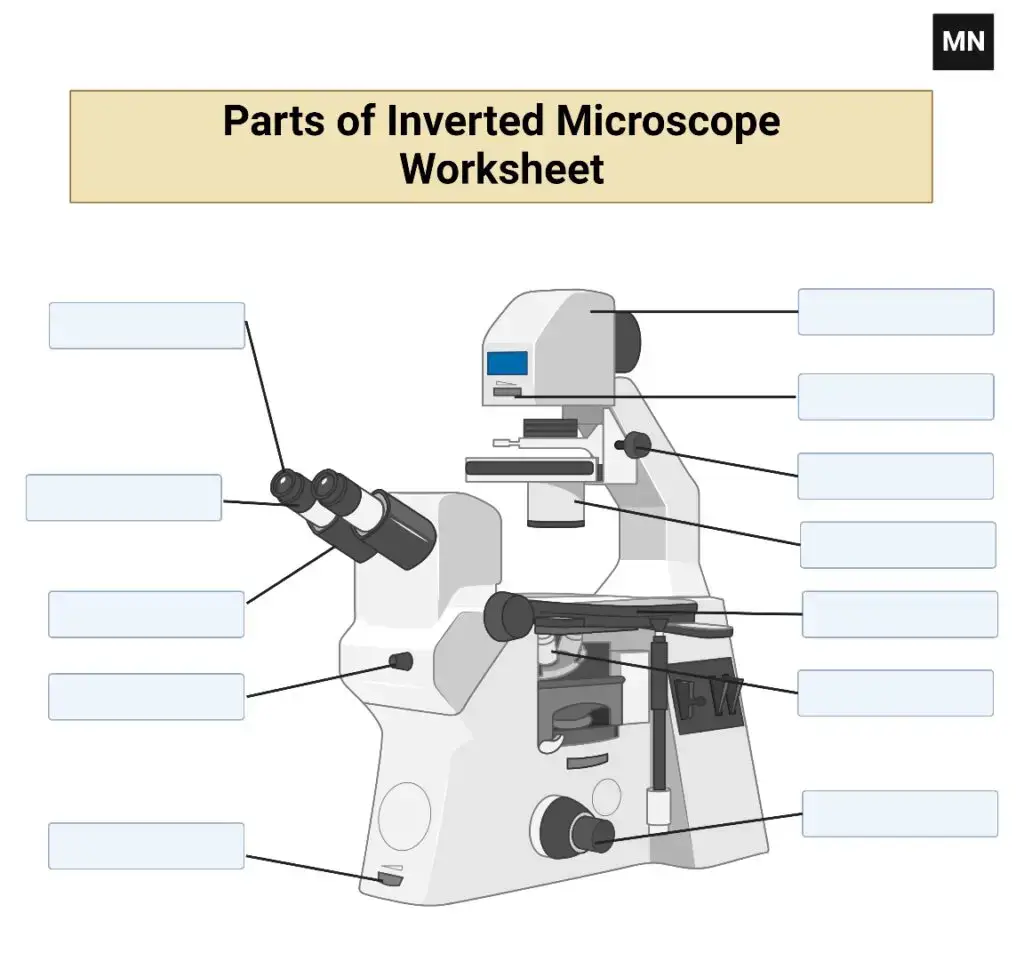
Inverted Microscope Free Worksheet

Inverted Microscope Worksheet

Inverted microscope Images
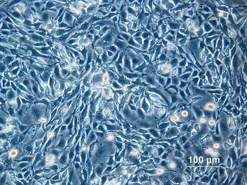
Inverted microscope Images – Cell Culture

Quiz

FAQ

What is an inverted microscope?

An inverted microscope is a type of microscope where the objective lenses are positioned below the stage, and the light source and condenser are located above the stage. This design allows for the observation of specimens from the bottom, making it suitable for viewing cells in culture dishes and other large containers.

What are the advantages of using an inverted microscope?

Some advantages of using an inverted microscope include the ability to view living cells in their natural environment, compatibility with large and thick specimens, and the convenience of accessing the specimen from the top for manipulation or introducing additional elements into the experimental setup.

What types of applications is an inverted microscope commonly used for?

Inverted microscopes are commonly used in cell biology, tissue culture, live-cell imaging, materials science, and metallurgy. They are also used in fields such as microbiology, nematology, and diagnostics of fungal cultures.

Can an inverted microscope be used with traditional microscope slides?

While inverted microscopes are primarily designed for viewing specimens in large containers, some models may have the capability to accommodate traditional microscope slides with the help of specialized accessories or stage adapters.

How is the image formed in an inverted microscope?

The image is formed by passing light through the specimen from above, which is then collected and magnified by the objective lenses located beneath the stage. The magnified image is then viewed through the eyepieces or captured digitally for further analysis.

Can inverted microscopes be used for fluorescence imaging?

Yes, inverted microscopes can be equipped with fluorescence capabilities, allowing for fluorescence imaging of labeled specimens. This is useful for studying cellular processes and protein localization.

Are inverted microscopes more expensive than upright microscopes?

In general, inverted microscopes tend to be more expensive than upright microscopes due to their specialized design and components. However, prices can vary depending on the specific model and brand.

Can inverted microscopes be used for quantitative analysis?

Yes, inverted microscopes can be used for quantitative analysis by integrating specialized software and image analysis tools. This allows for measurements, counting cells, tracking movements, and other quantitative assessments.

What are the limitations of inverted microscopy?

Some limitations of inverted microscopy include the higher cost compared to upright microscopes, limited availability from fewer manufacturers, and challenges in viewing specimens through thick glass vessels, which may require high-quality optics.

How do I choose the right inverted microscope for my application?

When choosing an inverted microscope, consider factors such as your specific research needs, budget, required magnification and resolution, compatibility with accessories and imaging techniques, and the reputation and support of the manufacturer. Consulting with experts or experienced users can also provide valuable insights and guidance in selecting the most suitable microscope for your application.

References

  1. Child, J. M. (1972). International Specifications for Technical Manuals. Journal of Technical Writing and Communication, 2(3), 189–197. https://doi.org/10.2190/hw02-644g-ekdm-7hua
  2. Oktaviyanthi, R., & Agus, R. N. (2021). Guided Worksheet Formal Definition of Limit: An Instrument Development Process. AL-ISHLAH: Jurnal Pendidikan, 13(1), 449–461. https://doi.org/10.35445/alishlah.v13i1.483
  3. Zimic, M., Velazco, A., Comina, G., Coronel, J., Fuentes, P., Luna, C. G., Sheen, P., Gilman, R. H., & Moore, D. A. J. (2010). Development of Low-Cost Inverted Microscope to Detect Early Growth of Mycobacterium tuberculosis in MODS Culture. PLoS ONE, 5(3), e9577. https://doi.org/10.1371/journal.pone.0009577
  4. Rines, D. R., Thomann, D., Dorn, J. F., Goodwin, P., & Sorger, P. K. (2011). Live cell imaging of yeast. Cold Spring Harbor protocols, 2011(9), pdb.top065482. https://doi.org/10.1101/pdb.top065482.
  5. Hickey, P. C., Swift, S. R., Roca, M. G., & Read, N. D. (2004). Live-cell imaging of filamentous fungi using vital fluorescent dyes and confocal microscopy. Methods in microbiology, 34, 63-87. https://doi.org/10.1016/S0580-9517(04)34003-1
  6. https://en.wikipedia.org/wiki/Inverted_microscope
  7. https://microbenotes.com/inverted-microscope/
  8. https://www.microscopemaster.com/inverted-microscope.html
  9. https://ibidi.com/content/212-inverted-and-upright-microscopy
  10. https://www.biocompare.com/25969-Inverted-Microscopes/
  11. https://www.microscopeworld.com/t-inverted_microscopes.aspx

Latest Questions

Start Asking Questions

This site uses Akismet to reduce spam. Learn how your comment data is processed.

⚠️
  1. Click on your ad blocker icon in your browser's toolbar
  2. Select "Pause" or "Disable" for this website
  3. Refresh the page if it doesn't automatically reload