Funaria is a moss you’ll often find thriving in damp, shady areas like forest floors or along stream banks. Unlike typical plants, it lacks true roots, stems, or leaves, absorbing water and nutrients directly through its surface. If you take a closer look, you’ll spot its vibrant green tufts topped with tiny, capsule-like structures—these hold spores that eventually scatter to grow new moss. Frequently featured in beginner biology classes, Funaria showcases how simpler plants adapt to moist habitats. Beyond the classroom, it quietly supports ecosystems by stabilizing soil and trapping moisture, creating better conditions for surrounding plants. Its straightforward structure makes it a go-to example for understanding how non-vascular plants survive and evolve.
What is Funaria?
- Comprising over 210 species, Funaria is a genus of mosses in the division Bryophyta; Funaria hygrometrica is the most often occurring and most researched species in this group.
- Derived from the Latin word “funis,” meaning “rope,” the genus name “Funaria” refers to the hygroscopic, twisted seta unique to its sporophyte, which responds to moisture variations by twisting or untwisting.
- Usually found on dirt, rocks, and rotting organic materials, funaria species are non-vascular, autotrophic plants that flourish in damp, shady conditions.
- Mostly gametophytic and displaying a dominating haploid stage, the plant body depends on the gametophy for sustenance and shapes the sporophyte.
- Often used as a model organism in bryological research because of its widespread occurrence and simplicity of cultivation, Funaria hygrometrica is renowned for its fast life cycle and completion in few months.
- The gametophyte comprises a protonema stage, then multicellular rhizoids with oblique septa and growth of an upright, leafy gametophore with spirally arranged leaves.
- Funaria uses both sexual and asexual reproduction; sexually, it is autoicous, generating both antheridia and archegonia on the same plant but on separate branches.
- The long, twisted seta of the sporophyte supports a capsule where spores are formed; the capsule has teeth that help spore distribution in response to variations in humidity.
- Ecologically, Funaria commonly colonises disturbed or nutrient-rich soils, notably those impacted by fire or human activity, therefore helping to stabilise soil and promote succession.
- Phylogenomic research show that the Funariaceae family—to which Funaria belongs—has seen fast diversification, therefore supporting the evolutionary knowledge of bryophytes.
Classification of Funaria
| Kingdom: | Plantae |
| Division: | Bryophyta |
| Class: | Bryopsida |
| Subclass: | Funariidae |
| Order: | Funariales |
| Family: | Funariaceae |
| Genus: | Funaria |
Habitat of Funaria
- Funaria hygrometrica, referred to as flaming moss or cord moss, is a global species seen on every continent except Antarctica.
- It flourishes in several habitats, encompassing urban environments, woodlands, grasslands, and disturbed locations.
- This moss frequently serves as one of the initial colonisers of bare soil, owing to its exceptional capacity for spore dispersion and rapid population establishment.
- Funaria hygrometrica is typically located in disturbed environments, including damp soil fissures, walls, pavements, adjacent to water taps, gutters, drains, and within soil in flower boxes and plant pots.
- It is frequently located in nurseries and greenhouses.
- In southern Africa, it is found at elevations between 20 and 2550 meters.
- Funaria hygrometrica is a prevalent greenhouse weed, often located in burnt areas with bare, disturbed soil that is nitrogen-rich, including gardens and neglected agricultural lands.
- It is renowned as a moss found on ancient bonfire locations and also thrives in scorched heathland.
- Funaria hygrometrica is a prevalent species among agricultural field ecosystems, but seldom in substantial numbers.
- It is a pioneering species that inhabits disturbed soils, establishing microhabitats and contributing significantly to ecosystem development.
- Funaria hygrometrica is a significant greenhouse weed, commonly located in scorched areas with bare and disturbed soil rich in nitrogen.
- It is a prevalent constituent of arable field communities, but seldom in substantial numbers.
- Funaria hygrometrica is a pioneer species that inhabits disturbed soils, establishing microhabitats and contributing significantly to ecosystem development.
Characteristics of Funaria
- Funaria shows a life cycle mostly controlled by the haploid gametophy, which sustains the dependent sporophyte by means of photosynthesis.
- Funaria’s mature gametophy grows as upright, thin, monopodially branching gametophores bearing spirally organised, sessile leaves one cell layer thick and with a highly defined midrib and complete border.
- From Funaria’s spore germination, a main protonema of filamentous, chlorophyllous cells forms from which secondary protonemal buds develop the leafy gametophore and fragmentation of protonema allows vegetative propagation.
- Arranging obliquely septate structures from gametophore base, multicellular rhizoids in Funaria serve in anchoring and absorption of water and dissolved nutrients.
- Funaria reproduces asexually by fragmentation of protonema, budding of secondary protonema, development of gemmae and bulbils enabling fast clonal establishment in suitable environments.
- Funaria is monoecious and autoicous; biflagellate sperm generated in antheridia on one branch and eggs in archegonia on another branch of the same gametophy with perichaetial leaves around archegonia.
- Released from Funaria antheridia, sperm travel through moisture films to reach archegonia where fertilisation produces a diploid zygote starting the sporophyte phase.
- The lengthy, hygroscopic seta of the Funaria sporophyte raises an asymmetric, oblique capsule with a double peristome of highly developed teeth regulating spore release under cover by a rostrate calyptra.
- An epidermis of chlorophyllous cells, a cortex of paredchymatous cells with chloroplasts and an inner conducting strand of dead, thin cells for water conduction and support, is shown by transverse slice of the Funaria gametophore axis
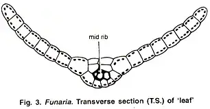
- A Funaria leaf’s transverse slice reveals a single layer of paredchymatous cells with conspicuous chloroplasts, a central midrib with a short conducting thread, and lateral wings devoid of conductive tissue.
- Funaria hygrometrica colonises disturbed, nutrient rich, wet shaded surfaces including soil, rock crevices and burnt areas helping to stabilise the ground and acting as a bioindicator of ambient moisture levels.
- From sea level to montane zones up to 4000 m typically dominating early successional bryophyte communities, Funaria is a global genus found in tropical to temperate zones worldwide.
- Organellar exome phylogenomic studies of 123 genes corroborate the monophyly of Funaria, expose fast radiation within Funariaceae beginning around 28 million years ago coincidental with Eocene cooling, and highlight bryophyte evolutionary dynamism.
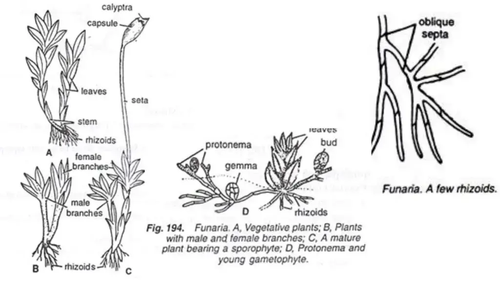
Morphology of Funaria
- Funaria has a gametophytic plant body with two separate stages: the protonema, which is young, and the green gametophyte, which is adult.
- It starts as a spore and grows into the green gametophyte. The protonema is a structure made of filaments and branches.
- The stem of the gametophyte is straight, thin, and branching. It can grow up to 3 cm tall.
- The stem goes around the plant and has many parts that grow axillary or extra-axillary, which means below the leaves.
- The leaves stay in place and are oblong to oval in shape, with full edges and pointed tips.
- Each leaf has a single midrib running through it, and the leaves are grouped spirally on the stem, usually in a 1/3 phyllotaxy that changes to a 3/8 phyllotaxy as the plant grows older.
- Rhizoids are made up of many cells, branches, and angled septa. They grow from the stem’s base and hold the plant in place on the ground.
- The sporophyte has a foot, a seta, and a capsule. The seta can absorb water and twist when it dries out, which helps the spores spread.
- The capsule is pear-shaped and not uniform, and it holds spores made by meiosis.
- Funaria is dioicous, which means that the male and female sexual parts of the plant grow on different branches.
- Antheridia are the male parts of the plant and archegonia are the female parts. They are found on different branches, which helps with fertilisation and the growth of the sporophyte.
Structure of Funaria (With Diagram)
A. External Features of Funaria
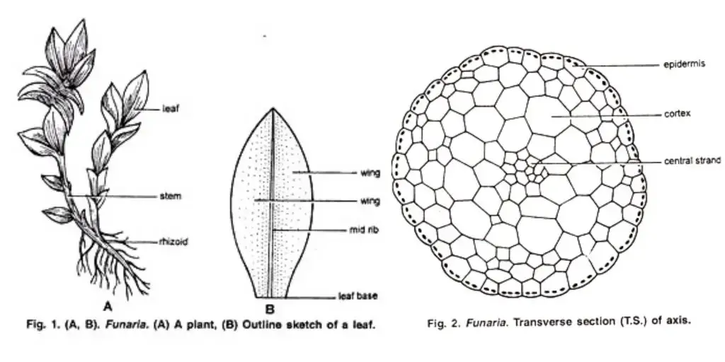
The height of the erect gametophytic plants is approximately one inch. There is an erect stem or leafy axis attached to the substratum via rhizoids. Radial axis, sometimes branched axillary and extra-axillary. The axis is covered in spirally arranged leaves that are closer to the apex, forming a roseette.
Eight leaves are actually arranged in three complete spirals, forming 3/8 of phyllotaxy. This corresponds to the three cutting faces that were used when they were young. The leaves are simple, straight, sessile and ovate with a pointed apex and smooth margins. They attach to the stem via a broad base. While mature leaves have midribs, younger leaves lack them.
The strong, multicellular, mu h-branched rhizoids have strong sys tems. They have oblique septa. The young rhizoids, which are colorless, turn brown or black as they mature. When exposed to sunlight, the branches of rhizoids produce chloroplasts. They are “absorbing and anchoring organs.”
B. Internal Features of Funaria
1. Stem:
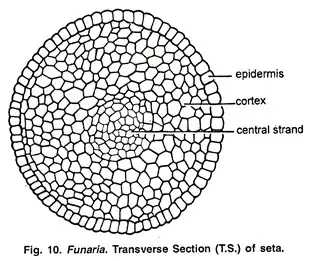
The mature stem’s transverse section shows three distinct regions: the outer epidermis and middle cortex.
- Epidermis: It is the outermost layer in the stem. The epidermal cells are free of pores and stomata, and contain chloroplasts.
- Cortex: It is located between the epidermis & the central cylinder. It is multilayered and contains large parenchymatous cell. When young, the cortex cells contain chloroplasts but these are lost as the stem ages. The cortical cells in a young stem look the same, but the cortex’s outer cells become thicker and more reddish brown as it matures. In the cortex, there are also isolated areas of cells in the peripheral area that represent the leaf traces. These traces are not connected to the central cylinder because they have blind ends.
- Central Cylinder: It is made up of colourless, long-walled cells that are thin-walled and have thin walls. These cells are dead and may not contain protoplasm.

2. Leaf
The transverse section (T. S. ) of the ‘leaf shows a clearly defined midrib and two lateral wings. The ‘leaf is made up of a single layer of parenchymatous, polygonal cells. Many prominent and large chloroplasts are found in these cells. The thick walls of cells in the central portion of the mid-rib have a narrow conducting strand. This helps with conduction.
Reproduction in Funaria
Funaria reproduces through vegetative and sexual methods.
1. Vegetative Reproduction
These are the methods that make it possible:
a. By multiplication of primary protonema
Funaria spores form a multicellular, branched structure upon germination. This is called primary protonema. Intercalary divisions create certain colourless separation cells. These cells die and split the protonema into many-celled fragments. These fragments become new protonemata that bear buds. Each bud becomes a leafy gametophore.
b. By secondary protonema
Secondary protonema occurs when protonema is created by other methods than the germination spore. It can be formed from any part of the gametophyte that is not spore-germinated, such as the’stem’ or ‘leaves, antheridium and archegonium paraphysis. It develops into a leafy gametophore similar to primary protonema.
c. By Gemmae
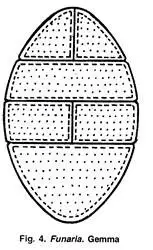
Unfavorable conditions cause the terminal cells of protonemal branches to divide by transverse and longitudinal divisions, forming green multicellular bodies with 10-30 cells. These cells are known as gemmae. Gemmae turn slightly reddish brown when they reach maturity. Gemmae will germinate new plants when they are exposed to favorable conditions.
d. By Bulbils
These structures, which look like gemmae, are created on the rhizoids within the substratum. They are called bulbils. These structures are not able to develop into leafy individuals, but they lack chloroplasts.
e. Apospory:
Apospory is the process of developing gametophyte out of sporophyte, without the formation of any spores. The sporophyte’s vegetative cells may develop green protonemal filaments that bear lateral buds. These buds eventually become leafy gametophores.
These gametophores are called diploid. The formation of tetraploid (4n), zygote results from sexual reproduction in these gametophores. Because they are incapable of producing spores, sporophytes derived from tetraploid are considered sterile.
2. Sexual Reproduction in Funaria
Oogamous reproduction is the preferred method of sexual reproduction. Antheridium is the name of male reproductive structure, while archegonium is the name for female. Funaria is monoecious, with male and female sex organs sharing the same thallus. It can also be autoicous (antheridia or archegonia grow on different branches of the same branch). Leafy gametophores are found in terminal clusters and sex organs are borne from these leafy gamestophores.
The main shoot of the leafy tree tophore bears antheridia, and acts as a male branch. The female branch is a lateral branch that grows from the male branch’s base and bears archegonia. It is taller than the male branch. Funaria is protandrous, meaning that archegonia mature before funaria. It allows cross fertilization.
Male Branch or Antheridiophore
L. S. of the male branch’s longitudinal section shows that its apex has an expanded and convex shape. In different stages of development, it bears a large number of orange or reddish-brown antheridia. The perigonial leaves are a rosette that surrounds the projected antheridia.
Perigonium is the antheridial cluster that surrounds perigonial leaves. Paraphyses are a large group of sterile hairs that look like clubs and are found in the antheridia. Paraphyses are water-storing structures that protect developing antheridia and aid in photosynthesis as well as dehiscence.

Structure of an Antheridium
Antheridium has a club-shaped shape. It can be divided into two parts:
(a) Short multicellular stalk
(b) Body of antheridium
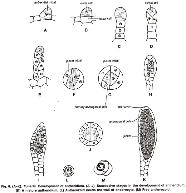
The body of antheridium is a single-layered, sterile jacket made of flattened polyhedral cells. The jacket’s cells contain chloroplasts, which at maturity turn orange-reddish brown. The jacket contains a lot of androcytes, which are mother cells from anantherozoid mothers.
The antheridium’s distal end bears one to two thick-walled, colourless cells known as operculum at maturity. The opercular cells are mucilaginous and absorb water. They swell and break down connections with neighbouring cells, and create a narrow pore. Through this pore, androcytes emerge as a viscous liquid.
Development of Antheridium
An antheridium cell is a single, slightly projecting cell. It is also known as the antheridial first. It is located at the apex the male branch. It divides through a transverse division, forming a basal and outer cell. The embedded portion of the stalk is formed by the basal cell. The entire antheridium is formed by the outer cell, also known as the antheridial mom cell. It divides through transverse divisions, forming a short filament of 2-3 cell.
The stalk of antheridium is made up of lower cells. A terminal cell of the filament is divided by two vertical intersecting walls. This differentiates an apical cells with two cutting faces.
Segments are cut in the apical cells in two rows, in an alternate order. This method leaves 5-7 segments. The apical cells are simultaneously dividing. Meanwhile, the third and fourth segments below the Apical Cells start dividing starting at the base by diagonal vertical walls.
The segment is divided by the first wall into two equal-sized cells. The jacket initial is a small cell. A larger cell divides periclinally to form an inner large primary,rogonial and outer jacket initial. A transverse section (T. S.), shows a triangular primary androgonial cells. This type of division occurs in all upper segments, except for the operculum-forming apical cells.
Anticlinal divisions are the only way to divide all jacket initials and form one layered wall made of antheridium. Primary androgonial cells split and re-divide to create the androcyte mom cells. The cells of the final cell generation are known as androcyte mother cells.
Each androcyte mother cells divides further to form two androcytes. Each androcyte can produce one spermatozoid, antherozoid, or biflagellate egg. Each antherozoid has a spirally coiled and bi-flagellated structure.
Female Branch or Archegoniphore
From the base of the male branches, the female branch grows. L. S. of the female branch shows that paraphyses and archegonia are interwoven at its apex. Paraphyses’ terminal cells are not swollen. A group of green leaves ‘leaves’ surrounds the archegonia cluster. Perichaetium is the name given to the archegonial cluster and its surrounding perichaetial foliage.
Structure of an Archegonium
The mature archegonium has a flask-shaped structure. It is attached to the female branch by an enormous stalk. It has a long, elongated neck and a basal globular portion called venter.
The neck is slightly tubular and twisty, single-layered, and contains six vertical rows neck cells. These cells enclose an abaxial row of 10 or more neck canal cells. Venter wall is two-layered and contains egg cell and venter canal cells. The venter canal cell is located just below the neck cells.

Development of Archegonium
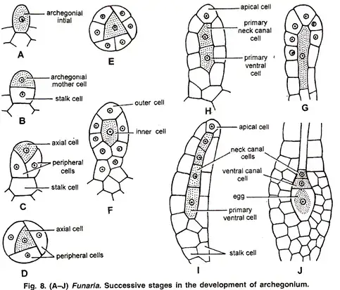
Archegonium is a result of a single supracellular cell, the archegonial initial. It divides at the apex, or female branch. The archegonial initial divides through transverse division to form the stalk cell or basal cell, and a terminal.
The stalk of the archegonium is formed by the basal cell. It divides and re-divides. The archegonial mother cells function as the terminal cell. It is divided by three intersecting walls, forming three peripheral cells that surround a tetrahedral-axial cell. The peripheral cells split anticlinally to form one layered venter wall, which becomes later two layered.
The axial cell splits transversely to create an outer primary cover and inner central cells. The outer primary cover cell acts as an aplical cell, with three lateral and one base cutting faces. It trims three lateral segments as well as one basal segment. Each lateral segment is divided by a vertical wall, so that six rows of cells make up the neck of archegonium. Each basal cell contributes to the neck canal cell.
Transverse division of the inner central cell results in a transverse division that creates an outer primary neck cell cell and an inner primaire ventr cell. Transverse divisions are performed on the primary neck canal cell to create a row of neck cells.
Funaria’s neck canal cells are therefore of double origin. The lower and middle neck canal cells are derived primarily from the primary neck cell, while the upper neck cells are derived mainly from the primary cover cells. Transverse division is used to create the primary ventr cell and the egg cell.
Fertilization in Funaria
- The female reproductive organ, the archegonium, which is situated at the tip of a female branch, is where fertilization takes place in Funaria.
- A tubular neck and a swelling ventr make up the archegonium; the neck has neck canal cells, and the ventr has an egg cell and a venter canal cell.
- The neck canal cells and the ventr canal cells break down as they mature, creating a mucilaginous material that swells and absorbs water.
- Because of the internal pressure created by this enlargement, the neck’s cover cells split apart, allowing sperm to pass through and reach the egg.
- The male reproductive organ, the antheridium, which is situated at the tip of a male branch, releases sperm (antherozoids).
- With the help of chemicals, mostly sugars, produced from the archegonium, the antherozoids are drawn to it through chemotaxis.
- Although several antherozoids penetrate the archegonium’s neck, only one of them unites with the egg cell to create a diploid zygote.
- The initial cell of the sporophyte generation, the zygote, divides to become the embryo after secreting a wall around itself right away.
- The sporophyte, which is divided into three parts—foot, seta, and capsule—develops from the embryo.

Sporophyte Development
- The gametophyte’s foot is a conical structure that acts as an absorption and anchoring organ.
- Water and nutrients are transferred from the gametophyte to the capsule by the seta, a long, thin stalk that supports the capsule.
- The pear-shaped capsule, which is first covered by a calyptra and then by an operculum, contains cells that produce spores.
- At the top of the capsule is an operculum, which resembles a cap and finally separates to release spores.
- There are three sections to the capsule:
- Apophysis: The lowest sterile portion that aids in sporophyte nourishment and is photosynthetic in nature.
- Theca: The core area that is encircled by spore sacs and contains sterile parenchyma cells (columella).
- Operculum: The cap-like structure that envelops the spore-producing area and finally separates to release the spores is called the operculum.
Spore Dispersal
- The peristome is revealed when the operculum separates, facilitating the spores’ controlled release.
- The exostome and endostome, two rings of triangular teeth that make up the peristome, react to variations in humidity to aid in the spread of spores.
- The life cycle is completed when spores are discharged into the environment, where they can germinate to create new gametophytes.
Sporophytic Phase
Zygote is a cell that forms the first phase of the sporophytic cycle. The venter of an archegonium is where sporophyte develops.
Structure of Sporophyte
Semi-parasitic, the sporophyte’s mature form can be divided into seta, foot, and capsule.
- Foot: This is the basal part of the sporogonium. It is a small, conical, dagger-like structure that is embedded at the apex female branch. It acts as an anchoring and absorption organ.
- Seta: This is a long, slim, stalk-like hygroscopic structure. It bears its capsule at the tip. It lifts the capsule from the leafy gametophore’s apex. Its internal structure is similar to the axis. The epidermis surrounds the axial cylindrical with thick walls. It serves a mechanical function and conducts water and nutrients to the developing capsule.
- Capsule: Capsule is the final part of the sporophyte. It is located at the apex the seta. It is usually green when it is young, but becomes bright orange as it matures. It is protected by a calyptra-like cap. The archegonium’s upper portion is where gametophytic tissue grows.
Internal Structure of the Capsule
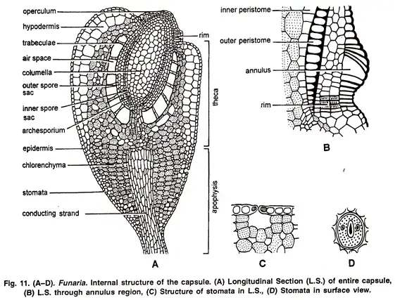
Longitudinal Section The capsule can be distinguished into three distinct regions: apophasis (theca), operculum and longitudinal Section (L.S.).
a. Apophysis
It is the base sterile portion of the capsule. It is bound by the single-layered epidermis, which is broken by stomata. The guard cells of the sotmata are shaped like a single ring. Below the epidermis, there is spongy paraenchyma. The central portion of the apophysis consists of long, thin-walled cells that form a conducting strand. It is also known as the neck of capsule. It connects seta and capsule.
b. Theca
It is located in the middle of the capsule, where the spore-bearing region is slightly bent. It is located between the operculum and apophysis.
Longitudinal section (L. S.) passing through the theca shows the following regions:
- Epidermis: The epidermis is the outermost layer. It can be single-layered or multilayered, with or without stomata.
- Hypodermis: The hypodermis is located below the epidermis. It is composed of two to three layers, each containing colourless cells that are compactly arranged.
- Spongy parenchyma: Spongy parenchyma is made up of two to three layers containing loosely organized chlorophyllous cells. It can be found between the hypodermis and innermost layer. These cells can produce their own food, but are dependent on gametophyte to obtain water and minerals. The sporophyte Funaria is therefore partially dependent upon gametophyte.
- Air spaces: These are located just below the spongy paraenchyma, and outside the spores. Green cells (chlorenchymatous cell) traverse these air spaces. Trabecular (elongated paraenchymatous cell) is another name for them.
- Spore sac: These are located below the air spaces on each side of the columella. It has a U-shaped shape and is broken at its base. It separates its two arms. It has an outer wall of 3-4 cells thickness and an inner wall of one cell. The cavity of the spores is located between the inner wall and outer wall. The cavity of the spore capsule is full of spore mother cells when it’s young. The spore mother cells divide through meiotic divisions at maturity and produce many haploidspores.
- Columella: This is the central region of the Theca region. It is composed of colourless, compactly arranged parenchymatous cells. It connects the central strand in apophysis by being wide above and narrow below. It aids in the conduction of water as well as mineral nutrients.
c. Operculum:
It is located in the top region of the capsule. It has four to five layers of cells and is dome-shaped. The epidermis, which is the thickest layer of cells, is the outermost. Operculum can be distinguished from theca by a clearly marked constriction. Below the constriction is a diaphragm or rim. It is made up of two to three layers radially elongated, pitted cells.
The annulus is located immediately above the rim and consists of five to six layers of cells superimposed on top. The upper cells of the annulus are thick, while the two layers below are thin. Annulus separates operculum and theca. The peristome is located below the operculum. It is located below the edge of your diaphragm. Two rings of peristomial peristomial teeth are made up of the peristome. Each ring contains sixteen teeth.
They are just the cuticle strips, and not cells. The outer ring’s teeth are prominent, with red transverse bands and thick, conspicuous teeth. However, the inner ring’s teeth are smaller, more delicate, without transverse bands, and are colourless. A mass of thin-walled parenchymatous cells lies between the inner and peristome teeth.
Development of Sporophyte
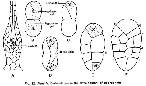
The zygote soon after fertilization forms a wall around itself and grows in size. It splits along a transverse wall, forming an epibasal and hypo basal cells. The epibasal cell is divided by two intersecting, oblique walls. It distinguishes an apical from an epibasal cell by using two cutting faces. The hypo basal cells also differentiate an apical.
These two apical cells are responsible for the differentiation of all sporophytes. The development of embryosporophyte therefore is bi-apical. The epibasal apical cells develop into capsule and the upper part of the seta, while the hypo basal cell becomes foot and the remaining portion of the seta. Both apical cell types cut out alternate segments to form the long, filamentous structure of youngsporogonium.
Development of Capsule
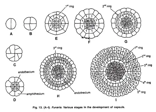
A cross-section of the upper portion shows two identical segments that divide at an angle at the last one to form a quadrant (4-souled stage).
Each quadrant cell divides by an anticlinal wall so that a smaller, almost triangular cell and larger, more or less rectangular cells are formed. Now, each rectangular cell divides by a periclinal division.
This results in the formation of four central cells, which are then surrounded by eight peripheral cells. The endothecium is the central tissue, while the amphithecium’s peripheral cells are the amphithecium. The next stage of development is achieved from these two groups. Different rings can be formed by anticlinal or periclinal divisions.
Periclinal division allows the amphithecium to divide into two concentric layers. The first ring is the inner layer with 8 cells. The outer layer’s cells divide by anticlinal divisions, forming 16 cells. This is followed in this layer by the periclinal divide.
This inner layer is known as the second ring. The outer layer of the two layers splits anticlinally to create 32 cells. This layer splits periclinally into two layers of 32 cells. The third ring is the inner layer. The inner layer is also formed by the periclinal divisions of fourth and fifth rings of 32 cells.
The four cells that make up the endothecium divide in a similar fashion to the amphithecium. The first division is anticlinal and curved. The second division is periclinal. This results in the formation a central group consisting of 4 endothecial cell groups, which is surrounded by 8 endothecial cell groups. These amphithecial rings, as well as endothecial cell development, further develop the tissues of the capsule region.
The fertile area in capsule is lined with outer and inner spores. Archesporium is endothecial. Its cells can undergo sub-divisions to become two layers thick spore mother cell cells. This meiosis forms a tetrad spores. Elaters are absent.
Dehiscence of the Capsule
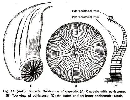
Funaria is a stegocarpous, or dehiscence-oriented moss. This is done by ‘breaking down’ the annulus. The capsule becomes inverted as it matures due to epinasty. The annulus’ thin walls are broken and the operculum is exposed.
Hygroscopic is the outer peristomial tooth (exostome). The inner peristomial (endostome), which do not exhibit any hygroscopic movements, acts as a sieve and allows only a few spores at a time. The dispersal of spores is made possible by the lengthening or shortening the outer peristomial tooth.
The exostome’s ability to absorb water and increase in length in high humidity conditions causes them to curve inwards. Dry weather causes the exostome teeth to lose water and bend inwards with jerky movements. This allows for the dispersal in small amounts of spores from a capsule. The seta develops jerky movements at maturity. The dispersal of spores is further assisted by the seta’s twisting and swinging in dry weather.
Structure and Germination of Spore
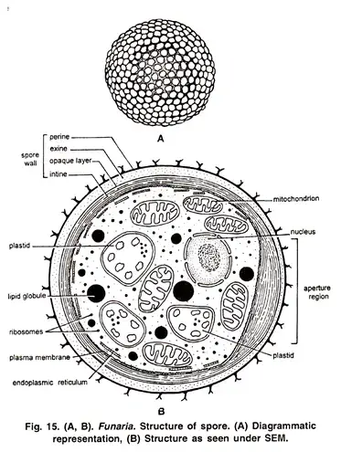
Spore is the initial cell in the gametophytic stage. Each spore measures 12-20 u in size and is surrounded by two layers of wall. The exosporium outer wall is thick, smooth and brown. The endosporium inner wall is thin and hyaline. Spore wall is made up of a single nucleus, a few chloroplasts, and many oil globules.
If there is sufficient moisture, spores can germinate under favorable conditions. Endosporium is formed as one or two germ tubes when exosporium bursts. Each germ tube has multicellular cells and green, with oblique septa. The germ tube divides by septa and grows in length to form primary protonema, a green algal filament-like structure.
Primary Protonema
This is the young (or juvenile) stage of the gametophyte that results from the germination spore. It can form two types of branches.
The majority of branches are horizontal and grow on the moist soil surface. They are known as chloronemal or positive phototrophic branches, which are thick, rich in chloroplast, and some branches are called rhizoidal, which are non-green, thin, and have oblique septa. If exposed to sunlight, these branches can produce chlorophyll.
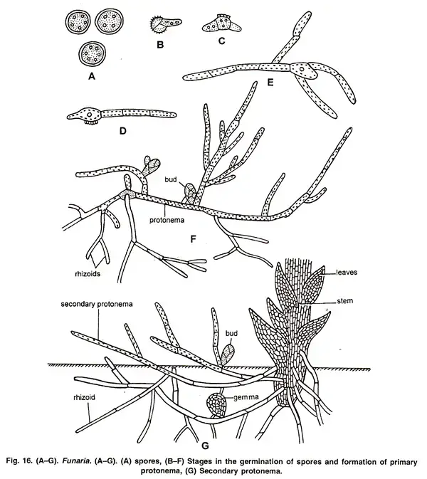
Chloronemal branches are anchoring and absorbing organs, while chloronemal branch develops tiny green buds behind cross walls that turn into leafy gametophores. Many moss plants can develop from one primary protonema, making them gregarious. The primary protonema is temporary.
Sirnoval (1947), protonema development under laboratory conditions can be divided into two stages: the chloronemal stage or caulonemal stage. The chloronemal stage features irregular branching and right angle colourless cross walls. There are also many evenly distributed chloroplasts.
Although it is phototropic, it does not produce buds. The caulonemal stage is formed shortly after the chloronemal stage has matured. This stage has regular branching brown cell walls, oblique crosses walls, and fewer chloroplasts. It produces buds that later turn into leafy gametophores. The base of a bud is where the rhizooids are.
Funaria Life Cycle
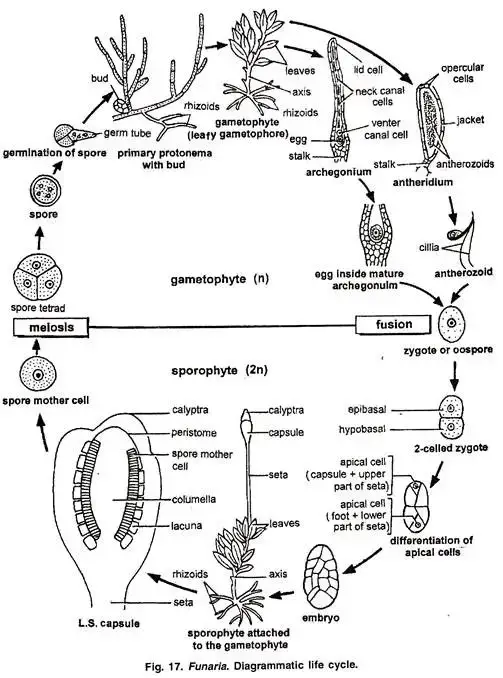
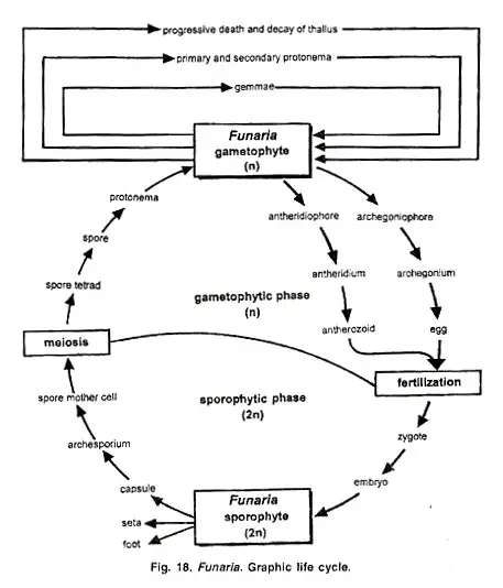
The life cycle of Funaria is characterized by an alternation of generations between the gametophyte (haploid) and sporophyte (diploid) stages, with the gametophyte being the dominant phase.
This moss exhibits a haplo-diplontic life cycle, where both the gametophyte and sporophyte are multicellular and morphologically distinct.
1. Spore Germination and Protonema Formation
- The life cycle begins when a haploid spore germinates in a suitable environment, such as moist, shaded areas.
- Upon germination, the spore develops into a filamentous structure known as the protonema.
- The protonema is green, branched, and often filamentous, serving as the juvenile stage of the moss.
- From the protonema, new gametophytes (leafy gametophores) arise, completing the transition to the adult stage.
2. Development of Leafy Gametophyte
- The protonema develops into the gametophore, which is the leafy adult form of the moss.
- The gametophore consists of an erect, slender stem with spirally arranged leaves.
- Leaves are sessile, oblong-ovate, with entire margins and pointed apices, each possessing a midrib.
- Rhizoids, which are multicellular and branched, arise from the base of the stem and anchor the plant to the substrate.
3. Sexual Reproduction
- Funaria is dioicous, meaning male and female reproductive organs are borne on separate plants.
- Male gametes (sperm) are produced in antheridia, while female gametes (eggs) are produced in archegonia.
- Fertilization occurs when sperm swim through a film of water to reach and fertilize the egg in the archegonium.
- The fertilized egg develops into a diploid zygote, which grows into the sporophyte.
4. Sporophyte Development
- The sporophyte consists of a foot, seta (stalk), and capsule.
- The foot anchors the sporophyte to the gametophyte and absorbs nutrients.
- The seta is a long, slender, and twisted structure that carries the capsule at its tip.
- The capsule is the fertile structure of the sporophyte and contains diploid archesporial cells.
- These archesporial cells undergo meiosis to produce haploid spores.
5. Spore Dispersal
- Spores are released from the capsule when environmental conditions trigger the operculum (lid) to open.
- The peristome, a structure surrounding the mouth of the capsule, aids in the controlled release of spores.
- Spores are dispersed by wind, water, or other external factors to new locations where they can germinate.
6. Vegetative Reproduction
- In addition to sexual reproduction, Funaria can reproduce vegetatively through several methods:
- Primary Protonema: Develops directly from spores and gives rise to new gametophytes.
- Secondary Protonema: Forms from fragments of the gametophyte and can develop into new gametophytes.
- Apospory: Development of gametophytes from diploid cells without meiosis.
- Bulbils: Small, bulb-like structures that can develop into new gametophytes.
- Gemmae: Asexual reproductive bodies that can develop into new gametophytes under favorable conditions.
7. Completion of Life Cycle
- The release of spores marks the completion of the life cycle.
- Spores germinate to form new protonemata, which develop into gametophytes, continuing the cycle.

- https://www.brainkart.com/article/Bryophytes_32870/
- https://biologyboom.com/type-funaria/
- https://www.biologydiscussion.com/bryophyta/features-of-funaria-with-diagram-bryophyta/54320
- https://en.wikipedia.org/wiki/Funaria
- https://www.biologydiscussion.com/botany/bryophytes/structure-of-funaria-with-diagram/46188
- https://www.biologydiscussion.com/botany/bryophytes/life-cycle-of-funaria-with-diagram-bryopsida/53931
- https://www.anbg.gov.au/bryophyte/photos-captions/funaria-hygrometrica-life-cycle.html