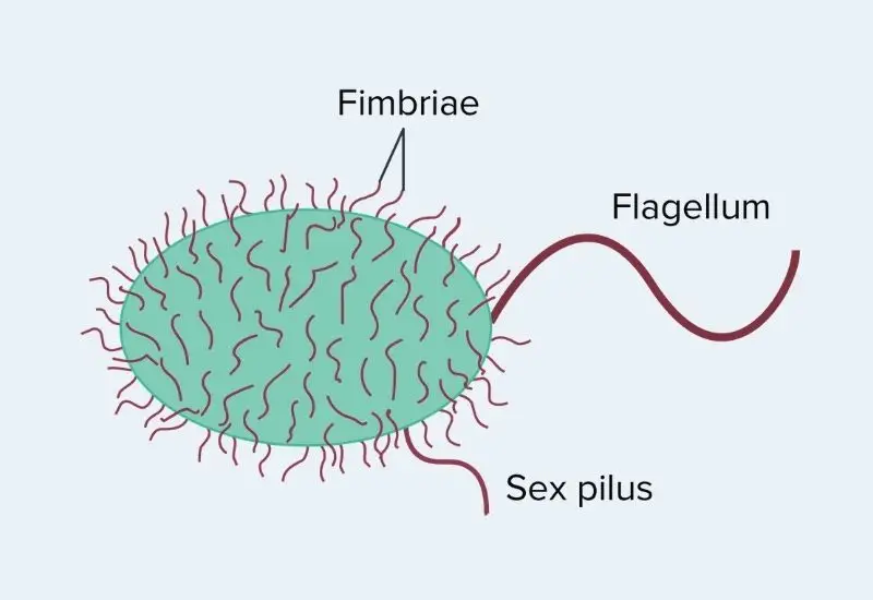
Fimbriae and pili are interchangeable words employed to indicate short, hair-like structures on the outsides of procaryotic cells. Same as flagella, they are made of protein.
Fimbriae are smaller and harder as compared to flagella, and lightly smaller in diameter. They emerge from the bacterial cell surface and involved in different functions such as adhesion, attachment, and assisting in genetic change.

Fimbriae Definition
- The fimbriae or fimbria (Singular) are bristle-like short fibers occurring on the surface of several gram-positive and gram-negative bacteria.
- It helps in attachment of bacterial cells on the surface of host cell and on some inanimate objects. For example, E. coli utilizes them to get attached to the mannose receptors.
- It is also known as “attachment pilus” and mainly found on Gram-negative and Gram-positive bacteria.
- A single bacteria may contain 1,000 fimbriae.
- They can be visualized by using only an electron microscope.
- Fimbriae enables the formation of pellicles or biofilms. Pellicles is a thin sheet of cell which is formed on the surface of liquid.
- Curli is an example of Fimbriae. It is a functional amyloid surface fibers of gram-negative bacteria and is made of curlins protein.
- Fimbriae is an important virulence factor of different bacteria such as E. coli, Bordetella pertussis, Staphylococcus, and Streptococcus.
- The main function of fimbriae is to help in the attachment of bacterial cells on a solid surface or host cell surface.
Fimbriae Types
There are present different types of Fimbriae such as;
1. Type I Fimbriae
- This type of fimbriae are mainly found on the surface of Gram-negative (E.g., E. coli) where it helps in adhesion.
- These are about 1 and 2 um long and about 7nm in diameter (width). The Type I Fimbriae are very much stiff and do not bend much like others.
- Composite structures are consist of a shorter and thin tip fibrillin which is located at the distal end of the rod.
- The helical orientation is also the characteristics feature of Type I Fimbriae. It has a right-handed helical orientation composed of 27 FimA subunits with about eight (8) helical turns.
- This types of fimbriae are also termed as mannose-sensitive or somatic fimbriae.
Functions of Type I Fimbriae
Type I fimbriae are commonly found in the family Enterobacteriaceae. They helps in the adherence of the bacteria to the cells of the host. However, this binding has been shown to be specific to glycoproteins that consist of one or N-linked high mannose structures. Hence, it assumed that the Type I fimbriae helps to promote infection of the lower urinary tract and some mucosal surfaces.
2. Type III Fimbriae
- The Type III Fimbriae are 0.5 and 2um long and 5nm in diameter.
- These are commonly found in the family Enterobacteriaceae and Klebsiella spp.
- The type III fimbriae assembly occurs through the chaperone/usher pathway. First of all, the subunits/building blocks are first transferred to the periplasm via the general secretory pathway where a chaperone encoded by the gene mrkB promotes the development and assembly of the fimbriae.
- The subunits (MrKA) of Type III fimbriae are polymerized in a helical orientation as like other pili and fimbriae.
- The C-terminal region of the subunits contain a Beta strands which provide structural support to the appendage.
Functions of Type III Fimbriae
The Type III Fimbriae helps in adhesion of bacteria to abiotic surfaces and also helps in formation of biofilm. bacteria get benefits from this, that it helps in pathogenesis by promoting antibiotic resistance among some species of bacteria.
3. Curli Fimbriae
- The Curli Fimbriae mainly found in Gram-negative bacteria such as Escherichia and Salmonella species.
- csgBA and csgDEFG operons encode proteins helps in biogenesis of the fimbriae in e. coli.
- It is extend from the outer membrane as like other fimbriae.
Functions of Curli Fimbriae
The Curli Fimbriae helps in biofilm formation. It is estimated that, this fimbriae helps in adherence of Salmonella enteritis to surfaces like stainless steel and as well as helps in formation of biofilm.
Structure of Fimbriae
- These are composed of a protein, known as fibrillin.
- Fimbriae are thinner and shorter as compared to flagella.
- Fimbriae has a molecular weight of 16,000 Daltons.
- They are 3–10 nanometers in diameter and are 0.03 to 0.14 micrometers in length.
Pili Definition
- Pili or Pillus (Plural) are hair-like appendages which are seen on the surface of many gram-negative bacteria and archaea.
- A special type of pili known as sex pili helps in bacterial conjugation.
- They are antigenic and also fragile and constantly replaced.
- These are visible under electron microscope.
- The length of a pill is ranges from 0.5- 2 um and the diameter ranges from 5-7
- The total number of pili in a single cell is limited, it is ranges from 1 – 10 per cell.
- These are only found in gram-negative bacteria.
- There are two types of Pili such as short attachment pili and long conjugation pili. The short one is known as fimbriae and the long one is known as “F” or sex pili.
Structure of Pili
- The length of a pill is ranges from 0.5- 2 um and the diameter ranges from 5-7.
- These are hollow and tubular structures.
- Pili is made of a protein known as pilin, which is an oligomeric protein.
Types of Pili
There are present two types of pili such as;
1. Conjugative Pili
- Conjugative Pili also known as the sex pili. It helps in transfer of genetic material from one cell to another during conjugation.
- Compared to the other pili, the F pilus (of F sex pilus) has been given more attention and is, therefore, better understood. Encoded by the F plasmid, the F pilus is found in “male” Gram-negative bacteria (F+).
- The microscopic studies shown that these appendages are dynamic and therefore elongate and retract continuously.
- The polymeric structure of the pilus is consist of protein pilin (VirB2) which is structurally similar to the F-like pilus pED208. Both of them are about 87 Å in diameter with an internal lumen which is about 28 Å in diameter.
- Based on the type of pili the arrangements of the proteins/building blocks can be in helical manner (five-start helical filaments) or in pentamer layers on top of each other.
- The bacterial pili contain phospholipids, and proteins, which form a protein-phospholipid complex.
- The F-pili measure about 20um in length.
Conjugative Pili Function
The Conjugative Pili helps in the transfer of DNA from one bacterial cell (male F+ ) to another (F-), that’s why they are known as sex pili. During the transfer, the single strands of DNA pass through the hollow lumen of the pilus and transported to the recipient cell. The Conjugative Pili also helps in the identification of the recipient.
Type IV pili
- Some of the pili is known as type IV pili (T4P), it generate motile forces.
- The exterior ends of the pili adhere to a solid substrate, either the solid surface to which the bacterium is appended or to another bacteria. Then, during the pili contract, they forced the bacterium ahead same as a grappling hook. Action generated by type IV pili is typically jerky, so it is termed as twitching motility, as exposed to other sorts of bacterial motility for example that generated by flagella.
- The structure of type IV pili is similar to the component flagellins of archaella (archaeal flagella) and both of them are associated to the Type II secretion system (T2SS).
- This type of pili mainly found in some Gram-positive bacteria (e.g., clostridia) and the majority of Gram-negative bacteria.
- Type IV pili have a tube-like structure, originated from the membrane.
- The polymeric structure of Type IV pili is consists of pilin protein.
- The pilin is classified into two categories such as; type IVa pilins (characterized by 5 to 7 amino acid signal peptides and phenylalanine) and type IVb pilins which consist of relatively longer peptides and a hydrophobic residue.
- The outer membrane of bacterial cells contains an oligomeric gated channel through which the pilus can pass. This channel is consists of secretin protein.
- PilN and PilO are pilus alignment subcomplex proteins which are associated with the inner membrane, also come in contact with secretin and channel the protein PilP which results in the formation of the periplasmic conduit so that the pilus can grow through.
- It also contains helical orientation with a diameter of about 6nm and 1um in length.
- Based on the type of species the pilin can be glycosylated or phosphorylated.
Type IV Pili Functions
There are different functions of Type IV Pili such as;
- Adhesion: The primary functions of Type IV pili is Adhesion. It helps in Adhesion of bacterial cell to different types of surfaces, the filament also allows the cell to adhere to other bacteria. The amino acid sequences of pilin play important role in bacterial cell adhesion.
- Motility: Motility is the another primary function of Type IV pili. The ability of the pili to retract make it possible for the bacterial cell to move along surfaces through a process known as twitching motility. This type of motility occurs in three different stages such as extension, tethering, and retraction.
- Biofilm formation: While type IV pili are involved in motility, allowing the cells to move to given sites, they have also been shown to play an important role in the formation of microcolonies and consequently biofilm maturation. They not only adhere to surfaces and cells, but also promote the close proximity between cells during biofilm formation.
- Other functions of pili: Pili also helps in Electrical conductivity, Adhesion of bacterial cells to eukaryotic cells (of a host), Protein production.
Type V Pili
- This is a unique type of pili which is mainly found in bacteria species of the class Bacteroidia.
- While the mechanism through which this structure is assembled is not clearly understood, researchers have suggested that it involves protease-mediated polymerization.
- Like Type IV pili, this pilus is also involved in adhesion and biofilm formation.
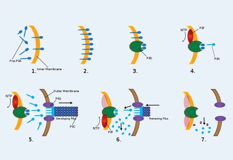
Mechanism of Twitching Motility
- Pre-PilA is occurred within the cytoplasm and then passes through the inner membrane.
- Pre-PilA is entered within the inner membrane.
- Next, a peptidase called PilD, separates a leader sequence, therefore producing the Pre-PilA shorter and into PilA, the central building-block protein of Pili.
- A NTP-Binding protein known as PilF supply energy for Type IV Pili Assembly.
- PilQ, a secretin protein, located on the outer membrane of the cell is required for the construction/enlargement of the pilus. PilC is the primary protein to create the pilus and is accountable for the overall affection of the pilus.
- Once the Type IV Pilus connects or associates with what it demands to, it starts to retreat. This happens with the PilT working to diminish the end parts of the PilA in the pilus. The processes of PilT is very related to PilF.
- Degradation of the pilus within the parts to be used and synthesized into PilA repeatedly.

Fimbriae vs Pili
| Characteristics | Fimbriae | Pili |
| Define | Fimbriae are tiny bristle-like fibers arising from the surface of bacterial cells. | Pili are hair like microfibers that are thick tubular structure made up of pilin. |
| Diameter | These are comparatively thinner in diameter. | These are thicker than fimbriae. The diameter is ranges from 5-7. |
| Length | The length id ranges from 0.03 to 0.14 um. | Length is ranges from 0.5 – 2um. |
| Number | The number of Fimbriae per cell ranges from 200 -400. | Number of pili per cell ranges from 1-10. |
| Composed of | Fimbriae is made up of Fimbrillin protein. | Pili is made up of Pilin protein. |
| Function | Helps in attachment of bacterial cell to the host cell surface or solid surface. | The sex pili Involve in bacterial conjugation. |
| Location | Fimbriae are mainly found in both gram positive and gram negative bacteria. | Pili is mainly found in gram negative bacteria and in some archae. |
| Formation | Is governed by bacterial genes in the nucleoid region. | Is governed by plasmid genes. |
| Rigidity | Fimbriae are Less rigid as compared to pili. | Pili are More rigid than fimbriae. |
| Motility | Don’t involve in Motility. | The type IV pili shows twitching type of motility. |
| Receptors | No receptors. | Functions as receptor for certain viruses. |
| Structure | These are solid structure. | These are hollow tubular structure. |
| Distribution of Cell Surface. | These are evenly distributed on the entire cell surface. | These are randomly distributed on surface of cell. |
| Examples | Salmonella typhimurium, Shigella dysenteriae. Shigella dysenteriae uses its fimbriae to attach to the intestine and then produces a toxin that causes diarrhea. | Escherichia coli, Neisseria gonorrhoeae. Neisseria gonorrhoeae, the cause of gonorrhea, uses pili to attach to the urogenital and cervical epithelium when it causes disease. |
References
- https://en.wikipedia.org/wiki/Fimbria_(bacteriology)
- http://textbookofbacteriology.net/structure_3.html
- https://bio.libretexts.org/Bookshelves/Microbiology/Book%3A_Microbiology_(Kaiser)/Unit_1%3A_Introduction_to_Microbiology_and_Prokaryotic_Cell_Anatomy/2%3A_The_Prokaryotic_Cell_-_Bacteria/2.5%3A_Structures_Outside_the_Cell_Wall/2.5C%3A_Fimbriae_and_Pili
- https://byjus.com/neet/fimbriae-and-pili
- https://www.easybiologyclass.com/difference-between-pili-and-fimbriae-of-bacteria-a-comparison-table/
- https://microbiologynotes.com/differences-between-fimbriae-and-pili/
- https://www.microscopemaster.com/pili-and-fimbriae.html
- Text Highlighting: Select any text in the post content to highlight it
- Text Annotation: Select text and add comments with annotations
- Comment Management: Edit or delete your own comments
- Highlight Management: Remove your own highlights
How to use: Simply select any text in the post content above, and you'll see annotation options. Login here or create an account to get started.