Fasciola hepatica – Classification
| Kingdom | Animalia |
| Phylum | Platyhelminthes |
| Class | Trematoda |
| Subclass | Digenea |
| Order | Echinostomida |
| Genus | Fasciola |
| Species | hepatica |
Fasciola hepatica
- Fasciola hepatica, also referred to as the common liver fluke or sheep liver fluke, is a parasitic trematode of the class Trematoda, phylum Platyhelminthes. It infects the livers of numerous mammals, including humans, and is transmitted worldwide by sheep and cattle.
- This neglected tropical disease is known as fasciolosis or fascioliasis. It is a form of helminthiasis.
- Currently, fasciolosis is classified as a plant/food-borne trematode infection, typically acquired by ingesting the parasite’s plant-encrusted metacercariae.
- F. hepatica, which is found worldwide, has been recognized for decades as a significant parasite of sheep and cattle, causing up to £23 million in economic losses in the United Kingdom alone.
- Due to its comparatively large size and economic significance, it has been the subject of numerous scientific studies and may be the most well-known species of trematode.
- Fasciola gigantica is the closest relative of F. hepatica. These two flukes are sister species; they share numerous morphological characteristics and can mate.
General Characteristics
- As an endoparasite, Fasciola hepatica is located in the bile duct of the liver of sheep.
- The body is dorso-ventrally compressed and resembles a leaf, measuring approximately 25-30 mm in length and 4-5 mm in width.
- The anterior extremity forms a conical protrusion known as the cephalic cone.
- The mouth is located anteriorly and ventrally, and it is encompassed by oral sucker.
- A muscular ventral sucker or acetabulum is located slightly behind the oral sucker.
- The posterior end is large and frontally more convex than it is posteriorly.
- The digestive system includes the esophagus, the pharynx, and the diverticulated intestine.
- The location of the excretory pore is at the posterior extremity.
- They are hermaphrodites, possessing both male and female reproductive organs.
- Between the ventral and oral suckers, there exists a median genital pore through which the ova exit.
Habitat of Fasciola hepatica
- Fasciola hepatica, also known as the common liver fluke, has a complex lifecycle that involves several different hosts and habitats. As an adult parasite, it primarily resides in the bile ducts of the liver of its definitive host, which can include a range of mammals, including humans, sheep, cattle, and other domesticated animals. The adult fluke feeds on blood and tissue fluids and reproduces by releasing eggs that pass out of the host through the feces.
- Once outside the host, the eggs hatch into larvae that infect a suitable intermediate host, typically a snail, where they develop into free-swimming cercariae that are released into the water. These cercariae then infect the definitive host through ingestion of contaminated water or vegetation.
- In addition to its hosts, F. hepatica can also be found in various environmental habitats, such as wetlands, marshes, and other areas with standing or slow-moving water, which are suitable for the snail intermediate hosts. The parasite has a wide distribution and can be found in temperate regions throughout the world, although it is most prevalent in areas with wet, marshy conditions.
Hosts of Fasciola hepatica
- Fasciola hepatica and F. gigantica are parasites of domestic and untamed ruminants (typically sheep, cattle, and goats; also camelids, cervids, and buffalo). Infections occasionally occur in equids, lagomorphs, macropods, and rodents, which are aberrant, non-ruminant herbivore hosts. Detection of Fasciola spp. ova in the feces of carnivores is likely an artifact of consumption of contaminated liver.
- The intermediary snail hosts for Fasciola species are members of the family Lymnaeidae, specifically Lymnaea, Galba, Fossaria, and Pseudosuccinea. At least 20 species of snail have been identified as intermediate hosts for one or more species of Fasciola. The suitability of snail species to function as intermediate hosts for F. hepatica versus F. gigantica may vary; research into the host ranges of both Fasciola spp. is ongoing.
History and distribution of Fasciola hepatica
- Fasciola hepatica, also known as the common liver fluke, is a parasitic flatworm that infects a range of mammals, including humans, as well as some birds. It is primarily found in temperate regions worldwide, with the highest prevalence in areas with wet, marshy conditions.
- The history of F. hepatica dates back to ancient times, with evidence of its presence found in the remains of human and animal mummies from ancient Egypt. The parasite has also been identified in the remains of prehistoric animals, such as the woolly mammoth. F. hepatica has been recognized as a veterinary and human pathogen since the early 19th century, with the first recorded case of human infection reported in the 1860s in Europe.
- Today, F. hepatica is found throughout much of the world, particularly in regions with high levels of livestock production. It is prevalent in Europe, Asia, Africa, and the Americas, and is responsible for significant economic losses in the agricultural sector due to decreased productivity and the cost of treating infected livestock. In humans, F. hepatica infection is most common in developing countries, particularly in areas where people consume raw or undercooked freshwater plants, such as watercress, that have been contaminated with the parasite.
Epidemiology of Fasciola hepatica
- When cyst-covered aquatic vegetation is consumed or when water containing metacercariae is consumed, infection begins. F. hepatica causes disease in ruminants frequently in the United Kingdom between March and December.
- By consuming watercress or the Peruvian beverage ‘Emoliente’, which contains drops of watercress liquid, humans become infected. When cattle and sheep consume the infectious stage of the parasite from low-lying, damp pasture, they become infected.
- Infections in humans have been reported from more than 75 countries worldwide. In Asia and Africa, both F. hepatica and F. gigantica cause human fasciolosis, whereas in South and Central America and Europe, only F. hepatica is responsible.
- Bovine tuberculosis detection can be hindered by the presence of F. hepatica. Cattle co-infected with F. hepatica have a weaker reaction to the single intradermal comparative cervical tuberculin (SICCT) test than those infected with M. bovis alone. Consequently, an infection with F. hepatica can make it difficult to detect bovine tuberculosis; this is, of course, a significant issue in the agricultural sector.
Morphology of Fasciola hepatica/Structure of Fasciola Hepatica
Shape, Size and Colour
- The body of F. hepatica is slender, leaf-shaped, dorsoventrally flattened, elongated, and oval. It is approximately 25 to 30 mm long and 4 to 12 mm wide.
- The maximum width occurs in the anterior third of the body, where the body tapers anteriorly and posteriorly; however, the anterior end is somewhat convex, whereas the posterior end is sharply pointed. The greatest breadth of F. indica occurs roughly in the middle of the body, and the tail is rounded. It is typically pinkish in color, but appears brownish due to the host’s bile.
External Morphology
- F. hepatica is an animal with a delicate, leaf-like body and bilateral symmetry. They range in length from 1 to 2.5 centimeters and width from 1 centimeter.
- The anterior end of the body is extended into a prominent conical projection known as the oral cone or head lobe, which bears at its apex a triangular opening, the mouth, and is surrounded by the oral or anterior sucker.
- On the ventral surface, slightly posterior to the head lobe, is a much larger sucker known as the ventral or posterior sucker (also known as the acetabulum). The genital orifice, through which the penis occasionally protrudes, is located between the two suckers and near the posterior sucker. The excretory aperture, which is a singular opening, is located at the body’s extreme posterior tip.
- The Laurel canal opens in the center of the dorsal surface. Spinules or papillae, which are extensions of the cuticle that surrounds the body, protrude from the body’s surface.
Internal Morphology
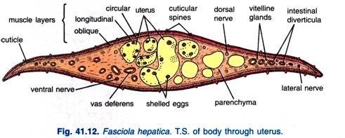
The architecture of F. hepatica’s body wall is unique and consists of the following sequence of layers:
- the external layer of the skin, the epidermis (formerly known as the cuticle), from which spinules originate;
- A subterranean membrane;
- Circular, longitudinal and oblique or diagonal muscle fibres; and
- A collection of unicellular gland cells that contain protoplasmic processes. Interorgan spaces are densely populated with parenchyma cells. Numerous ectodermal dells are sunk into the parenchyma and linked to the cuticle by protoplasmic projections.
Body Wall of Fasciola Hepatica
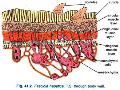
In contrast to turbellarians, the body wall of F. hepatica is devoid of an epidermal cellular layer. However, it is composed of a dense layer of cuticle, a thin basement membrane, and layers of muscle beneath the mesenchyme.
1. Cuticle
- A tough, resistant cuticle composed of a uniform layer of scleroprotein protects the fluke from the host’s digestive fluids. It is covered with tiny spines, spinules, or scales. The spinules secure the fluke to the biliary duct of the host, offer protection, and aid in locomotion.
- F. indica has broad, robust, and angular scales on its cuticle. The epidermis has been lost during the cercaria stage’s development. However, special mesenchymal cells located beneath muscle layers secrete the cuticle. It was believed that these cuticle-secreting cells were recessed epidermal cells.
2. Basement Membrane
- The basement membrane is the thinnest and most vulnerable layer of the cuticle. It marks the boundary between the epidermis and the muscle layer.
3. Muscle Layer
- Following the basement membrane is the sub-cuticular musculature. It consists of an outer layer of circular muscle fibers, a middle layer of longitudinal muscle fibers, and an interior layer of diagonal muscle fibers, which are more developed in the frontal half of the body. Every muscle is fluid. In the suckers, the muscles form robust bundles of radial fibers.
4. Mesenchyme
- Below the muscles is parenchyma (mesenchyme) composed of numerous loosely arranged uninucleate and bi-nucleate cells with a fluid-filled syncytial network of fibres.
- Some of these cells are enormous and have large processes that extend to the base of the cuticle, where they are said to secrete. In fact, the mesenchyme acts as filler between the muscle layer and internal organs. It facilitates the transport of nutrients and wastes.
- The body wall serves an important role in fluke physiology. It provides protection, is the site of gaseous exchange, diffuses out various nitrogenous wastes, and aids in the assimilation of amino acids to some extent.
5. Structure of Body Wall Under Electron Microscope
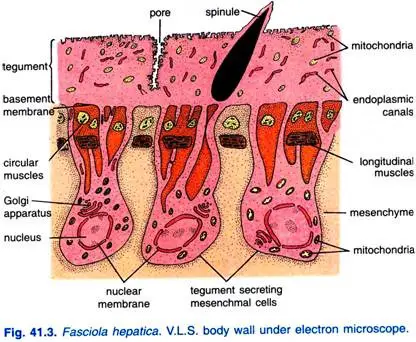
- Threadgold (1963), Bils (1966), and Martin (1966) determined via electron microscopy that the cuticle of F. hepatica is a syncytial layer of protoplasm containing mitochondria, endoplasmic canals, vacuoles, and pinocytic vesicles. Cuticle is now referred to as the integument due to its metabolic activity. The tegument is continuous with tegument-secreting cells located within the mesenchyme.
- In order to facilitate the absorption of the host’s fluid, the outer surface of the integument is dissected into numerous fine projections. Additionally, the tegument has numerous minute capillary canals through which dissolved substances are absorbed into the mesenchyme.
Digestive System of Fasciola Hepatica
1. Alimentary Canal
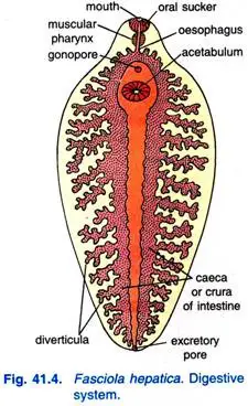
- The oral sucker encloses a ventral mouth that leads to a funnel-shaped mouth cavity, followed by a muscular, round pharynx with robust walls and a small lumen. There are pharyngeal glands in the pharynx. From F. indica’s short, muscular pharynx originates an oral pouch that is roughly half the size of the pharynx.
- A short, slender oesophagus connects to an intestine that divides into two branches or intestinal caeca or crura, each running to the posterior end and terminating blindly. As there is no circulatory system, the intestinal caeca release a number of branching diverticula to transport food to all regions of the body. The median diverticula are brief, whereas the lateral diverticula are long and branching. The absence of an anus.
- The inner portion of the alimentary canal up to the oesophagus is lined with cuticle and functions as a suctorial foregut; the intestine is lined with endodermal columnar epithelial cells. The caceal epithelium contains glandular cells.
2. Food, Feeding and Digestion
- It consumes bile, blood, lymph, and dead cells. Together, the oral sucker and pharynx comprise an efficient sucking apparatus. Extracellular digestion occurs in the intestine. As this animal lacks a circulatory system, the digested food material is distributed to all body regions via branching diverticula of the intestine. Consequently, the digestive system serves as a gastrovascular system.
- Through intestinal diverticula, digested nutrients are transferred to the parenchyma; from there, they are diffused to the various organs of the body.
- In the parenchyma, dietary reserves, primarily in the form of glycogen and fats, are stored. However, monosaccharide carbohydrates such as glucose, fructose, etc. are directly diffused into the body of the fluke from the surrounding fluid of the host via the general body surface. If there are any, the indigestible remnants of the food are likely expelled through the pharynx.
Respiration of Fasciola Hepatica
The respiration process is anaerobic or anoxybiotic. In reality, glycogen is metabolized into carbon dioxide and fatty acids, releasing heat-based energy.
The procedure consists of the subsequent steps:
- Glycogen undergoes anaerobic glycolysis in order to produce pyruvic acid.
- Pyruvic acid is decarboxylated in order to produce carbon dioxide and an acetyl group.
- The acetyl group then combines with coenzyme A to form acetyl coenzyme A, and (iv) acetyl coenzyme A is then transported to the mitochondria.
- The acetyl coenzyme A is then reduced and condensed to produce fatty acids.
- Thus, carbon dioxide is diffused out through the body’s general surface, and fatty acids are excreted via the excretory system.
Excretory System of Fasciola Hepatica
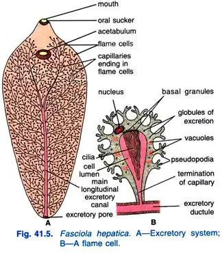
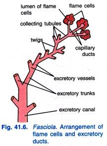
Both excretion and osmoregulation are priorities for Fasciola hepatica’s excretory system. It is made up of several flame cells, flame bulbs, or protonephridia that are connected by a network of excretory channels.
1. Flame Cells
- The flame cells, which are thought to be modified mesenchymal cells, are numerous, irregularly shaped bulb-like bodies distributed throughout the mesenchyme of Fasciola’s body. The distribution pattern of flame cells conforms to a particular pattern known as the “flame cell pattern” (Faust, 1919).
- Each flame cell has a thin elastic wall with pseudopodia-like processes, a nucleus, and an intracellular cavity with a large number of long cilia originating from basal granules; these characteristics are defining. The cilia in a living organism vibrate like a wavering flame, hence the name flame cell.
2. Excretory Ducts
- There is an excretory pore at the posterior end from which arises a longitudinal excretory canal, from which arise four main branches, two dorsal and two ventral, that subdivide into numerous small capillaries that anastomose; the capillaries continue into the intracellular cavity of flame cells. The longitudinal excretory canal is not lined with cilia, but the capillaries are.
3. Process of Excretion
- The excretory byproducts, which are typically fatty acids and ammonia, diffuse from the surrounding mesenchyme into the flame cells, where they are subsequently accumulated within their intracellular cavities. By hydrostatic pressure, impurities move from the intracellular cavities of flame cells into the excretory ducts, then into the main excretory canal, and finally to the exterior through the excretory pore, as a result of the vibrating movement of the cilia.
- This excretory system of flame cells and canals or ducts of various orders with no internal opening and leading to an excretory pore that opens to the exterior is referred to as a protonephridial system, whose primary function is to regulate the amount of fluid within the animal’s body.
Nervous System of Fasciola Hepatica
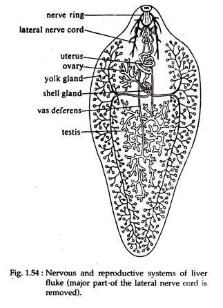
- The oesophagus is surrounded by a nerve ring, which contains a pair of cerebral ganglia dorsolaterally and a ventral ganglion beneath the oesophagus. The ganglia distribute small nerves anteriorly. Three pairs of longitudinal nerve cords arise posteriorly from the ganglia: a dorsal, lateral, and ventral pair.
- The lateral nerve fibers are the most developed and extend to the back. Nerve cords are joined by transverse commissures and give off numerous minor branches, of which some form plexuses. The majority of nerve cells are bipolar. Fasciola adults lack sensory organs due to their parasitic lifestyle.
Reproductive System of Fasciola Hepatica
Fasciola hepatica is hermaphrodite, but cross-fertilization typically occurs. The reproductive organs are complex and well-developed.
1. Male Reproductive System of Fasciola Hepatica
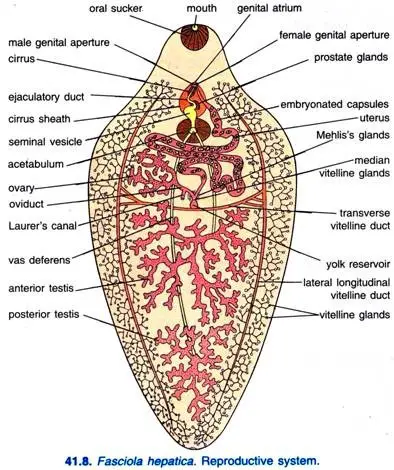
Testes, vasa deferens, seminal vesicle, ejaculatory duct, cirrus or penis, prostate glands, and genital atrium make up the male reproductive system.
a. Testes
- These are two in number, highly ramified tubular structures located one behind the other (i.e., in tandem) in the body’s posterior middle region. In fact, they occupy a considerable amount of space behind the middle portion of Fasciola’s body. The cells that line the testicular wall give birth to spermatozoa.
b. Vasa Deferentia
- Each testis produces a vas deferens or sperm duct that travels forward and is slender and narrow.
c. Seminal Vesicle
- The two vasa deferentia unite near the acetabulum (ventral sucker) and enlarge to form the vesicula seminalis, a muscular, elongated, broad, bag-like seminal vesicle. It provides the function of sperm storage.
d. Ejaculatory Duct
- The seminal vesicle continues anteriorly into the ejaculatory duct, a very narrow and convoluted duct.
e. Cirrus
- The ejaculatory duct emerges into the muscular and elongated cirrus of the penis. The cirrus opens through the male genital orifice in a shared genital atrium. F. indica’s cirrus is coated in small spines.
f. Prostate Glands
- A large number of unicellular prostate glands encircle the ejaculatory duct. The alkaline secretion of these glands, which opens into the ejaculatory duct, facilitates the free migration of sperm during copulation.
g. Genital Atrium
- The genital atrium is a chamber shared by male and female genital openings; it is externally accessible via a gonopore located ventrally in front of the acetabulum. During copulation, the cirrus can be expelled through the gonopore. The penis, seminal vesicle, and prostatic organs are encased in a cirrus sheath or sac.
2. Female Reproductive System of Fasciola Hepatica
The female reproductive system consists of the ovary, the oviduct, the uterus, the vitelline glands, Mehlis’ glands, and Laurer’s canal.
- Ovary: The singular, tubular, and highly branched ovary is located anterior to the testes on the right side of the anterior one-third of the body.
- Oviduct: All ovarian branches terminate in a short, slender conduit known as the oviduct. The oviduct descends perpendicularly and connects to the median vitelline duct.
- Uterus: From the confluence of the oviduct and median vitelline duct arises a large, convoluted uterus containing fertilized eggs or capsules. The uterus opens into the common genital atrium on the left side of the male genital aperture via the female genital aperture. The uterus is relatively small and located in front of the testes and ovaries. The terminal portion of the uterus, known as the metraterm, has muscular walls that eject the ova and sometimes receive the cirrus during copulation.
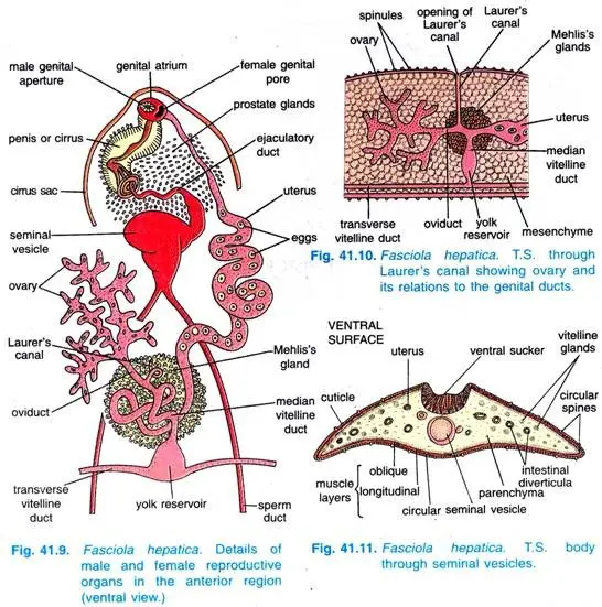
- Vitelline Glands: On both lateral sides and behind the testes are numerous follicles that make up the vilellaria, yolk glands or vitelline glands, which produce albuminous egg yolk and egg shell material. Each side of the vitelline glands opens via minute conduits into a longitudinal vitelline duct. The two longitudinal ducts are joined by a transverse vitelline duct located above the body’s midline. The center of the transverse vitelline duct is enlarged to form the yolk reservoir or vitelline reservoir. A median vitelline duct emerges from the yolk reservoir and travels forward to join the oviduct.
- Mehlis’s Glands: Around the junction of the median vitelline duct, oviduct, and uterus, there is a cluster of numerous unicellular Mehlis’s glands. The secretion of Mehlis’ glands lubricates the passage of eggs through the uterus and presumably hardens the egg shells; it also likely stimulates spermatozoa. According to some authorities, the junction of the oviduct and the median vitelline duct is enlarged to form an ootype in certain flukes, such as F. indica, in which the egg parts are assembled and the eggs are shaped, but an ootype is absent in F. hepatica.
- Laurer’s Canal: The oviduct gives rise to a narrow Laurer’s canal that ascends vertically. This canal temporarily opens on the dorsal side during reproductive season and serves as a vestigial vagina and copulation canal.
Life Cycle of Fasciola Hepatica
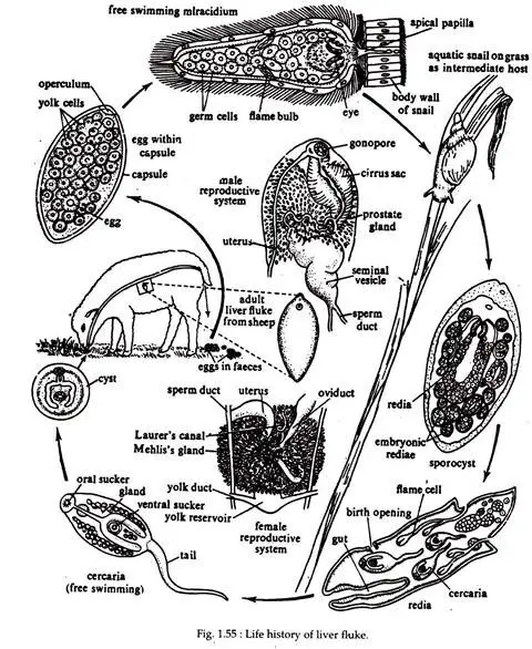
F. hepatica undergoes indirect development, involving four varieties of free-swimming and parasitic larval stages. Fasciola is digenetic, and its life cycle consists of at least two infective stages at all times. Two or more hosts are infected prior to the completion of its life cycle. The definitive or primary host is a vertebrate (sheep), whereas the intermediate host is a gastropod (Lymnaea, Planorbis, etc.).
Fertilization
- Self-fertilization is uncommon even though Fasciola is hermaphrodite. During copulation, there is a mutual exchange of sperm, and cross-fertilization is the norm. The site of copulation is the biliary duct of the host. During copulation, the cirrus or penis of one worm is inserted into the uterus of another via the gonopore.
- Additionally, Laurer’s canal copulation has been reported. Thus, sperms are ejaculated. The prostate organ produces sperm-sustaining semen. The sperm then ascend the uterus via the ootype and are retained in the seminal receptacle.
Release of Fertilized Eggs from Primary Hosts (sheep) Body
- The embryos are fertilized either in the oviduct or within the ootype after being released from the ovary. The yolk cells and vitelline secretions supply each egg with a sufficient amount of yolk. It is eventually encased in a proteinaceous shell or capsule, which is secreted by the shell glands.
- The carapace becomes rigid upon entering the uterus. The hardening is brought about by quinone. One pole of the egg shell contains a small cap or operculum for the future larva’s egress. Thus, the embryo becomes mature and spends a brief time in the uterus.
- Eventually, the egg exits the fluke’s body through its gonopore and travels through the sheep’s bile ducts into the intestine, where it is expelled along with the feces. The embryo can only survive if it lands on moist soil.
Miracidium Larva
- At this stage, active development within the zygote commences. The egg shell opens at the operculum after three to six weeks, depending on the temperature, and the miracidium larva emerges. The body of the miracidium larva is somewhat conical and covered in vibrating cilia.
- At the broad extremity, a distinct head lobe or apical papilla is located. Behind the head lobe are two pigmented patches, the eye spots. The mesenchyme consists of delicate layers of circular and longitudinal muscle fibers located just beneath the epidermis.
- There is also a pair of flame cells with ducts and an intestinal sac. The remainder of the interior is full with a mass of infectious organisms. The cranium lobe contains glands for penetration. The miracidium larva swims freely in water or crawls over a moist surface for a period of time before dying if it does not encounter a specific water snail (preferably Limnaea truncatula).
Infection to Secondary Host (Snail) of Fasciola Hepatica
Sporocyst
- The miracicium larva, upon encountering the snail, enters through the snail’s penetration ducts and travels to the internal organs, particularly the pulmonary sac. It transforms into a sporocyst by shedding its ciliated covering.
- The sporocyst has an internal cavity containing germ balls and is enclosed by a layer of cells with remnants of eye spots and flame cells. Eventually, the germ balls endure cleavage, which results in the formation of redia larva (first generation).
Redia Larva
- Redia has a mouth, pharynx, and a rudimentary intestine, as well as an excretory vessel system. Near the anterior end, a circular ridge or collar is formed by the thickening of the body wall. Redia’s mature body is elongated and exhibits a pair of short, muscular projections on its posterior side.
- In the body’s interior are undifferentiated germ balls, which, depending on the season, give birth to the second generation of redia or cercaria larva, respectively. In the wall of the redia, an opening near the collar known as the birth pore allows the cercaria to exit.
Cercaria Larva
- The cercaria have a lengthy tail as well as oral and ventral suckers. The well-developed alimentary canal consists of the mouth, pharynx, and a bifid intestine. There are also paired excretory tubules with flame cells, germ spheres, and peripheral cyst-forming cells.
- They use their tail to move actively and force their way out of the snail’s body. Then, without a tail, they become encysted. The encysted cercaria is referred to as metacercaria larva.
Infection to New Primary Host (Sheep) of Fasciola Hepatica
- The metacercaria remains attached to grass stalks or other herbaceous plant leaves. When a ruminant consumes these infected green leaves, metacercaria enters its digestive tract. The young fluke emerges from the cyst and bores its way through the intestinal wall, passes over the viscera, and penetrates the liver and bile duct, where it reaches sexual maturity and grows swiftly.
- After successfully invading the intermediate host (snail), it is evident from the life cycle of F. hepatica that Fasciola’s developmental stages endure two rounds of asexual division. Once via the formation of one or two generations of numerous redia, then via the formation of numerous cercariae.
- This significantly increases their population, thereby improving their chances of completing their life cycle. Thus, a single embryo is capable of producing dozens of sexual adults in the definitive host.
Fasciolosis
- F. hepatica and F. gigantica are both capable of causing fasciolosis. Depending on whether a disease is acute or chronic, a person may display a variety of symptoms. During the acute phase, immature worms invade the gut, causing fever, nausea, liver enlargement (caused by Fh8), skin rashes, and severe abdominal discomfort.
- When the worms mature in the bile duct, they enter the chronic phase, which can cause intermittent pain, jaundice, and anemia. Classic indications of fasciolosis in cattle and sheep include persistent diarrhea, chronic weight loss, anemia, and decreased milk production.
- Some continue to lack symptoms. Due to internal hemorrhaging and liver damage, F. hepatica can cause sudden mortality in both sheep and cattle.
- Fasciolosis is a significant cause of economic and production losses in the dairy and livestock industries. The prevalence has increased over time and is likely to continue doing so in the future.
- Flukicides, which are chemicals that are toxic to flukes and include bromofenofos, triclabendazole, and bithionol, are commonly used to treat livestock. Ivermectin, which is commonly used to treat many helminthic parasites, and praziquantel have low efficacy against F. hepatica.
- The type of human control depends on the environment. The strict regulation of the growth and distribution of consumable water plants, such as watercress, is a vital method. This is especially crucial in highly endemic regions. Some farms are irrigated with contaminated water; therefore, vegetables grown on these farms must be thoroughly cleansed and cooked before consumption.
- The best method to prevent fasciolosis is by reducing the population of lymnaeid snails or isolating livestock from areas containing these snails.[39] These two methods are not always the most effective, so it is common practice to treat the herd before they become potentially infected.
Clinical Presentation
- Infection with Fasciola spp. in humans occurs in two phases, which may or may not be accompanied by symptoms or other clinical manifestations.
- During the early phase of the infection (typically referred to as the acute phase; also known as the migratory, invasive, hepatic, parenchymal, or larval phase), when the fluke larva migrates from the intestines to the liver parenchyma, inflammation, tissue destruction, and toxic/allergic reactions may occur.
- It is possible to develop nonspecific symptoms and indications (such as abdominal pain, nausea, vomiting, hepatomegaly, malaise, fever, and cough) and laboratory abnormalities (such as peripheral eosinophilia and elevated transaminase levels). On occasion, fluke larvae migrate to ectopic locations, including the lungs, subcutaneous tissue, pancreas, genitourinary tract, eyes, and brain.
- During the chronic phase of the infection (also known as the biliary or adult phase), clinical manifestations, if any, may develop months to years after exposure and may include intermittent inflammation or occlusion of the bile ducts or gallbladder (e.g., cholangitis, cholecystitis). Pancreatic inflammation may also occur.
Diagnosis
- The presence of yellow-brown eggs in the faeces can be used as a diagnostic indicator. They are identical to the eggs of Fascioloides magna, despite the fact that the eggs of F. magna are rarely passed by sheep, goats, or cattle.
- If a patient has consumed infected liver and the eggs travel through the body and out through the feces, a false-positive test result is possible. This false diagnosis will be unmasked by daily examinations during a liver-free diet.
- The preferred diagnostic test is the enzyme-linked immunosorbent assay (ELISA) test. ELISA is commercially available and can detect antihepatica antibodies in serum and milk; new assays designed to detect antihepatica antibodies in feces are being developed.
- ELISA is more precise than Western blotting and Arc2 immunodiffusion. F. hepatica-secreted proteases have been used experimentally to immunize antigens.
Parasitic Adaptations of Fasciola Hepatica
As an endoparasite that resides in the liver and bile ducts of sheep, Fasciola hepatica is well adapted to its parasitic lifestyle. In fact, the adult fluke displays a number of adaptive characteristics, and its various life stages also account for a number of adaptive characteristics.
Therefore, Fasciola hepatica’s adaptive characteristics can be discussed under the following two headings:
1. Adaptations of the Adult Fluke
- Its dorsoventrally compressed, leaf-shaped body increases the body’s surface area for increased diffusion of substances through the body’s fluid.
- Its body is clothed with a thick cuticle that protects it from the antitoxins of its host.
- Cilia are absent in adult flukes.
- Well-developed adhesive organs such as suckers (anterior sucker and ventral sucker) that provide a firm attachment to the recipient tissue. Numerous cuticular spines on its body erode the host tissue that serves as its sustenance and also prevent the fluke from being expelled from the bile ducts.
- It has an anteriorly located mouth and a muscular pharynx for drawing nutrients from the host body. Since it nourishes on pre-digested and digested substances from the host, its alimentary canal and digestive glands are not well developed.
- Since there is no digestion, there is no anus; consequently, there is no circulatory system, as the various organs of the alimentary canal (intestine and its various branches) do not distribute the already digested food substances to the various portions of the body.
- Because it resides in an oxygen-depleted environment, anaerobic respiration occurs; respiratory organs are wholly absent.
- As flukes are endoparasites, its nervous system is very rudimentary and its sense organs are completely absent.
- There are no locomotory organs in flukes because they live in a protected environment.
- The excretory system is comprised of a complex network of branched tubules to facilitate the collection of the body’s various metabolic byproducts.
- The reproductive system is highly developed and optimally tailored to the parasitic lifestyle of the organism.
- Since the adult fluke resides in the body of the sheep, it may perish along with the animal. Therefore, a secondary host is required for the transmission of the parasite from one host to another, so that the parasite’s race can continue. The snail is therefore the secondary host.
2. Adaptations in Life History
Fasciola hepatica’s numerous parasitic adaptation traits can be traced back to the following stages of the parasite’s life cycle:
- One solution to the problem of eggs being wasted during transfer is to produce an extremely high volume of eggs.
- The fertilized eggs are protected from the enzymes of the host by a chitinous covering, the shell, which allows them to travel down the bile duct into the gut of the sheep and subsequently out with its feces. Eggs with a protective shell are known as capsules.
- The first larvae to emerge from the capsules are called miracidia, and they are perfectly suited to a life of free swimming (thanks to their ciliated bodies and the development of eye spots) and to entering the body of the secondary host, Limnaea, Planorbis, etc. (thanks to their penetration glands).
- The sporocyst is a parasite whose body is wrapped in a cyst-like structure to avoid being digested by the snail’s digestive enzymes. The sporocyst’s germ balls develop into rediae, which can then give rise to either more rediae or cercariae.
- Five, rediae (lappets or procruscula) and cercariae (tail) may travel and find their way into the fresh tissues of the snail thanks to their locomotory organs.
- Six, after escaping the snail’s body, the cercariae have a brief period of freedom before returning to a cyst they’ve secreted on plants.
- Due to their herbivorous diet, sheep are a perfect host for cysted cercariae termed metacercariae that live on plant matter.
- The cyst provides the metacercariae with protection from the elements, allowing them to wait for admission into the sheep’s body for an extended period of time.
- The larvae’s parthenogenetic mode of reproduction also guarantees the survival of their species.
- The rapid reproductive rate, specialized larvae, sexual and asexual reproduction, and morphological and physiological adaptations of the fluke all serve a single purpose: to guarantee the species’ continued existence.
Life Cycle of Fasciola Hepatica Summery
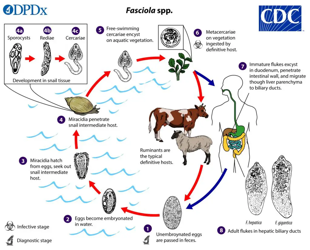
- The biliary ducts secrete immature eggs, which are then excreted in the feces. Over the course of around two weeks, eggs in freshwater develop into embryos, and the miracidia they produce infect a snail intermediate host.
- There are various stages of parasite development in the snail (images of sporocysts, rediae, and cercariae).
- As metacercariae, the cercariae leave the snail and encyst on aquatic plants or other surfaces. Consumption of infected plants (like watercress) is the primary route of infection for humans and other mammals.
- A metacercariae’s first stop after ingestion is the duodenum, where it will excyst and then go on to the peritoneal cavity.
- The juvenile flukes enter the biliary ducts via the liver parenchyma and develop into adult flukes that lay eggs.
- It takes about 3 months for human hosts to develop from metacercariae to adult flukes; F. gigantica may take longer to mature than F. hepatica.
FAQ
What is Fasciola hepatica?
Fasciola hepatica, also known as the common liver fluke, is a parasitic flatworm that infects a range of mammals, including humans, as well as some birds.
How is F. hepatica transmitted?
F. hepatica is transmitted through the consumption of contaminated water or vegetation, particularly freshwater plants such as watercress, that have been contaminated with the parasite.
What are the symptoms of F. hepatica infection in humans?
Symptoms of F. hepatica infection in humans can include abdominal pain, diarrhea, fever, and fatigue. In severe cases, the infection can lead to liver damage, jaundice, and other serious complications.
How is F. hepatica diagnosed in humans?
F. hepatica infection is typically diagnosed through a combination of clinical evaluation, laboratory testing, and imaging studies, such as ultrasound or CT scans, to detect evidence of liver damage or the presence of the parasite.
What is the treatment for F. hepatica infection?
The primary treatment for F. hepatica infection in humans is a course of antiparasitic medications, such as triclabendazole, which is effective against the adult parasite in the liver.
Can F. hepatica infection be prevented?
F. hepatica infection can be prevented through a range of measures, including avoiding consumption of contaminated water or vegetation, thoroughly cooking meat, and practicing good hygiene and sanitation.
What is the economic impact of F. hepatica in livestock?
F. hepatica infection can have a significant economic impact on the agricultural sector, particularly in areas with high levels of livestock production, due to decreased productivity and the cost of treating infected animals.
What is the lifecycle of F. hepatica?
The lifecycle of F. hepatica involves several different hosts and habitats, with the adult parasite residing in the bile ducts of the liver of its definitive host, while the intermediate hosts are typically snails.
Is F. hepatica infection common in humans?
F. hepatica infection is relatively uncommon in humans, particularly in developed countries, although it remains a significant public health concern in many developing countries.
Can F. hepatica infection be fatal?
In rare cases, F. hepatica infection can lead to serious complications, including liver damage and other life-threatening conditions, particularly in individuals with weakened immune systems or underlying medical conditions.
References
- https://www.cdc.gov/dpdx/fascioliasis/index.html
- https://byjus.com/biology/fasciola-hepatica-diagram/
- https://en.wikipedia.org/wiki/Fasciola_hepatica
- https://www.biologydiscussion.com/invertebrate-zoology/phylum-platyhelminthes/fasciola-hepatica-habitat-structure-and-life-history/28888
- https://www.notesonzoology.com/animals/study-notes-on-fasciola-hepatica-platyhelminthes/1735
- https://www.notesonzoology.com/zoology/distinguish-between-taenia-solium-and-fasciola-hepatica/3116
- Text Highlighting: Select any text in the post content to highlight it
- Text Annotation: Select text and add comments with annotations
- Comment Management: Edit or delete your own comments
- Highlight Management: Remove your own highlights
How to use: Simply select any text in the post content above, and you'll see annotation options. Login here or create an account to get started.