What is Dark Field Microscopy?
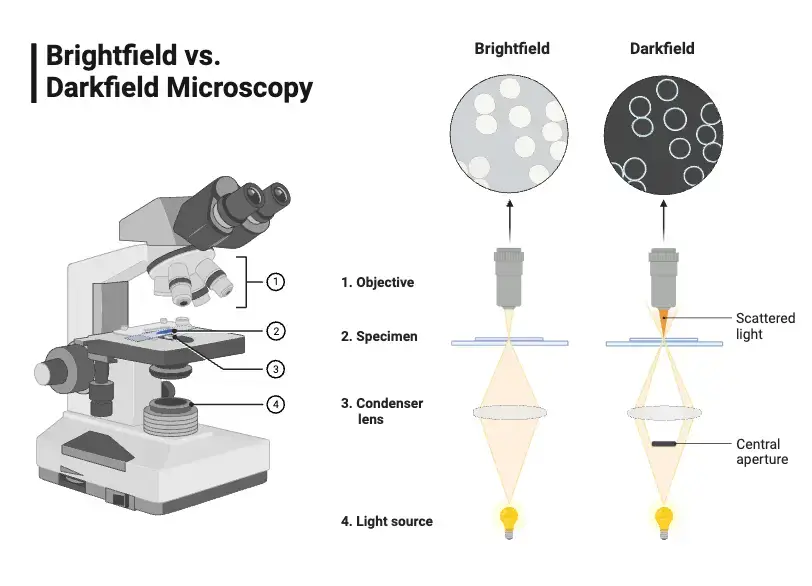
Darkfield microscopy is a form of light microscopy that facilitates detection by contrast. For example, brightfield microscopy uses a light source that shines light directly through the sample in order to illuminate the sample from below; darkfield microscopy uses a condenser that shines light at an oblique angle so that when the light is scattered and reflected off the sample, it can be seen as bright in a dark field. Thus, darkfield microscopy is more effective for viewing live, unstained samples because even the smallest details can be noted as the experiment continues.
Although abbreviated microscopes have been around since the 1600s, the first darkfield microscopy was used in 1673 by Antoni van Leeuwenhoek, although its components are much more sensitive and detailed today. Additionally, today’s dark-field microscopy has become digital with multilayered computerized imaging and automation processes for improved resolution and real-time views of sensitive, living biological entities. This is crucial for microbiology and educational settings to observe microbes that would otherwise go unnoticed or be too small to remain still long enough to be observed.
Definition of Dark Field Microscope
A darkfield microscope is a type of optical microscope that uses oblique illumination to light the specimen, causing it to appear bright against a dark background. This technique enhances the visibility of transparent and living specimens by excluding the unscattered light from the image.
Principle of Dark Field Microscope (How does dark field microscopy work?)
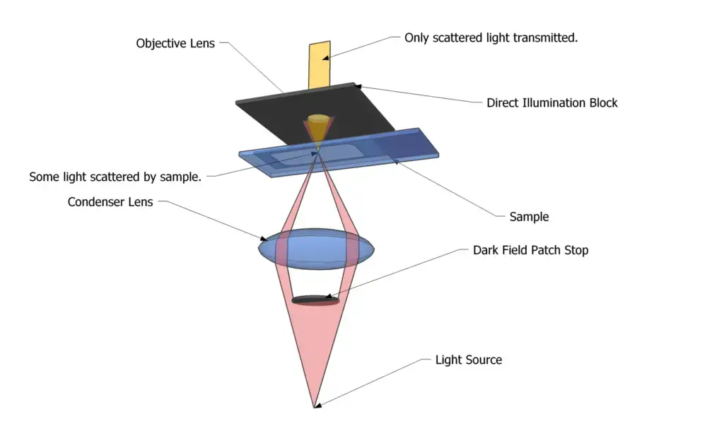
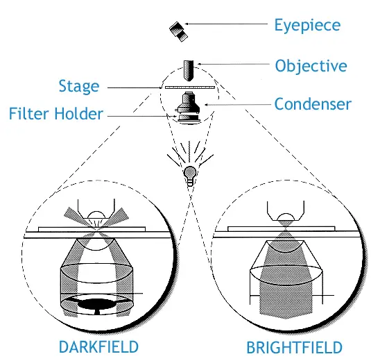
Darkfield microscopy is a high-resolution microscopy due to the contrast by light scattering, which allows for a bright sample on a black background. Basically, the condenser of the light source has a diaphragm with a central circular opening so that the central light rays are blocked and a “cone of light” is projected with larger angles. For example, more light rays are projected from many angles to hit the sample. Therefore, when one wants to see the transparent sample, the sample will scatter, refract, or reflect the light rays directed towards it; only the deflected ray will enter the objective. This creates the bright sample on the black background. Therefore, this transparent microscopy works best with transparent samples or organisms; unstained living samples can be examined as long as their refractive index is the same as that of the surrounding fluid. Therefore, it works best with unstained organisms because it makes them look better; It does not use transmitted light, which allows living or delicate organisms to be observed.
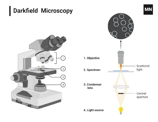
Parts of Dark Field Microscope
The parts of a darkfield microscope include the following:
- Light source – As with any other type of microscopy, it is necessary to visualize the sample; however, a darkfield microscope, in particular, requires a more intense light source to provide the oblique and intensified light required for this specific process.
- Condenser with central circular stop- This is necessary to make any microscope that uses darkfield illumination. A central opaque disk resides that prevents direct light from hitting the sample (controlled by the aperture); it allows only oblique rays to hit the sample. Therefore, the objective lens receives only the light diffracted by the sample itself and thus, a bright image on a dark background is produced.
- Objective lens– Collects the light that has been scattered by the sample to form the image. In darkfield microscopy, the objective lens is positioned to collect not the incident light from the condenser but the light that is scattered after interacting with the sample.
- Condenser Lens- Focuses all the light coming from below and shines it upward toward the specimen.
- Stage – The horizontal plane apparatus where the specimen slide is positioned for observation.
- Focusing Mechanism – Usually coarse and fine adjustments that move the stage or the position of the objective lenses to bring the specimen into focus.
- Eyepiece (ocular lens) – Apparatus through which the observer looks down to view the magnified image.
- Arm and Base – Structural and supporting elements of the microscope.
| Component | Description |
|---|---|
| Dark Field Condensers | – General Overview: Ensures the objective lens does not receive direct light. Captures light reflected, refracted, or diffracted by the specimen. – Low Magnification: Standard bright field condensers with an opaque disc in the front focal plane. – High Numerical Aperture: Uses a parabolic glass reflector to produce a hollow cone of light. Modern versions may have an iris diaphragm. |
| Eyepiece | The lens allowing observers to view magnified images of the specimens. |
| Light Source | Essential for magnification. In electron microscopy, high-accelerating electrons serve as the light source. |
| Objective Lens | Located within the dark recesses of the apex cone, this lens captures and magnifies the specimen’s image. |
| Arm | The curved upper part connecting the base to the eyepiece, providing support. It’s also the typical holding point when carrying the microscope. |
| Rotating Nosepiece | Positioned below the eyepiece, it holds multiple objective lenses. It can be rotated to switch between different objective lenses for varying magnifications. |
| Dark Disc | An opaque disc in the condenser that blocks direct light, ensuring only oblique light rays reach the specimen. |
| Course and Fine Adjustment Knobs | Used to adjust the focus of the specimen. The coarse knob makes large changes in focus for initial viewing, while the fine knob makes smaller, precise changes for sharp focus. |
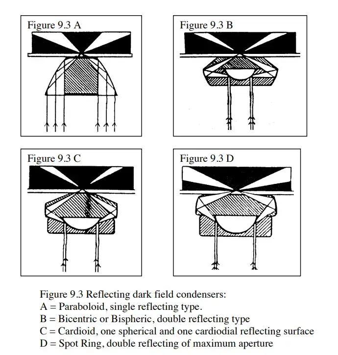
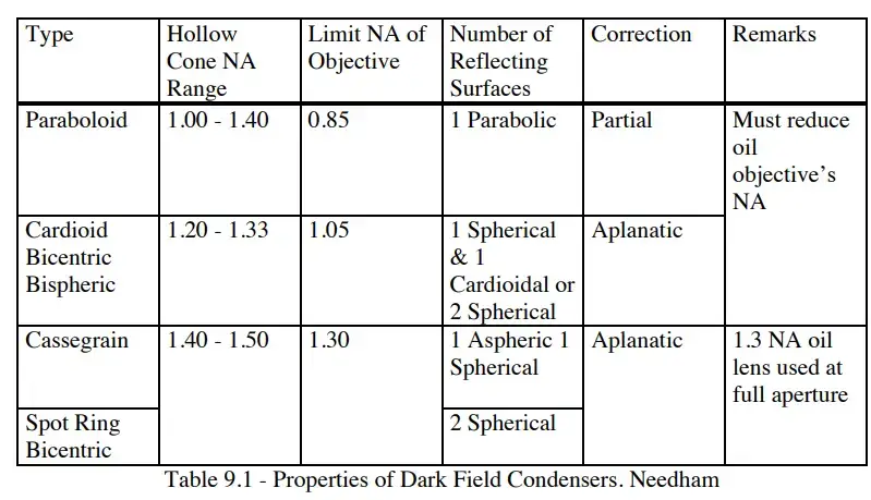
The light’s path in Dark Field Microscope
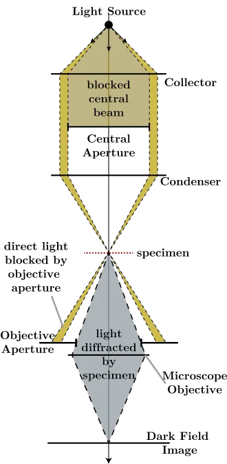
- Light passes through the microscope and falls on the slide.
- The aperture ring ensures that the light does not pass completely through the microscope, but forms a ring around the outside.
- The condenser lens focuses the light onto the slide.
- Most of the light passes directly through the slide, but some is refracted.
- The objective lens captures the refracted light as a disk that blocks normal light from passing through.
- The refracted light is what makes up the photograph of the slide, with the slide being white and the negative space being black.
Methods for Darkfield Microscope (Operating Procedure of Darkfield Microscope)
- Place the slide on the microscope stage and secure it with stage clips. Insert the darkfield condenser, either an annular aperture or a central opaque stop.
- Ensure that the light source is at maximum power and intensity.
- Open the aperture diaphragm to admit as much light as possible.
- Adjust the condenser diaphragm to the numerical aperture of the objective being used.
- Select an objective lens with a numerical aperture of 0.65 or higher.
- After focusing with a coarse adjustment, use a fine adjustment for finer focus clarity.
- Observe the specimen, which should appear bright on a dark background.
- Darkfield illumination relies on optical components, a condenser, and lenses. Remember that thick specimens do not show up well in dark field.
Application of Dark Field Microscope
- Filamentous Bacteria—Enables detection of filamentous bacteria invisible under brightfield.
- Untreated Live Organisms—Essential for viewing live, unstained cells and microbes.
- Inculturative Bacteria—Enables viewing of bacteria that cannot be grown in the lab.
- Microbial Movement—Important for studying microbes while they’re moving.
- Blood and Algae—Allows for viewing of blood and algae.
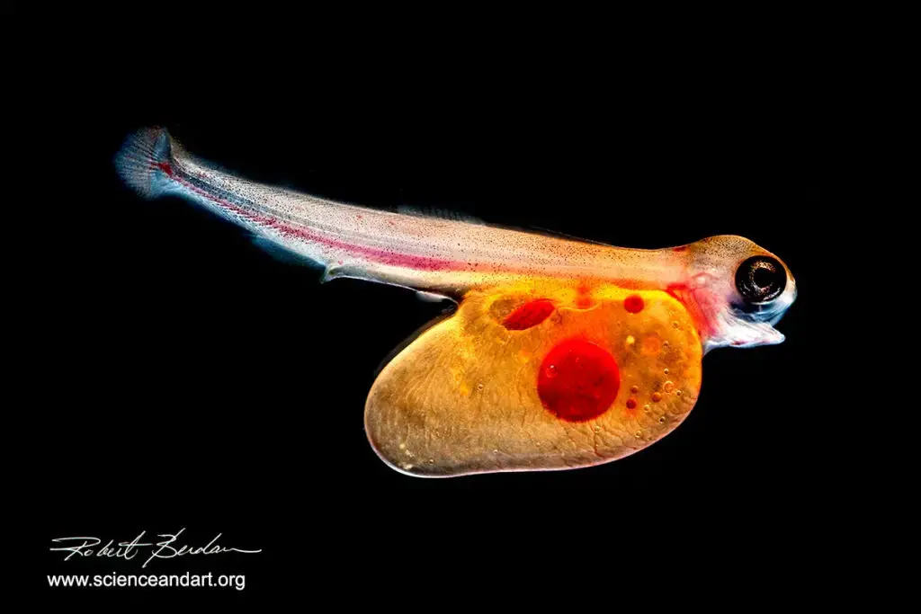
- Invertebrate—Important for viewing invertebrates like squids and their activity.
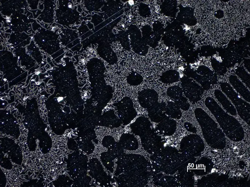
- Scientific Applications In Materials— This would be useful for materials studies; for example, the grain boundaries of a given material.
Types of specimens used for Dark Field Microscope
- Easier to see in brightfield or without staining- Objects that are easier to see without staining and have a higher refractive index or clarity. For example, living cells and non-pigmented microorganisms are quite visible without staining.
- Microorganisms – The ability to see some types of microorganisms such as bacteria and protozoa is a problem in brightfield, but the darkfield condenser provides excellent contrast and is easy to see.
- Histological sections- Specimens prepared for histology and very thin sections (or with silver staining) can be viewed to reveal more intricate details of the structure.
- Specimens with opaque backgrounds or materials behind transparent objects- Specimens with inclusions or refractive materials are ideal for darkfield microscopy because the invisible background material creates a much more effective contrast with the refracted light.
Advantages of Dark Field Microscope
- Enhanced Contrast – Provides high contrast images of transparent or unstained specimens.
- No Need for Staining – Can observe live cells and microorganisms without the need for staining.
- Observation of Fine Structures – Allows detailed observation of small, fine structures like bacteria, protozoa, and algae.
- Live Specimens – Ideal for observing live specimens without altering or damaging them.
- Improved Visibility – Can reveal features of specimens that are invisible under standard light microscopy.
- Better Resolution – Helps achieve better resolution for thin or transparent samples that would otherwise be difficult to see clearly.
Limitations of Dark Field Microscope
- Low light levels can make images appear dim and require sensitive detection.
- Strong illumination is needed, which may damage delicate specimens.
- Dust, dirt, or air bubbles can distort the image.
- Not ideal for precise quantitative measurements.
- Limited depth of field makes it difficult to focus on thicker specimens.
- Intense light can cause glare and image distortion.
- Artifacts can occur due to improper specimen preparation.
Challenges of Using Dark Field Microscope
Here are the challenges of using dark field microscopy:
- Specimen preparation must be precise to avoid image degradation.
- Clean slides are crucial, as any debris can distort the image.
- Requires specialized and often costly equipment.
- Sensitive to contaminants like dust or air bubbles.
- Limited depth of field makes focusing on thicker specimens difficult.
- Strong illumination can damage delicate samples.
- Improper specimen preparation can cause artifacts in the images.
Why Is Dark Field Microscope a Good Imaging Technique?
Here’s why dark field microscopy is a good imaging technique:
- It provides high contrast images, making fine details easier to see.
- No need for staining, allowing observation of live cells and microorganisms.
- It is effective for observing small, fine structures like bacteria and protozoa.
- Ideal for studying live specimens without damaging them.
- It can reveal features that are invisible under standard light microscopy.
- Helps achieve better resolution for thin or transparent samples.
Difference between Dark field and Bright field microscopy – Bright Field vs Dark Field
| Topics | Bright Field | Dark Field |
|---|---|---|
| Image Type | Dark image against a bright field. | Bright image against a dark background. |
| Specimen Types | Stained, fixed, and live specimens. | Living and unstained cells. |
| Image Formation | Only scattered lights enter the objective. | Light reflected or refracted by specimen. |
| Specimen Color | Dark or blue (depends on stain). | Appears as bright. |
| Background Color | Bright. | Dark. |
| Type of Condenser Used | Abbe, Variable focus, Achromatic. | Abbe, Paraboloid, Cardioid. |
| Specimen Structure | Morphological and internal structure. | External structure in detail. |
| Cost Implication | Inexpensive. | Expensive. |
| Specimen Preparation | Complex and lengthy staining process. | No staining required. |
| Opaque Disc | Absent. | Present. |
| Usability | Easy to use. | More intricate operation. |
| Study on Metals and Minerals | Not suitable for metals and minerals. | Suitable for minerals, crystals, polymers, etc. |
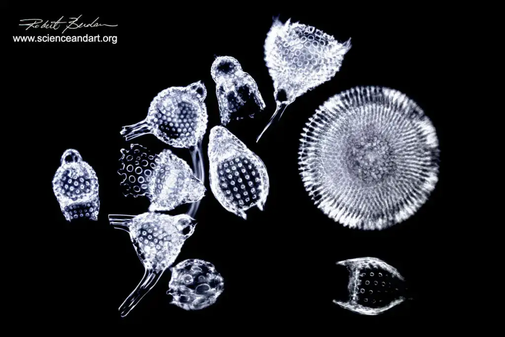
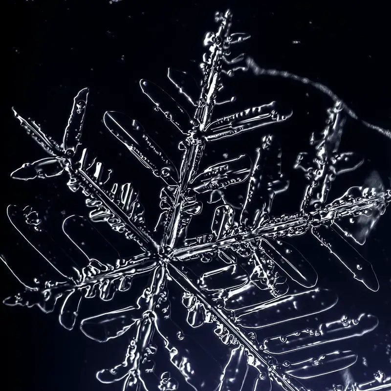
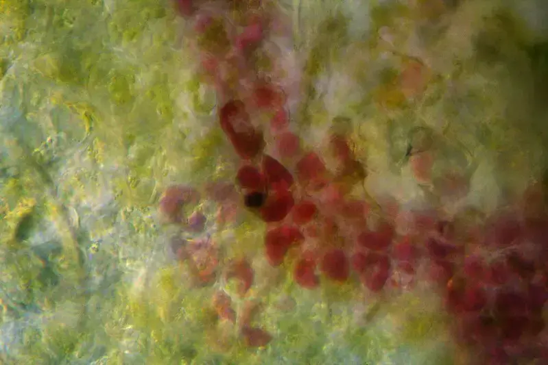
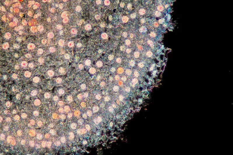
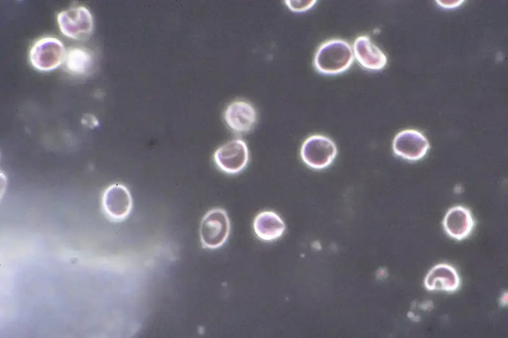
Dark Field Microscope Images

Quiz
FAQ
What is Darkfield Microscopy?
Darkfield microscopy is an optical technique used to enhance the contrast of transparent specimens by illuminating them against a dark background. It’s particularly useful for observing unstained or low-contrast samples.
How does Darkfield Microscopy work?
Darkfield microscopy works by using a specialized condenser to direct oblique or angled illumination onto the specimen. This causes scattered light to enter the objective lens, creating a bright image against a dark background.
What types of specimens are suitable for Darkfield Microscopy?
Darkfield microscopy is ideal for transparent specimens, such as live cells, microorganisms, and small organisms like plankton. It’s also used for viewing particles, fibers, and thin sections.
What are the advantages of Darkfield Microscopy?
Darkfield microscopy provides enhanced contrast, improved resolution, and allows for the observation of live, unstained samples. It’s versatile and can be used for various types of specimens.
Are there any limitations to Darkfield Microscopy?
Darkfield microscopy has limitations, including the requirement for strong illumination, potential sample damage from intense light, and sensitivity to dust and debris on slides.
How is Darkfield Microscopy different from Brightfield Microscopy?
In brightfield microscopy, specimens are viewed against a bright background, while darkfield microscopy illuminates specimens against a dark background, providing better contrast.
Is special staining necessary for Darkfield Microscopy?
Darkfield microscopy does not require staining, making it suitable for live samples. However, some samples may benefit from staining for specific purposes.
Can Darkfield Microscopy be used with high-magnification objectives?
Darkfield microscopy can be used with high-magnification objectives, but it requires careful condenser alignment and proper slide thickness to maintain image quality.
What is the critical angle in Darkfield Microscopy?
The critical angle is the angle beyond which light is totally internally reflected at the interface of the specimen and the surrounding medium. It’s crucial for proper darkfield illumination.
Can Darkfield Microscopy be adapted for other types of microscopes?
Yes, darkfield condensers can be added to various types of microscopes, including compound and stereo microscopes, to enable darkfield illumination.
References
- Madigan Michael T, Bender, Kelly S, Buckley, Daniel H, Sattley, W. Matthew, & Stahl, David A. (2018). Brock Biology of Microorganisms (15th Edition). Pearson.
- Procop, G. W., & Koneman, E. W. (2016). Koneman’s Color Atlas and Textbook of Diagnostic Microbiology (Seventh, International edition). Lippincott Williams and Wilkins.
- Tille, P. (2017). Bailey & Scott’s Diagnostic Microbiology (14 edition). Mosby.
- Willey, Joanne M, Sherwood, Linda M, & Woolverton, Christopher J. (2016). Prescott’s Microbiology (10 edition). McGraw-Hill Education.
- https://www.microscopemaster.com/dark-field-microscope.html
- https://en.wikipedia.org/wiki/Dark-field_microscopy
- https://www.microscopyu.com/techniques/stereomicroscopy/darkfield-illumination
- https://www.slideshare.net/abhishekindurkar/dark-field-microscopy-81857005
- https://bio.libretexts.org/Bookshelves/Microbiology/Book_Microbiology_(Boundless)/3_Microscopy
- https://www.ruf.rice.edu/~bioslabs/methods/microscopy/dfield.html
- Text Highlighting: Select any text in the post content to highlight it
- Text Annotation: Select text and add comments with annotations
- Comment Management: Edit or delete your own comments
- Highlight Management: Remove your own highlights
How to use: Simply select any text in the post content above, and you'll see annotation options. Login here or create an account to get started.