Little, single-celled creatures living practically everywhere are bacteria. They’re prokaryotic, meaning they lack a real nucleus. They differ from eukaryotic cells in that they lack membrane-bound organelles.
Shapes vary; spherical (cocci), rod-like (bacilli), spiral (spirilla), or comma-shaped (vibrios). Their survival in hostile environments depends on this diversity. Some twist like corkscrews, designated as spirochaetes.
There are beneficial bacteria. They promote digestion, create vitamins, and guard the body from dangerous germs. Not all strain, nevertheless, are friendly. Some kinds bring diseases, including throat infections or tuberculosis.
Their procreate? A basic process termed binary fission. One cell divides to produce two exactly identical progeny. In good settings, their numbers rise swiftly.
Though microscopic, microorganisms play tremendous responsibilities, altering life in apparent and subtle ways.
Characteristics of Bacteria
Morphology Features
Bacteria exhibit diverse shapes and arrangements, identifiable through basic wet-mount or staining techniques. These methods show structure traits such as Gram staining qualities, acid-fastness, movement, and cellular inclusions like spores or capsules. Different species can also be told apart by the way their flagella are arranged. Such microscopic reviews often narrow down recognition to a genus or group, streamlining further research.
Growth Dynamics
Bacteria are either aerobic, anaerobic, facultative, or microaerophilic based on how much oxygen they need. This difference is very important when growing these microorganisms because things like the amount of oxygen, the temperature of the incubator, and the pH all have a big effect on growth. Pathogens like Campylobacter jejuni do best in warmer (42°C) and more antibiotic-rich environments. Yersinia enterocolitica, on the other hand, likes cooler temperatures, about 4°C. Some, such as Legionella and Haemophilus, demand specific growth factors, contrasting with the ease of media needed by E. coli.
Antigenic Properties and How They Interact with Phages
The cell wall (O antigens), flagella (H antigens), and capsules (K antigens) are some of the parts of cells that are used for serotyping and classifying. In epidemiological settings, this knowledge is very useful for finding pathogenic forms of E. coli or highly contagious types of Vibrio cholerae (O1). Molecular methods have mostly replaced phage susceptibility testing as a way to keep an eye on pathogens like Staphylococcus aureus and Salmonella Typhi. However, phage susceptibility testing still shows how certain bacteria interact with viruses.
Profiling of biochemicals
Biochemical behavior, which is measured by a number of standard tests, is a key part of classifying bacteria. These tests look at metabolic processes, like carbohydrate fermentation, and enzyme functions, like oxidase or nitrate reduction. Some tests, like the coagulase test, are species-specific, which makes it easier to find specific bugs like Staphylococcus aureus.
Even though bacteria are very small, they have a huge number of characteristics that help us identify and group them. Each of these characteristics gives us more information about how they work and how they affect health or illness.
Classification Of Bacteria On The Basis Of The Cell Wall And Staining Reaction

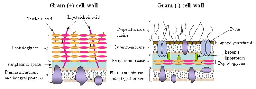
1. Gram-positive bacterial organisms – Under Gram staining, gram-positive bacteria have a strong cell wall mostly composed of a thick peptidoglycan layer that tightly holds the crystal violet dye. Under a microscope, this quality makes them clearly blue or purple. Their walls, along with peptidoglycan, include teichoic acids, which are vital for structural integrity and control of cell development. As a defence mechanism, the thick wall also enables these microorganisms to survive in difficult environments.
2. Gram-negative bacterium – By contrast, Gram-negative bacteria have a more complicated cell membrane. Two membranes placed between a quite thin peptidoglycan layer inhibits crystal violet retention after staining. Under magnification, these bacteria pick up the counterstain, safranin, looking pink or red. Rich in lipopolysaccharides, the outer membrane not only increases their pathogenicity but also acts as a barrier against several drugs. This resistance underlines the complexity of controlling infections produced by Gram-negative bacteria.
3. Variable Gram-Based Bacteria– Not all bacteria fit exactly the above divisions. Gram-variable species show a mixture of staining results, with purple and pink cells within the same sample. This mismatch generally comes from variations in cell wall design at distinct development stages. Though difficult to classify, these bacteria still offer important new perspectives on cellular mechanisms.
4. Gram-Indeterminate Bacteria – Some bacteria completely resist accepted Gram-staining techniques. Often referred to as Gram-indeterminate bacteria, they need other methods for consistent identification. Often requiring sophisticated diagnoses, examples include those with unusual cell walls or inadequate structural elements to successfully hold stains.
Classification Of Bacteria On The Basis Of Oxygen Requirements
- Obligate Aerobes– Obligate aerobes are bacteria that survive only in oxygen. Using oxygen as the terminal electron acceptor in their metabolic activities, many species rely on aerobic breathing. They cannot flourish without it. These bacteria grow in settings containing around 20% oxygen, typical of atmospheric conditions. Examples include Pseudomonas, a frequent infection seen in hospital settings, and Mycobacterium tuberculosis, the causative agent of TB. For many species, oxygen is not only a need; it’s their lifeline.
- Microaerophiles. – Conversely, microaerophiles also need oxygen but in very smaller amounts. Usually, they thrive in conditions with oxygen levels between 1% and 10%, significantly less than what the atmosphere offers. Given their sensitivity to strong doses, too much oxygen can be detrimental to them. Two such include Helicobacter pylori and Campylobacter. Famously linked to stomach ulcers, the latter flourishes in the acidic environment of the human stomach, where oxygen levels are somewhat low.
- Facultative Anaerobes – Facultative anaerobes are quite flexible. They may develop in the presence or absence of oxygen, alternating between aerobic respiration and anaerobic respiration or fermentation, depending on the availability of oxygen. One famous gut bacteria that fits this is Escherichia coli. These species are flexible and strong in many metabolic contexts since they can adapt to many surroundings.
- Aerotolerant Anaerobes- Aerotolerant anaerobes differ from facultative anaerobes in their metabolic activities not using oxygen. They can, nonetheless, live with it. These bacteria rely exclusively on fermentation for energy production, regardless of oxygen availability. One falls into this category: lactobacillus, used in the making of yogurt. Oxygen doesn’t help them develop, yet they can coexist with it without suffering.
- Obligate Anaerobes – Obligate anaerobes cannot withstand oxygen. Oxygen is toxic to them and can even be fatal, so stopping their development. These bacteria depend on anaerobic respiration or fermentation and flourish only in oxygen-starved surroundings. One such a perfect example is the pathogen causing botulism, Clostridium botulinum. These species have evolved to live in conditions like the intestines or deep wounds where oxygen is either rare or nonexistent.
Classification of Bacteria on The Basis of pH Requirements
Bacteria, like all living organisms, have specific environmental conditions under which they thrive. Among these factors, pH is absolutely important for bacterial development. The capacity of bacteria to grow at different pH levels helps one to classify them. Acidophiles, alkaliphiles, and neutrophiles form three main groups.
- Acidophiles– Acidophiles are bacteria that survive in acidic conditions, often with a pH below 7. Their cytoplasm is naturally acidic; their cellular processes and metabolic activities are suited for an acidic environment. At high temperatures, some acidophiles—also known as thermoacidophiles—can also flourish. These bacteria have evolved mechanisms to maintain their internal pH despite external acidity. Thiobacillus thioxidans, Thiobacillus ferroxidans, and Sulfolobus are well-known examples of acidophilic organisms. Fascinatingly, these bacteria are essential for industrial activities such sulfur compound’ oxidation and bioleaching.
- Alkaline – In contrast, alkaliphiles prefer conditions with a high pH, often over 7. These species have evolved to flourish in simple, alkaline habitats. An example is Vibrio cholerae, the causative agent of cholera, which has an ideal growth pH of about 8.2. Often by controlling the pH within their cells, these bacteria have evolved strategies to offset the demanding conditions of alkalinity. Alkaliphilic bacteria are of great importance in biotechnology, since they are utilized in the manufacturing of detergents, as well as in the food and beverage sector.
- Neustrophiles – Conversely, neutrophiles like neutral pH settings, generally falling between 6.5 and 7.5. This category comprises many of the bacteria that are regularly found in the human body, such as Escherichia coli. Neutrophilic bacteria are well-suited to situations where the pH is near to that of water, making them the most abundant form of bacteria on Earth. Most bacteria fit this group since neutral pH is usually ideal for cellular operations and enzyme performance.
The evolutionary changes these species have made to their surroundings define bacterial pH tolerance. Research on these species not only clarifies microbial survival mechanisms but also creates opportunities for useful applications in several spheres like environmental management, industry, and medicine.
Classification of Bacteria on The Basis of Temperature Requirements
By grouping bacteria according to their temperature requirements, one can get amazing new understanding of their adaptation. Key in nature, temperature determines the limits of growth and survival for these microorganisms, each with developed strategies to survive in different thermal conditions.
- Psychrophiles- Cold-loving bacteria, known as psychrophiles, may thrive at or below 0°C; their ideal growth temperature is between 15°C and so. Their capacity to keep cell membrane fluidity helps them to be resilient at low temperatures. They integrate polyunsaturated fatty acids into their membranes, which prevents it from becoming stiff. It is hardly surprising, then, that psychrophiles are frequently found in very cold conditions, such deep-sea waters or polar areas. A few well-known examples are Vibrio psychroerythrus and Polaromonas vaculata.
- Psychrotrophs – Especially, there is a subgroup known as facultative psychrophiles or psychrotrophs. Though their ideal temperature for development falls between 20 and 30 degrees Celsius, they can survive at 0°C. These bacteria show fascinating adaptability, moving between states more suited for moderate conditions and cold-adapted ones.
- Mesophiles – Mesophiles, on the other hand, grow in mild settings. Their ideal growing range is 25°C to 40°C; the best temperature usually falls around 37°C. These are the temperature-loving organisms that contain most of the germs that infect humans. E. coli, Salmonella, and Staphylococci are typical examples of mesophilic bacteria, thriving within the body temperature of humans.
- Thermophiles- Thermophiles have a penchant for heat. Usually surviving in hot springs or geothermal vents, these bacteria are suited to conditions above 45°C. Their cell membranes contain saturated fatty acids, which under high temperatures aid to maintain structural integrity. Stenothermophiles, or obligatory thermophiles, are those thermophiles that are stringent heat lovers while some are facultative, able to develop within the mesophilic range. Two of the best are Bacillus stearothermophilus and Streptococcus thermophilus.
- Hyperthermophiles- We discover the Hyperthermophiles at the very extreme end of the spectrum. These bacteria are suited to flourish at temperatures exceeding eighty-degree Celsius. Usually found in deep-sea hydrothermal vents or other harsh habitats, they comprise archaebacteria such Thermodesulfobacterium and Pyrolobus fumarii. Their cell membranes have an unusual structure, with a monolayer membrane that offers additional temperature tolerance. Their capacity to live at such extremes makes them a major focus of biotechnology research, especially in relation to industrial enzyme manufacture.
Classification of Bacteria on The Basis of salt and tolerance
Bacteria have varied adaptations when it comes to their tolerance and liking for salt in their environment. Different kinds of bacteria depending on salt tolerance expose different survival strategies in reaction to different salinity levels.
- Non-halophiles – Non-halophiles are bacteria unable of surviving in high-salt conditions. Usually less than 0.2M (about 1% NaCl), these species flourish in freshwater habitats with low salt concentrations. They are vulnerable to salty environments and do not possess mechanisms to endure such stress.
- Halophilic bacteria– Conversely, halophiles—that is, halophilic bacteria—flourish in settings with plenty of salt. These bacteria have evolved strategies that let them not only survive but also flourish in settings with appreciable salt concentrations. The concentration of NaCl they can tolerate determines the many groups for their tolerance and demands.
- Halotolerant bacteria- Within the halophiles, a special category are halotolerant bacteria. These species thrive in low or none at all of salt, although they can grow in settings with moderate levels. For example, Staphylococcus aureus can survive salt levels of up to 5-10%; nonetheless, its development is not dependent just on it. Actually, these bacteria are rather flexible and may even flourish at reduced salinity levels.
- Slight halophiles- Slight halophiles like low to moderate salinity settings. Salt levels between 0.2 and 0.85M (1-5% NaCl) show their ideal development. Typical example Halomonas finds its place in these saline environments and has evolved systems to collect nutrients and preserve cellular integrity in spite of salt presence.
- Moderate halophiles- More specialized are moderate halophiles, which can develop best between 0.85 and 3.40M (5-20% NaCl). These bacteria, including Bacillus and Marinococcus, need more salt, hence even if they may exist without it, their growth is best observed when salt is present. Their cell architectures are particularly suited to preserve osmotic equilibrium under these circumstances.
- Extreme halophiles- Extreme halophiles, the most extreme category, flourish in very high salt concentrations, between 3.4 and 5.1M (20–30% NaCl). Prime examples of bacteria that not only survive but also need very high salt concentrations include Halobacterium, Haloarcula, and Halococcus. These species have developed unique metabolic processes, frequently including the manufacture of suitable solutes, to safeguard their physiological activities under severe osmotic pressure.
Classification of Bacteria on the Basis of Mode of Nutrition
Bacteria’s nutritional methods are centered around how they obtain carbon and energy, and help to classify bacteria. This classification consists of four general groups: phototrophs, chemotrophs, autotrophs and heterotrophs. All these groups are further subdivided, which gives more detailed knowledge about bacterial metabolism.
- Phototrophs– Phototrophs, an interesting organism, obtain their energy from light. The source of electrons helps to classify them. Phototrophs absorb light and utilize reduced inorganic molecules, such as H2S, as an electron source. A typical representative of this group is, for example, Chromatium okenii. Conversely, photoorganotrophs use organic substances, such as succinic acid, as an electron source, even if they also capture light energy. In ecosystems where light-driven activity is essential, these bacteria are essential participants.
- Chemotrophs– Chemotrophs obtain their energy from chemicals. Unlike phototrophs, chemotrophs do not depend on photosynthesis. Chemotrophs are also divided into two major categories based on the electron donor: chemolithotrophs and chemoorganotrophs. Chemoolithotrophs obtain their energy by oxidizing inorganic compounds such as NH3, which are used as an electron source. Nitrosomonas is one such category. Conversely, chemoorganotrophs use organic substances such as glucose and amino acids to power their energy activities. One commonly used example is Pseudomonas pseudoflava.
- Autotrophs– Autotrophic bacteria have the remarkable ability to use carbon dioxide (CO2) as their main carbon source. These organisms play a key role in carbon fixation. Depending on how they obtain energy to assimilate CO2, autotrophs are divided into photoautotrophs and chemoautotrophs. Photoolithotrophs (using inorganic electron donors) and photoorganotrophs (using organic electron donors) are two further classification systems based on the electron source. Photoautotrophs use light energy. Conversely, chemoautotrophs use compounds to obtain the energy they need, thus allowing them to assimilate CO2 by chemical means.
- Heterotrophs– Bacteria known as heterotrophs depend on organic molecules for carbon. Heterotrophs cannot repair CO2 like autotrophs can. This group includes many harmful microorganisms found in humans. The diverse group of heterotrophs also includes some that have very low dietary requirements. These are the simple heterotrophs. However, other bacteria require more specialized nutrients and are therefore demanding heterotrophs. Due to their stringent dietary requirements, it is difficult to grow these bacteria in a laboratory environment.
Classification of Bacteria on The Basis of Shape And Arrangement

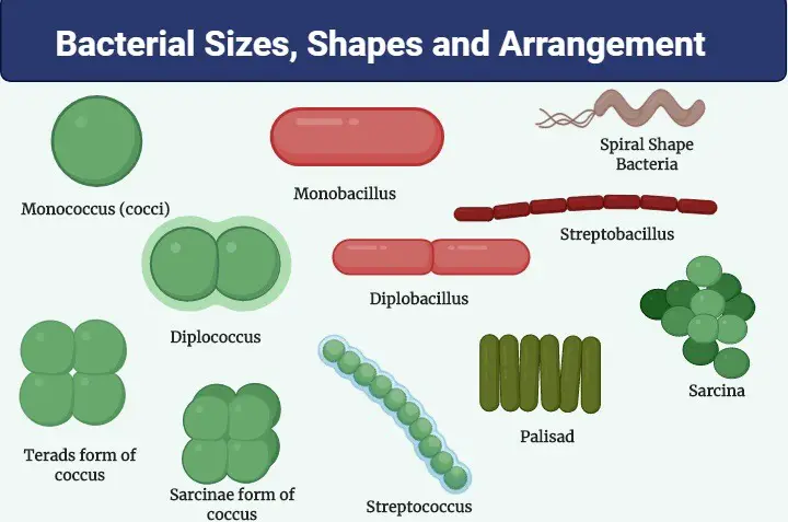
By use of their structure and organization, the categorization of bacteria offers an unambiguous approach to distinguish and identify various microorganisms. Our knowledge of bacterial shape began in 1872 when the scientist Cohn identified four main kinds of bacterial forms. These are cocci, bacilli, vibrios, and spirilla. Every kind shows different structural elements that determine its look and development trends.
- Cocci– Cocci are spherical or elliptical shaped microorganisms. They can either be single cells or group into particular configurations. There is variation in the clustering of cocci; each arrangement has great diagnostic value.
- Monococcus – Often known as Micrococcus, monococcus is a singular, round cell that seems as a separate entity. One often used example is Micrococcus flavus.
- Diplococcus: Once in a given plane, diplococcus divides and the resultant cells remain attached to create pairs. One well-known instance of this arrangement is Diplococcus pneumoniae.
- Streptococcus: This kind repeatedly divides in one plane to produce a chain of cells. One such a well-known culprit for strep throat is Streptococcus pyogenes.
- Tetracoccus: These four-cell group forms after splitting in two planes at right angles to one another. Gaffkya tetragena is a representative of this group.
- Staphylococcus: Dividing in several directions, staphylococcus produces clusters like bunches of grapes. Within this group is the common pathogen Staphylococcus aureus.
- Sarcina: This arrangement produces a cube-like form made of eight or sixteen cells by means of divisions in three planes. Sarci lutea exhibits this configuration.
- Bacilli – Bacilli are cylindrical or rod-shaped, bacterial cells. Usually, they are encountered either alone or in couples. Often improving their survival chances, their elongated form helps them fit to particular surroundings. One usual example of a bacillus is Bacillus cereus.
- Vibrio– Curved, comma-shaped bacteria belonging to a single species are vibrios. Their unusual form distinguishes them from other varieties. A famous example is Vibrio cholerae, the bacterium responsible for cholera epidemics.
- Spirilla– Spirilla show many curvatures and a spiral or spring-like form. Many times, these bacteria have terminal flagella that help them to move. One of this spiral-shaped bacteria is Spirillum volutans.
Classification of Bacteria on The Basis of Flagella

Depending on how flagella are arranged on a bacterial cell, they are grouped into distinct categories.
- Atrichous– The simplest in terms of motility, atrichous bacteria lack flagella totally. They are non-motile so absence indicates this. A well-known example of this type is Corynebacterium diphtheriae, which causes diphtheria. These bacteria survive and spread using other means without flagella.
- Monotrichous– Conversely, monotrichous bacteria have one flagellum usually found at one pole of the bacterial cell. Directive movement is made possible by this single framework. One monotrichous bacterium is the pathogen causing cholera, Vibrio cholerae. The flagellum acts as a propeller, pushing the bacterium forward through liquids.
- Lophotrichous– Though not scattered over the cell, lophotrichous bacteria have many flagella. Instead, these flagella are gathered together at one end, providing a tuft-like look. This type of arrangement helps in more powerful swimming, often in a specific direction. For example, Pseudomonas shows this feature and amazing motility.
- Amphitrichous -Flagella seen in amphitrichous bacteria exist at both ends of the cell. These bacteria have flagella at both poles, often arranged in tufts. More complicated movement patterns made possible by the dual flagellar arrangement help to improve navigation across different surroundings. One such a typical species displaying this arrangement is Rhodospirillum rubrum.
- Peritrichous – Finally, Peritrichous bacteria feature flagella dispersed throughout the whole surface of their cells. This dispersion gives these bacteria with a diverse mode of mobility, enabling them to migrate in all directions. Common genus Bacillus is one where flagella are distributed over the bacterial cell surface, so supporting a more fluid and omnidirectional mobility.
Classification of Bacteria on The Basis of Capsule
A bacterium falls into either non-capsulated or capsulated depending on whether or not it has a capsule.
Non-Capsulated Bacteria
Non-capsulated bacteria—well, their outer layer lacks protection. Usually, this lack leaves people susceptible, especially in relation to immunological reactions. When phagocytes (those immune cells meant to engulf and destroy infections) find these germs, they’re more likely to succeed in eradicating them. A couple of familiar examples include Mycobacterium tuberculosis, the cause of tuberculosis, and Shigella spp., which are notorious for causing dysentery. These bacteria are more vulnerable to outside hazards without the capsule, usually depending on alternative means of survival.
Capsulated Bacteria
Conversely, capsulated bacteria have a clearly defined, thick capsule that protects them in several respects. The capsule helps bacteria elude the immune system—especially phagocytosis—by making them difficult to identify and eat. Beyond immunity evasion, the capsule also aids in preserving moisture and fighting desiccation. Moreover, it can help create biofilms, which are basically sticky coatings allowing bacteria to adhere to surfaces, such as tissue or medical equipment, therefore causing ongoing infections.
Classic examples of capsulated bacteria include Streptococcus mutans, known for its role in dental plaque, Klebsiella pneumoniae, a cause of pneumonia, and Bacillus anthracis, the infamous anthrax pathogen. In these species, the capsule’s presence is directly correlated with their pathogenicity and capacity to generate disease.
Whether a bacterium is capsulated or not effects much more than simply its outward appearance—it affects how it interacts with its environment, how it spreads, and, ultimately, how it causes harm or evades discovery. The larger picture of bacterial infection and immune response depends much on this structural element.
Classification of Bacteria on The Basis of Ability to form spores

Spore forming bacteria
Endospore-forming and exospore-forming bacteria are the two main types of spore-forming bacteria distinguished by where the spores are formed. These spore-forming bacteria show great adaptation in conditions that would otherwise be hostile to non-spore-producing organisms.
- Endospore-forming bacteria are those that produce spores inside of their own cells. Often quite resistant to chemicals, drying, and heat, these spores act as a survival mechanism in less than ideal circumstances. Nestled inside the bacterial cell, the spore can lie dormant for extended periods of time until germinating when conditions are better. Among the well-known genera of endospore-forming bacteria are Sporosarcina, Clostridium, and Bacillus. The capacity to create endospores has enabled these bacteria to live in severe environments—whether it’s great heat or nutritional deprivation.
- By contrast, exospore-forming bacteria create their spores outside of a cell. These spores are discharged into the surrounding environment, affording the bacterium another mechanism of persistence. Methylosinus, a genus that forms exospores, is an example. Though they might not be as robust as endospores, these exospores can resist environmental pressures.
Non-sporing bacteria
Then, there are the non-sporing bacteria, which simply don’t form spores. Usually more sensitive to unfavorable environments, these bacteria rely on alternative survival tactics. Escherichia coli and Salmonella, which may be found in both ambient and host contexts, are examples of non-sporing bacteria. They lack the latent, extremely resistant form that endospores or exospores give, making them less robust in harsh environments.
Whether endospores or exospores, the capacity of forming spores gives bacteria a survival edge in changing surroundings. While non-sporing bacteria must adapt by other methods, spore-forming species have a strong mechanism to survive hostile environments. This capacity to create spores has substantial ramifications in domains like medicine, where spore-forming bacteria are of particular concern due to their resistance to sterilization methods and their propensity to cause illness.
Classification of Bacteria on The Basis shape of Spores
- Oval and central spore E.g., Bacillus spp.
- Oval and sub-terminal spore: E.g., Clostridium spp. except C. tetani (round and terminal) and C. bifermentans (oval and central)
FAQ
What is the basis for classifying bacteria?
Bacteria are classified based on various factors such as shape, cell wall composition, nutritional requirements, oxygen requirements, presence of flagella, and genetic makeup.
What are the main shapes of bacteria?
The primary shapes of bacteria are cocci (spherical), bacilli (rod-shaped), and spirilla (spiral-shaped).
How are bacteria classified based on oxygen requirements?
Bacteria can be classified as obligate aerobes (require oxygen), obligate anaerobes (cannot survive in the presence of oxygen), facultative anaerobes (can live with or without oxygen), and microaerophiles (require low levels of oxygen).
What are Gram-positive and Gram-negative bacteria?
Based on the Gram staining technique, bacteria are classified as Gram-positive (retain the violet stain) and Gram-negative (do not retain the violet stain). This classification is based on the differences in their cell wall composition.
What is the significance of bacterial spore formation?
Some bacteria can form spores, which are dormant and resistant structures, to survive in unfavorable environmental conditions. When conditions become favorable, the spore can germinate and revert to its vegetative form.
How are bacteria classified based on their nutritional requirements?
Bacteria can be classified as autotrophs (synthesize their own food) and heterotrophs (rely on external sources for food). Autotrophs can further be classified as photoautotrophs (derive energy from light) and chemoautotrophs (derive energy from chemical reactions).
What are capsulated bacteria?
Capsulated bacteria possess an outer covering called a capsule, which provides protection against the host’s immune system and helps in adherence to surfaces.
How do flagella help in classifying bacteria?
Based on the presence and arrangement of flagella, bacteria can be classified as monotrichous (single flagellum), lophotrichous (cluster of flagella at one end), amphitrichous (flagella at both ends), and peritrichous (flagella all over the surface).
What is the role of genetic studies in bacterial classification?
Genetic studies, especially DNA sequencing, have revolutionized bacterial classification by providing insights into the evolutionary relationships between different bacterial species.
Why is it important to classify bacteria?
Classification helps in understanding the relationships between different bacterial species, predicting their behavior, and devising strategies for their control, especially in the case of pathogenic bacteria.
References
- Pitt TL, Barer MR. Classification, identification and typing of micro-organisms. Medical Microbiology. 2012:24–38. doi: 10.1016/B978-0-7020-4089-4.00018-4. Epub 2012 May 24. PMCID: PMC7171901.
- Smith, N. W., Shorten, P. R., Altermann, E., Roy, N. C., & McNabb, W. C. (2019). The Classification and Evolution of Bacterial Cross-Feeding. Frontiers in Ecology and Evolution, 7. doi:10.3389/fevo.2019.00153
- Schleifer, K. H. (2009). Classification of Bacteria and Archaea: Past, present and future. Systematic and Applied Microbiology, 32(8), 533–542. doi:10.1016/j.syapm.2009.09.002
- Al-mohanna, Moshtaq & H., quine. (2016). MORPHOLOGY AND CLASSIFICATION OF BACTERIA.
- Classification of Bacteria. In: Carroll KC, Hobden JA, Miller S, Morse SA, Mietzner TA, Detrick B, Mitchell TG, McKerrow JH, Sakanari JA. eds. Jawetz, Melnick, & Adelberg’s Medical Microbiology, 27e. McGraw Hill; 2019. Accessed October 15, 2022. https://accessmedicine.mhmedical.com/content.aspx?bookid=1551§ionid=94105368
- THE CLASSIFICATION OF BACTERIA. (1922). The Lancet, 199(5140), 444. doi:10.1016/s0140-6736(01)31846-9
- Bacterial Taxonomy: Meaning, Importance and Levels. (n.d.). BiologyDiscussion.com. Retrieved from https://www.biologydiscussion.com/bacteria/bacterial-taxonomy/bacterial-taxonomy-meaning-importance-and-levels/54679
- IAS ABHIYAN. (2017, May 8). Classification Of Bacteria. IAS Abhiyan. https://www.iasabhiyan.com/classification-of-bacteria/
- [Author Unknown]. (Date Unknown). Bacterial Taxonomy: Meaning, Importance, and Levels. Biology Discussion. https://www.biologydiscussion.com/bacteria/bacterial-taxonomy/bacterial-taxonomy-meaning-importance-and-levels/54679
- [Author Unknown]. (Date Unknown). General Bacteriology. RVSKVV. http://www.rvskvv.net/images/General-Bacteriology_23.04.2020.pdf
- [Author Unknown]. (Date Unknown). Prokaryote Classification. CK-12. https://flexbooks.ck12.org/cbook/ck-12-biology-flexbook-2.0/section/7.2/primary/lesson/prokaryote-classification-bio/
- [Author Unknown]. (Date Unknown). [Title Unknown]. COGEM. https://cogem.net/app/uploads/2019/07/CGM-2011-07-Bijlage-I-Algemene-text.pdf
- THE CLASSIFICATION OF BACTERIA. (1909). JAMA. Retrieved from https://jamanetwork.com/journals/jama/article-abstract/428898
- Biology Wise. (n.d.). Different Types of Bacteria. Retrieved from https://biologywise.com/different-types-of-bacteria
- The Pharmapedia. (n.d.). Classification of Bacteria on the basis of Nutrition, oxygen requirement, growing temperature & pH. Retrieved from https://thepharmapedia.com/classification-of-bacteria-nutrition-oxygen-requirement-growing-temperature/pharmacy-notes/
- DUHS Library. (n.d.). Academic Script. Retrieved from https://duhslibrary.ac.in/Content/919_35_Mooc4_Mod23_Academic%20script200225040402025555.pdf
- Kaggle. (n.d.). Bacteria Classification at the Genus Level. Retrieved from https://www.kaggle.com/c/bacteria-classification-at-the-genus-level
- GPAT India. (n.d.). Classification and Characteristics of Bacteria. Retrieved from https://gpatindia.com/classification-and-characteristics-of-bacteria/
- Wiley Online Library. (1940). [Title not provided]. Retrieved from https://onlinelibrary.wiley.com/doi/pdf/10.1002/j.2050-0416.1940.tb06032.x
- Taylor & Francis Online. (1969). [Title not provided]. Retrieved from https://www.tandfonline.com/doi/abs/10.1080/00021369.1969.10859443
- Inspirit VR. (n.d.). Bacteria – Structure and Classification Study Guide. Retrieved from https://www.inspiritvr.com/general-bio/prokaryotes-and-viruses/bacteria-structure-and-classification-study-guide
- NCBI. (n.d.). [Title not provided]. Retrieved from https://www.ncbi.nlm.nih.gov/pmc/articles/PMC7156943/
- Text Highlighting: Select any text in the post content to highlight it
- Text Annotation: Select text and add comments with annotations
- Comment Management: Edit or delete your own comments
- Highlight Management: Remove your own highlights
How to use: Simply select any text in the post content above, and you'll see annotation options. Login here or create an account to get started.