Cheek cells, fascinating eukaryotic entities, grace the lining of our mouths. These microscopic wonders boast a distinct structure that encapsulates a world of biological marvels within their tiny boundaries. Thanks to their ease of shedding, acquiring them for observation has become a simple yet enlightening endeavor.
Within the confines of these small entities, a realm of essential components operates in harmony to sustain life. The cell membrane, acting as the guardian, stands as the outer boundary, selectively allowing the passage of substances in and out. Nestled within the cell membrane lies the cytoplasm, a fluidic medium that facilitates various cellular processes. At the very core of the cell, a potent controller resides—the nucleus, overseeing and orchestrating the functions of the cell as a master conductor guides an orchestra. Furthermore, organelles, such as the mitochondria, play distinct roles akin to specialized cell structures. These organelles bear specific functions that contribute to the overall harmonious symphony of life within the cell.
Unlocking the secrets held by these cells requires the assistance of biological stains, such as methylene blue. With the aid of this remarkable dye, the cellular landscape becomes an open book, unveiling its inner workings for our perusal. As the stain selectively colors certain parts of the cell while leaving others untouched, a mesmerizing tapestry of colors unravels before our eyes. The cell membrane emerges as a defining boundary, painting itself a vibrant hue. The cytoplasm reveals its fluidic nature, enveloped in a delicate azure. At the core of it all, the nucleus commands attention, donning a shade that sets it apart from the rest. The organelles, each with a specific mission, present themselves, showcasing their unique hues and functional significance.
In this microscopic theater of life, cheek cells demonstrate the beauty and complexity of the biological world. Though small, their significance cannot be underestimated, as they grant us a glimpse into the fundamental building blocks of living organisms. Through careful observation and the aid of biological stains, we uncover the mysteries housed within these unassuming entities.
So, let us marvel at the elegance of cheek cells—their eukaryotic wonder, their easy accessibility, and their readiness to disclose their innermost secrets. As we peer through the lens of science, we are reminded of the astounding intricacies that shape life on a cellular level, perpetuating the symphony of existence in which we all play our part.
Requirements for Observing Cheek Cells Under a Microscope
- Sterile Cotton Swab: The quest begins with a sterile cotton swab. This simple yet indispensable tool serves as the gateway to the microscopic universe within our mouths. By gently swabbing the inside of the cheek, we collect a sample of cells, allowing us to proceed with our investigation.
- Clean, Sterile Microscope Slides: The cheek cell expedition necessitates a pristine surface on which to place our sample. Clean and sterile microscope slides provide the perfect canvas for these tiny specimens to reveal themselves. Ensuring that the slides are free from contaminants guarantees accurate observations.
- Microscope Cover Slips: As we lay the swab-extracted sample on the microscope slide, it’s essential to add another layer of precision. Microscope cover slips act as shields, protecting the sample while providing a flat and uniform surface for microscopy.
- Methylene Blue Solution (0.5% to 1%): To uncover the intricate details of cheek cells, a biological stain such as methylene blue proves invaluable. By gently applying this solution to the sample, certain cell structures are accentuated, enhancing visibility and revealing the cellular landscape in greater clarity.
- Dropper: Precision and control are vital when dealing with stains, and a dropper is a trusty companion for this task. It enables us to add just the right amount of methylene blue to the cheek cell sample, ensuring optimal staining without overwhelming the delicate structures.
- Blotting Paper/Tissue Paper: Tidiness plays a key role in the microscopic world. Blotting paper or tissue paper allows us to gently remove excess liquid from the slide after staining, preventing unwanted smudging or distortion of the cheek cells.
- Microscope: The heart of the expedition lies in the microscope itself. Equipped with lenses and a light source, this instrument magnifies the cellular landscape, revealing the hidden wonders of cheek cells. Adjusting the focus and fine-tuning the settings on the microscope unveils intricate details that might otherwise escape the naked eye.
Preparation of Wet Mount of Cheek Cells
- Clean Microscopic Slide: We begin by placing a drop of physiological saline solution at the central part of a clean microscopic slide. The saline serves as a gentle medium to preserve the cells’ natural environment during the observation.
- Applying the Cheek Cells: Taking the previously collected cheek cells on the cotton swab, we gently smear them onto the central part of the slide containing the saline drop. This step should take approximately 4 seconds, ensuring a sufficient number of cells are transferred to the slide’s center.
- Adding Methylene Blue Solution: To enhance the visibility of the cheek cells, we apply a drop of methylene blue solution to the smear. This biological stain selectively colors certain cell structures, making the cells more distinguishable and detailed under the microscope.
- Placing the Cover Slip: With the methylene blue solution and the cheek cells beneath it, we carefully and gently place a cover slip on top of the smear. The cover slip acts as a protective shield, securing the cells and stain in place for observation.
- Removing Excess Solution: To avoid any interference during the observation process, any excess solution can be removed. Gently touch one side of the slide with a paper towel or blotting paper to absorb the surplus liquid without disturbing the cells or the stain.
- Observation under the Microscope: The prepared slide is now ready for observation under the microscope. Initially, we use a low-power objective, such as the 4x or 10x, to locate the cheek cells on the slide. Once found, we can gradually increase the magnification to explore the finer details and intricacies of these eukaryotic entities.
- Note – As a responsible practice, used cotton swabs and cotton towels should be safely discarded in the trash and not left lying on the working table. This ensures proper disposal and minimizes the risk of cross-contamination.
Staining the Cells
The hidden intricacies of cells come to life through staining, a transformative process that reveals the previously unseen details of these microscopic wonders. Within a cell, certain parts possess the remarkable ability to absorb stains, rendering them chromatic and distinctly visible under the microscope, setting them apart from the rest of the cell.
The art of staining is indispensable in the world of microscopy. Without this technique, cells would appear nearly transparent, obscuring the boundaries between their various components and making it arduous to discern the individual parts that constitute the cell’s structure.
Methylene blue, a versatile biological stain, holds a particular affinity for both DNA and RNA. Upon encountering these fundamental cellular molecules, a captivating metamorphosis occurs. The stain seeps into the cell, producing a darker hue that becomes clearly discernible under the microscope.
As we focus our attention on cheek cells, we find the nucleus occupying the central stage. The nucleus, housing the precious genetic material, DNA, stands out as the focal point of interest. With the introduction of a single drop of methylene blue, this vital center is marvelously stained, allowing it to emerge from the surrounding cytoplasm and be distinctly observed under the microscope.
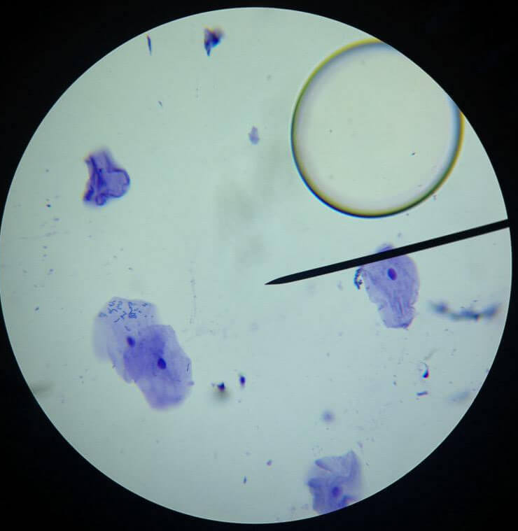
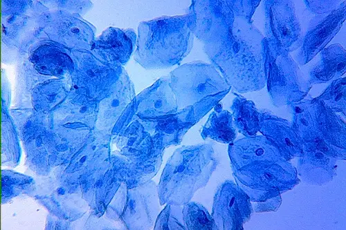
The result is a breathtaking sight. While the entire cell takes on a light blue tint, the nucleus, wrapped in a darker shade, shines like a guiding star amidst the celestial landscape of the cell. This stark contrast is the key to identification, as it enables scientists and enthusiasts alike to pinpoint the nucleus and delve into its significance in the cell’s overall functioning.
Indeed, staining the cheek cells with methylene blue uncovers a whole new world of cellular elegance. It transforms seemingly unremarkable structures into breathtaking masterpieces, unraveling the hidden complexity that resides within these tiny eukaryotic entities. As we peer through the lens of the microscope, we gain insight into the mysteries of life at the cellular level, reminding us of the astounding wonders that lie at the heart of every living organism.
Observation of the Cheek Cells
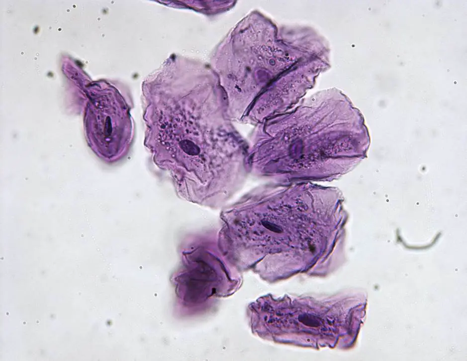
As we mount the wet slide, the microscopic world of cheek cells unfurls before our eyes, revealing a captivating tapestry of cellular structures. Our attentive gaze is met with remarkable features that provide insight into the intricacies of these eukaryotic entities.
The cheek cells exhibit a unique and irregular shape, defying uniformity as they sprawl across the slide. Each cell stands out with a distinct cell membrane, a defining boundary that separates the cellular interior from the external milieu. The cell membrane acts as a selective gatekeeper, regulating the exchange of substances and preserving the delicate equilibrium within the cell.
At the heart of each cheek cell, a focal point of significance captures our attention—the nucleus. With a striking dark blue hue, the nucleus assumes its identity as the epicenter of genetic information. Sheltered within this vital organelle lies the DNA, the blueprint that governs the cell’s functions and hereditary traits. As we explore further, the nucleus appears as a commanding presence, overseeing the cell’s activities and orchestrating the symphony of life within.
Surrounding the nucleus is the lightly stained cytoplasm, a fluidic medium that engulfs various organelles and facilitates essential cellular processes. Although pale in comparison to the intense blue of the nucleus, the cytoplasm plays a crucial role in supporting the cell’s functions, providing a stage for various cellular operations to transpire.
With each observation, the cheek cells bestow upon us a glimpse into the remarkable complexity of life at the cellular level. Their large and irregular shapes, distinguished cell membranes, prominent nuclei, and delicately stained cytoplasm beckon us to appreciate the elegance of nature’s building blocks.


FAQ
What are cheek cells, and why are they commonly observed under a microscope?
Cheek cells are eukaryotic cells found in the lining of our mouths. They are easily accessible and serve as a convenient and informative sample for microscopic observation due to their shedding nature.
How do I collect cheek cells for microscopic observation?
To collect cheek cells, gently scrape the inside of your mouth using a clean and sterile cotton swab.
What is the purpose of staining cheek cells before observation?
Staining with a biological dye, such as methylene blue, enhances the visibility of cellular structures, making it easier to distinguish different parts of the cell under the microscope.
Which cellular structures are typically visible when observing cheek cells?
Under the microscope, you can observe large, irregularly shaped cells with distinct cell membranes, a central and darkly stained nucleus, and a lightly stained cytoplasm.
How does methylene blue staining work on cheek cells?
Methylene blue has a strong affinity for DNA and RNA, causing the nucleus of cheek cells to appear darker, making it stand out from the surrounding cytoplasm.
What is the function of the nucleus in cheek cells?
The nucleus serves as the command center of the cell, containing genetic material (DNA) that regulates cellular activities and inheritance.
Can I observe cheek cells with a basic microscope, or do I need an advanced one?
Cheek cells can be observed using a basic light microscope. Even entry-level microscopes with 4x and 10x objectives are sufficient for initial observations.
How can I differentiate cheek cells from other types of cells under the microscope?
Cheek cells are unique in their irregular shape and are often larger than some other cell types, making them relatively easy to distinguish during microscopic observation.
What safety precautions should I take when handling cheek cells for observation?
Ensure you are working in a clean environment, wear clean gloves to avoid contamination, and dispose of used cotton swabs and towels safely to prevent cross-contamination.
Why are cheek cells commonly used in educational settings for microscopic observations?
Cheek cells are readily available and easy to collect, making them an ideal and non-invasive sample for students to explore basic cell biology concepts and practice their microscopy skills.