- Basidiomycetes are a group of fungi which belong to the division Basidiomycota.
- It is the process of sexual reproduction where spores are formed on a club-shaped structure called basidium.
- The sexual spores produced are known as basidiospores and these are formed externally on the basidium.
- These fungi are filamentous in nature and the body is made up of hyphae forming a mycelium.
- The mycelium generally shows a dikaryotic condition where two nuclei is present in each cell.
- Clamp connections are present in many basidiomycetes helping in maintaining dikaryotic stage.
- These are commonly referred to as higher fungi and include mushrooms, puffballs, bracket fungi, rusts and smuts.
- Some members are microscopic and yeast-like in nature such as Cryptococcus.
- Basidiomycetes plays an important role in decomposition of organic matter especially lignin present in wood.
- Many members form symbiotic association with plant roots known as ectomycorrhizae.
Habitat of Basidiomycetes
- Basidiomycetes are commonly found on plant surfaces where they live as parasites.
- Many species occur on leaves stems and reproductive parts of vascular plants such as wheat maize and barley.
- Some basidiomycetes are host specific and grow only on particular plants like rust fungi on wheat and barberry.
- Certain members are found parasitizing ferns and mosses in moist habitats.
- These fungi are also present in soil where they form association with plant roots.
- It is the process of symbiosis where basidiomycetes form ectomycorrhiza with roots of forest trees like pine oak and birch.
- The mycelium is commonly found in forest soil and forms underground networks.
- Many basidiomycetes grow on dead and decaying organic matter such as wood logs stumps and fallen trees.
- These are commonly seen on leaf litter and forest debris acting as decomposers.
- Some species inhabit animal hosts including insects where fungal mats are formed.
- Certain yeast forms of basidiomycetes are found in humans causing disease under suitable conditions.
- Basidiomycetes are also present in freshwater and marine habitats.
- Some members occur in damp buildings wastewater and other man made environments.
- The spores of basidiomycetes are commonly present in air and spread through atmosphere.
Characteristics of Basidiomycetes
- Basidiomycetes are fungi in which sexual reproduction takes place by the formation of basidium.
- The basidium is a club shaped structure where karyogamy and meiosis is completed.
- Sexual spores are known as basidiospores and these are produced externally on the basidium.
- Usually four haploid basidiospores is formed on each basidium.
- The mycelium is well developed and composed of septate hyphae.
- The septa of hyphae show a special type of pore known as dolipore septum.
- Clamp connections are present in many members and help in maintaining dikaryotic condition.
- The vegetative body mainly remains in dikaryotic stage where two nuclei is present in each cell.
- Rhizomorphs are formed in some basidiomycetes for conduction of food materials.
- Spores are discharged forcibly by a mechanism known as ballistospory.
- The life cycle is complex and in some members different spore stages are seen.
- These fungi are important decomposers of wood and are capable of degrading lignin.
- Many species form ectomycorrhizal association with roots of higher plants.
- Some basidiomycetes are parasitic and cause diseases like rust and smut in plants.
- Certain members also include pathogenic yeasts affecting humans.
- Based on characters basidiomycetes are divided into Agaricomycotina Pucciniomycotina and Ustilaginomycotina.
Morphology of Basidiomycetes
- Basidiomycetes are mostly filamentous fungi. The vegetative body is a well developed mycelium.
- The mycelium consists of septate hyphae. The septa are regularly present and show a central pore.
- Most Basidiomycetes show a dikaryotic condition. Each cell of the hypha contains two haploid nuclei (n + n) for a long period of life cycle.
- The septa are of dolipore type. The septal pore is barrel shaped and surrounded by a swollen rim.
- On either side of the dolipore septum, curved membrane structures are present. These are called parenthesomes.
- The dolipore–parenthesome septum allows cytoplasmic continuity but restricts the movement of nuclei.
- Clamp connections are commonly present in dikaryotic hyphae. These are hook like outgrowths formed during cell division.
- Clamp connections help in proper distribution of nuclei and maintenance of dikaryotic condition.
- In some Basidiomycetes, hyphae aggregate to form thick cord like structures called rhizomorphs. These help in conduction of water and nutrients.
- The reproductive structure is the basidium. It is club shaped and develops on the fertile layer.
- Each basidium bears usually four basidiospores. These spores are produced exogenously on sterigmata.
- Basidia may be non septate (holobasidium) or septate (phragmobasidium). Septate basidia are common in rusts and smuts.
- Basidiospores are haploid and usually forcibly discharged. After dispersal, they germinate to form primary mycelium.
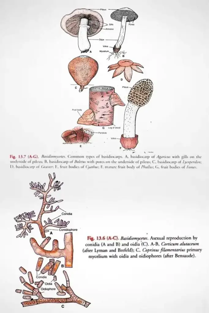
- Many Basidiomycetes form large fruiting bodies called basidiocarps. These may be mushrooms, brackets, puffballs or jelly like forms.
- In rusts and smuts, basidiocarps are generally absent. Spores are produced in masses within host tissues.
- The presence of dikaryotic mycelium, dolipore septum with parenthesomes, clamp connections and basidium are important morphological features of Basidiomycetes.
Examples of Basidiomycetes
- Agaricus bisporus – common edible mushroom.
- Lentinula edodes – shiitake mushroom.
- Pleurotus ostreatus – oyster mushroom.
- Pleurotus eryngii – king oyster mushroom.
- Marasmius oreades – fairy ring mushroom.
- Coprinopsis cinerea – ink cap mushroom.
- Hypsizigus marmoreus – beech mushroom.
- Flammulina velutipes – winter mushroom.
- Puccinia graminis – causal organism of stem rust of wheat.
- Gymnosporangium juniperi-virginianae – causes cedar apple rust.
- Cronartium ribicola – causes white pine blister rust.
- Ustilago maydis – corn smut fungus.
- Tilletia caries – causes bunt of wheat.
- Phakopsora pachyrhizi – soybean rust fungus.
- Polyporus squamosus – bracket fungus growing on wood.
- Trametes versicolor – turkey tail fungus.
- Ganoderma lucidum – medicinal bracket fungus.
- Auricularia auricula – jelly fungus found on decaying wood.
- Tremella mesenterica – yellow jelly fungus.
- Cryptococcus neoformans – pathogenic yeast in humans.
- Sporobolomyces salmonicolor – mirror yeast.
- Septobasidium – parasitic fungus on insects.
Classification of Basidiomycetes
Based on modern phylogenetic analysis the phylum Basidiomycota is divided into three major subphyla. In earlier systems these fungi was grouped under Homobasidiomycetes and Heterobasidiomycetes but this classification is now considered obsolete. The modern system is more acceptable as it is based on molecular and evolutionary relationships.
Subphylum Agaricomycotina
It is the largest group of Basidiomycetes and includes the typical mushroom forming fungi. These fungi generally possess holobasidia which are unicellular and non-septate. The basidiocarps are well developed in most members.
Some of the important classes are–
- Class Agaricomycetes. It includes most of the common mushrooms shelf fungi puffballs polypores corals and chanterelles. These are saprophytic or parasitic in nature.
- Class Dacrymycetes. These are jelly fungi with gelatinous basidiocarps. The basidia are typically forked.
- Class Tremellomycetes. It includes jelly fungi and several yeast forms. The basidia are septate and irregular in shape.
- Class Wallemiomycetes. This group includes xerophilic fungi and sometimes treated as a sister group of Agaricomycotina.
Subphylum Pucciniomycotina
This subphylum mainly includes rust fungi insect parasites and different yeasts. The basidia are phragmobasidia which are septate. Most members show complex life cycles with more than one host.
The important classes are–
- Class Pucciniomycetes. It is the largest class and includes rust fungi under order Pucciniales and also Septobasidiales.
- Class Microbotryomycetes. These include plant pathogenic fungi and mirror yeasts.
- Class Cystobasidiomycetes. It contains yeasts and dimorphic fungi.
- Class Agaricostilbomycetes. These fungi possess stilboid fruiting bodies.
- Class Atractiellomycetes. These are auricularioid fungi with special organelles called symplechosomes.
- Class Classiculomycetes. These are aquatic fungi found mainly in freshwater habitats.
- Class Cryptomycocolacomycetes. These are mycoparasitic fungi infecting ascomycetes.
- Class Mixiomycetes. It includes a single fern parasite Mixia osmundae.
Subphylum Ustilaginomycotina
This group consists mainly of smut fungi and related plant parasites. Like rust fungi they possess phragmobasidia. The basidiocarps are usually reduced and spores are produced in large numbers.
The major classes are–
- Class Ustilaginomycetes. These are the true smut fungi parasitic on flowering plants.
- Class Exobasidiomycetes. It includes plant parasites such as Exobasidium.
- Class Entorrhizomycetes./ These are root associated fungi and sometimes treated as a separate lineage.
Traditional (Obsolete) Classification
Although no longer used in modern taxonomy these terms are still used in a descriptive sense.
- Homobasidiomycetes. These included mushrooms and puffballs having non-septate basidia.
- Heterobasidiomycetes. These included rusts smuts and jelly fungi characterized by septate basidia and complex life cycles.
Economic Importance of Basidiomycetes
Negative Economic Importance
- Basidiomycetes include rusts and smuts which are destructive plant pathogens causing heavy loss in agriculture.
- Cereal rusts like Puccinia graminis and Puccinia striiformis attack wheat barley and oats resulting in severe reduction of crop yield.
- Smut fungi such as Ustilago maydis and Tilletia caries infect grains and produce malformed reproductive structures leading to poor grain quality.
- Rust fungi also infect crops like soybean and coffee where yield loss may reach very high levels.
- Wood destroying basidiomycetes cause decay of timber railway sleepers and wooden structures.
- Brown rot fungi degrade cellulose and hemicellulose making wood brown and brittle.
- White rot fungi degrade lignin cellulose and hemicellulose causing spongy and bleached wood.
- Sudden outbreak of rusts and smuts may disturb food supply and increase economic burden on agriculture based countries.
- Some basidiomycetes like Cryptococcus neoformans cause serious human diseases and increase medical cost.
- Airborne spores of certain basidiomycetes act as allergens and cause respiratory problems.
Positive Economic Importance
- Many basidiomycetes are edible and are used as food throughout the world.
- Common edible mushrooms include Agaricus bisporus Pleurotus spp. and Lentinula edodes.
- These mushrooms are rich in proteins vitamins and bioactive compounds and are considered as functional food.
- Corn smut (Ustilago maydis) is used as a food delicacy in some countries.
- Basidiomycetes form ectomycorrhizal association with roots of forest trees like pine oak and birch.
- Mycorrhizal association helps in absorption of minerals like phosphorus nitrogen and zinc from soil.
- These fungi increase tolerance of plants against drought salinity heavy metals and soil pathogens.
- Mycorrhiza is essential for successful reforestation and growth of trees in poor soils.
Industrial and Pharmaceutical Importance
- Basidiomycetes produce many secondary metabolites with antibiotic and antifungal properties.
- Strobilurins isolated from Strobilurus tenacellus are used as agricultural fungicides.
- Some basidiomycetes act as biological control agents against plant pests.
- Pleurotus ostreatus produces toxins that kill nematodes present in soil.
- Extracts of fungi like Pycnoporus sanguineus show insecticidal activity.
- Medicinal mushrooms such as Ganoderma lucidum possess antitumor antiviral and antioxidant properties.
- White rot fungi produce lignin degrading enzymes useful in paper industry and biofuel production.
- Basidiomycetes are used in bioremediation and waste water treatment processes.
- Fungal mycelium is used for preparation of eco friendly construction and packaging materials.
Mycelium of Basidiomycetes
The mycelium of Basidiomycetes is the vegetative body which is made up of a mass of branched hyphae. It is well developed and grows within the substratum for absorption of nutrients. The mycelium is usually long lived and shows different structural and functional modifications.
Structural Characteristics
The mycelium consists of septate hyphae. These hyphae are tubular and branched and the septa are regularly present. Each septum has a central pore which allows continuity of cytoplasm.
The septa are of a special type known as dolipore septa. It is the process in which the septal pore shows a barrel shaped swelling. On both sides of the pore, membranous structures called parenthesomes are present. These structures help in movement of cytoplasm but restrict the movement of nuclei and large organelles.
Clamp connections are commonly present in the mycelium. These are hook like outgrowths formed during cell division. It helps in equal distribution of nuclei and maintaining the dikaryotic condition of the hyphae.
In some basidiomycetes, the hyphae are aggregated to form thick rope like structures called rhizomorphs. These structures help in conduction of water and nutrients over long distance. The rhizomorphs are often covered by a protective outer layer.
The mycelium when seen in mass appears white or sometimes yellowish or orange. It may form fan shaped or cottony growths on the substratum.
Developmental Stages of Mycelium
Primary Mycelium
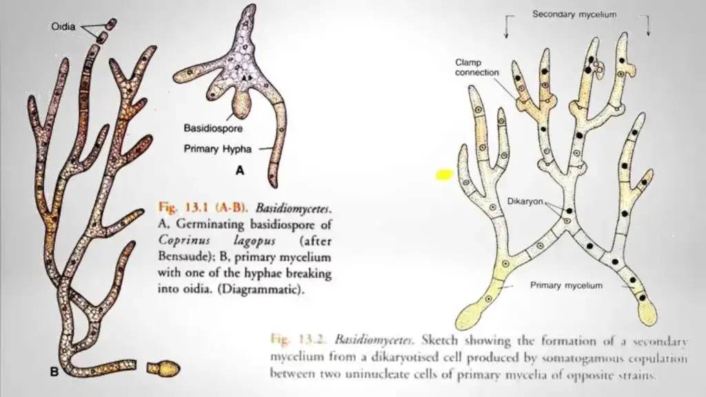
It is developed from the germination of basidiospore. The hyphae are monokaryotic and contain a single haploid nucleus in each cell. This mycelium is usually short lived.
Secondary Mycelium
It is formed by fusion of two compatible primary mycelia. This fusion is referred to as plasmogamy. The secondary mycelium is dikaryotic (n + n) and each cell contains two unfused nuclei. It is the dominant and vegetative phase of the life cycle.
Tertiary Mycelium
The tertiary mycelium forms organized tissues which give rise to the basidiocarp or fruiting body. It is formed under suitable environmental conditions from the secondary mycelium.
Ecological Forms
In ectomycorrhizal forms, the mycelium forms a sheath around the roots of higher plants. The hyphae penetrate between cortical cells forming a network known as Hartig net. This association helps in exchange of minerals and nutrients.
In parasitic forms such as rusts and smuts, the mycelium grows intercellularly within host tissues. Special absorbing organs called haustoria are formed which help in absorption of nutrients from living host cells.
Importance
The mycelium of Basidiomycetes plays an important role in decomposition of organic matter. It also helps in nutrient cycling and forms beneficial symbiotic associations with plants. The structural specialization of the mycelium helps in survival and wide distribution of these fungi.

Clamp Connections
- Clamp connection is a special hook like structure formed in the dikaryotic hyphae of Basidiomycetes. It is concerned with the maintenance of dikaryotic condition in secondary mycelium.
- When the dikaryotic cell begins to divide, a pouch like outgrowth arises from the lateral wall. It develops midway between the two nuclei of the dikaryon.
- The two closely associated nuclei divide simultaneously. This type of nuclear division is referred to as conjugate division.
- Out of the four daughter nuclei formed, one nucleus migrates into the pouch like outgrowth. A septum then appears at the base of the pouch and cuts it off from the parent cell. This pouch is now known as clamp cell.
- The clamp cell elongates and bends over the parent hypha forming a hook like structure. The tip of the clamp cell gets attached to the lateral wall of the parent cell and acts as a bridge. This structure is called clamp connection.
- A vertical septum is formed below the bridge. It divides the parent cell into two daughter cells. The terminal daughter cell contains two nuclei.
- The basal daughter cell initially contains only one nucleus. The fourth nucleus remains inside the clamp connection.
- The nucleus present in the clamp connection migrates into the basal daughter cell. Thus both daughter cells become binucleate.
- The clamp connection therefore functions as a bypass. It ensures proper distribution of sister nuclei into the two newly formed daughter cells.
- Clamp connections are usually formed on terminal cells of the hyphae of secondary mycelium. They generally persist on older dikaryotic hyphae.
- The presence of hook like clamp connections is a reliable criterion for distinguishing secondary (dikaryotic) mycelium from primary (monokaryotic) mycelium.
- Some mycologists consider clamp connections to be homologous to the hooks of ascogenous hyphae of Ascomycetes. This view is supported by the presence of clamp connections at the base of basidium.
- Clamp connections along with dikaryotic mycelium, basidium formation and dolipore septum with parenthesomes are regarded as important diagnostic features of Basidiomycetes.

What is Diploidisation or Dikaryotisatton?
- Diploidisation or dikaryotisation is the process by which a dikaryotic condition is established in Basidiomycetes. It is the process of formation of dikaryotic mycelium from monokaryotic mycelium.
- In this process, the first step is the establishment of a dikaryon in the fusion cell. The two compatible haploid nuclei remain unfused and lie together in the same cell.
- By repeated divisions and formation of clamp connections, the dikaryotic condition is maintained and spread throughout the hyphae.
- As a result of this repeated dikaryotic division, a well developed secondary mycelium is formed in which each cell possesses two nuclei (dikaryon).
Methods of Diploidisation
- Hyphal fusion– In this method, the vegetative cells of two adjacent hyphae belonging to opposite sexual strains fuse with each other. The fusion results in a binucleate cell which gives rise to dikaryotic mycelium.
- Conjugation of basidiospores– In this method, basidiospores of two opposite strains meet and conjugate. The binucleate basidiospore thus formed germinates and develops into a secondary mycelium.
- Fusion of germinating oidium with primary mycelium– Diploidisation may take place by fusion of a germinating oidium of one strain with a cell of the primary mycelium of the opposite strain. The binucleate cell formed elongates and divides with clamp connections to form secondary mycelium.
- Fusion of germinating basidiospore with basidial cell– In some forms, diploidisation occurs when a germinating basidiospore fuses with a haploid cell of the basidium. For example, Ustilago violacea.
- Fusion of haploid basidial cells– Diploidisation can also occur when two haploid cells of opposite strains of the basidium fuse. For example, Ustilago carbo.
- Fusion of basidia formed from smut spores– In certain cases, the basidia produced by the germination of two smut spores fuse with each other resulting in dikaryotic condition.
- Basidia are always produced from binucleate cells of the secondary mycelium.

Basidiomycetes life cycle or Reproduction

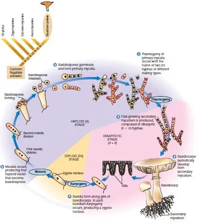
- The life cycle of Basidiomycetes is characterised by a long lived dikaryotic phase. Sexual reproduction is well developed and takes place by means of basidia and basidiospores.
- Vegetative body is a mycelium which shows three distinct phases namely primary, secondary and tertiary mycelium.
Primary Mycelium
- The life cycle begins with the germination of a basidiospore.
- Each basidiospore is haploid and on germination it gives rise to a primary mycelium.
- The primary mycelium is monokaryotic and each cell contains a single haploid nucleus.
- This mycelium is short lived and does not produce fruiting bodies.
Secondary Mycelium
- Secondary mycelium is formed after plasmogamy.
- Plasmogamy takes place by fusion of two compatible primary mycelia or by other methods of dikaryotisation.
- As a result, a dikaryotic mycelium is formed in which each cell contains two nuclei (n + n).
- This is the dominant and long lived vegetative phase.
- Clamp connections are usually formed to maintain the dikaryotic condition during cell division.
Tertiary Mycelium and Basidiocarp Formation
- Under favourable environmental conditions, the secondary mycelium becomes organised into compact tissues.
- This organised mycelium is called tertiary mycelium.
- The tertiary mycelium gives rise to the basidiocarp or fruiting body such as mushroom, bracket fungus or puffball.
- The basidiocarp bears a fertile layer known as hymenium.
Sexual Reproduction
- Sexual reproduction takes place in the basidium which is the characteristic reproductive structure of Basidiomycetes.
- The basidium develops on the hymenial layer of the basidiocarp.
- In each basidium, the two haploid nuclei fuse. This process is known as karyogamy.
- As a result, a diploid nucleus is formed. This diploid phase is very short lived.
- The diploid nucleus immediately undergoes meiosis and produces four haploid nuclei.
- These nuclei move into four outgrowths called sterigmata.
- Each nucleus develops into a basidiospore. Thus, four basidiospores are formed exogenously on each basidium.
- The basidiospores are forcibly discharged and dispersed. After reaching a suitable substratum, they germinate to produce new primary mycelia.
Asexual Reproduction
- Asexual reproduction is generally absent in Basidiomycetes.
- In some forms, it may occur by fragmentation of mycelium or by formation of oidia or conidia, especially in rusts and smuts.
Special Life Cycles
- In rusts, the life cycle is very complex and may involve two hosts and several types of spores.
- In smuts, thick walled resting spores called teliospores are formed which later germinate to produce basidia and basidiospores.
- Formation of basidium, basidiospores, dikaryotic mycelium and clamp connections are the important diagnostic features of Basidiomycetes.

FAQ
Q1. What are Basidiomycetes?
A. Basidiomycetes are a class of fungi in which sexual reproduction takes place by means of a special structure called basidium. They are characterised by the formation of basidiospores and a well developed dikaryotic mycelium.
Q2. What are the characteristics of Basidiomycetes?
A.
- Vegetative body is a well developed, septate mycelium.
- Mycelium shows a prolonged dikaryotic condition.
- Septa are of dolipore type with parenthesomes.
- Clamp connections are usually present.
- Sexual reproduction occurs by basidia and basidiospores.
Q3. What is the ecological role of Basidiomycetes?
A. Basidiomycetes play an important role as decomposers of wood and organic matter. They help in recycling of nutrients in nature and also form symbiotic associations with higher plants.
Q4. How do Basidiomycetes reproduce?
A. Basidiomycetes reproduce mainly by sexual method through formation of basidia and basidiospores. Vegetative reproduction may occur by fragmentation of mycelium.
Q5. Do Basidiomycetes reproduce both sexually and asexually?
A. Yes, Basidiomycetes reproduce sexually by basidiospores. Asexual reproduction is rare but may occur by oidia, conidia or fragmentation in some forms like rusts and smuts.
Q6. What are basidia?
A. Basidia are club shaped reproductive structures found in Basidiomycetes. Karyogamy and meiosis take place in the basidium resulting in formation of basidiospores.
Q7. What are basidiocarps?
A. Basidiocarps are fruiting bodies formed from the tertiary mycelium of Basidiomycetes. They bear basidia on their fertile surface.
Q8. What are some examples of Basidiomycetes?
A. Common examples of Basidiomycetes are Agaricus, Polyporus, Puccinia, Ustilago, Coprinus and Lycoperdon.
Q9. What is the difference between Basidiomycetes and Ascomycetes?
A.
- In Basidiomycetes, spores are formed on basidia, while in Ascomycetes spores are formed inside asci.
- Basidiomycetes show long lived dikaryotic mycelium, whereas it is short lived in Ascomycetes.
- Clamp connections are present in Basidiomycetes but absent in Ascomycetes.
Q10. How do Basidiomycetes contribute to nutrient cycling?
A. Basidiomycetes decompose cellulose and lignin present in dead plant material. This releases minerals back into soil and maintains nutrient balance in ecosystem.
Q11. Are Basidiomycetes edible?
A. Yes, many Basidiomycetes are edible. For example, Agaricus (mushroom) and Pleurotus are commonly consumed as food.
Q12. Are there toxic Basidiomycetes?
A. Yes, some Basidiomycetes are poisonous. Examples include Amanita phalloides and Amanita muscaria which are highly toxic.
Q13. Do Basidiomycetes form mycorrhizal associations?
A. Yes, many Basidiomycetes form ectomycorrhizal associations with roots of higher plants. This association helps both fungus and plant.
Q14. What are the different types of basidiocarps?
A.
- Mushrooms
- Bracket or shelf fungi
- Puffballs
- Jelly fungi
Q15. What is the life cycle of Basidiomycetes?
A. The life cycle of Basidiomycetes includes formation of primary monokaryotic mycelium from basidiospore, development of secondary dikaryotic mycelium by plasmogamy, formation of basidiocarp, and production of basidiospores after karyogamy and meiosis in basidium.
- Aime, M. C., Matheny, P. B., Henk, D. A., Frieders, E. M., Nilsson, R. H., Piepenbring, M., McLaughlin, D. J., Szabo, L. J., Begerow, D., Sampaio, J. P., Bauer, R., Weiß, M., Oberwinkler, F., & Hibbett, D. (2006). An overview of the higher level classification of Pucciniomycotina based on combined analyses of nuclear large and small subunit rDNA sequences. Mycologia, 98(6), 896–905. https://doi.org/10.3852/mycologia.98.6.896
- Delahaut, K. (n.d.). Mycorrhizae. Wisconsin Horticulture, Division of Extension. Retrieved from https://hort.extension.wisc.edu/articles/mycorrhizae/
- everythingapartfromofficialkaj. (n.d.). What is dolipore septum. Scribd. Retrieved from https://www.scribd.com/document/947487970/What-is-Dolipore-Septum
- GeeksforGeeks. (2023, July 4). Difference between rust and smut. https://www.geeksforgeeks.org/biology/difference-between-rust-and-smut/
- Godfray, H. C. J., Mason-D’Croz, D., & Robinson, S. (2016). Food system consequences of a fungal disease epidemic in a major crop. Philosophical Transactions of the Royal Society B: Biological Sciences, 371(1709), 20150467. https://doi.org/10.1098/rstb.2015.0467
- Goodell, B., Qian, Y., & Jellison, J. (2008). Fungal decay of wood: Soft rot – brown rot – white rot. In T. P. Schultz, H. Militz, M. H. Freeman, B. Goodell, & D. D. Nicholas (Eds.), Development of commercial wood protection systems: Issues and strategies (pp. 9–31). American Chemical Society. https://composites.umaine.edu/publication/fungal-decay-of-wood-soft-rot-%C2%96-brown-rot-%C2%96-white-rot-1/
- Legner, E. F. (n.d.). Basidiomycota & Heterobasidiomycetes. University of California, Riverside. Retrieved from https://faculty.ucr.edu/~legneref/fungi/basidiomycota.htm
- Lumen Learning. (n.d.). Basidiomycota: The club fungi. Biology for Majors II. Retrieved from https://courses.lumenlearning.com/wm-biology2/chapter/basidiomycota/
- Riley, R., Salamov, A., Brown, D. W., Nagy, L. G., Floudas, D., Held, B., Levasseur, A., Lombard, V., Morin, E., Otillar, R., Lindquist, E., Sun, H., LaButti, K., Schmutz, J., Jabbour, D., Luo, H., Baker, S. E., Pisabarro, A., Walton, J. D., … Grigoriev, I. V. (2014). Extensive sampling of basidiomycete genomes demonstrates inadequacy of the white rot/brown rot paradigm for wood decay fungi. Proceedings of the National Academy of Sciences, 111(27), 9923–9928. https://doi.org/10.1073/pnas.1400592111
- Sivanandhan, S., Khusro, A., Paulraj, M. G., Ignacimuthu, S., & Al-Dhabi, N. A. (2017). Biocontrol properties of Basidiomycetes: An overview. Journal of Fungi, 3(1), 2. https://doi.org/10.3390/jof3010002
- Stolze-Rybczynski, J. L., Cui, Y., Stevens, M. H. H., Davis, D. J., Fischer, M. W. F., & Money, N. P. (2009). Adaptation of the spore discharge mechanism in the Basidiomycota. PLoS ONE, 4(1), e4163. https://doi.org/10.1371/journal.pone.0004163
- The phylum Basidiomycota: Structure, biogeochemistry, and global economic impact—An expert review. (n.d.). [Review Article].
- Wikipedia contributors. (2025, February 18). Basidiomycota. Wikipedia, The Free Encyclopedia. https://en.wikipedia.org/w/index.php?title=Basidiomycota&oldid=1318477350
- Wikipedia contributors. (2025, January 19). Clamp connection. Wikipedia, The Free Encyclopedia. https://en.wikipedia.org/w/index.php?title=Clamp_connection&oldid=1291986665
- Wikipedia contributors. (2025, March 2). Mycorrhiza. Wikipedia, The Free Encyclopedia. https://en.wikipedia.org/w/index.php?title=Mycorrhiza&oldid=1326693958
- Text Highlighting: Select any text in the post content to highlight it
- Text Annotation: Select text and add comments with annotations
- Comment Management: Edit or delete your own comments
- Highlight Management: Remove your own highlights
How to use: Simply select any text in the post content above, and you'll see annotation options. Login here or create an account to get started.