- Automated Cell Counter refers as an instrument used for counting of cells automatically without using manual hemocytometer.
- The device mainly used by laboratories for accurate and faster determination of cell concentration, viability, and size distribution in suspension.
- Counting is done by different principles like optical imaging / electrical impedance / fluorescence method etc.
- In this equipment, cell images are captured by camera sensor and analyzed by computer algorithm for identifying live and dead cells.
- The counting process is usually performed by loading small amount of sample (mostly 10–20 µL) into special slide or chamber.
- Manual error which occur by human observation is largely reduced by this automated technique.
- The data obtained are displayed directly on screen with graphs and numeric values, sometimes exported for further analysis.
- Accuracy of counting depend by calibration, sample mixing, and cleanliness of slide used.
- This device are commonly used in cell culture, biomedical research, clinical diagnostics etc.
- Compared with traditional hemocytometer, it saves more time, improve reproducibility, and provide sturdy and hardy performance in busy lab setup.
- In some models viability is determined by dyes like Trypan Blue, which stain dead cells only.
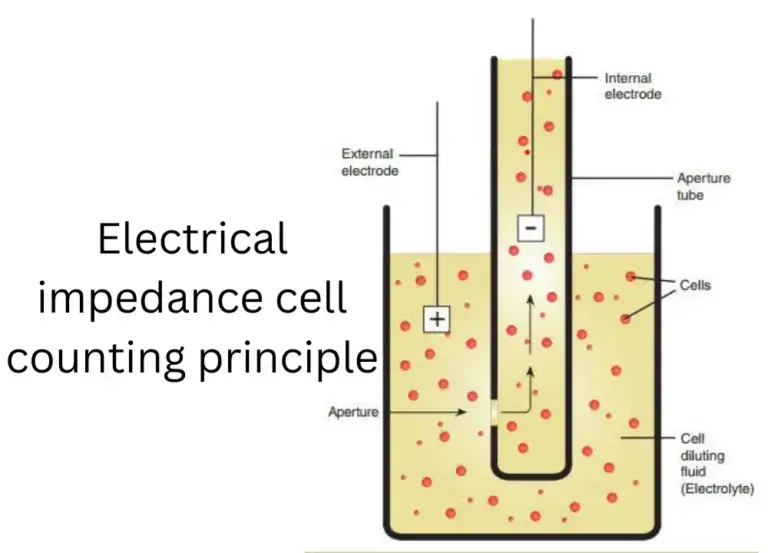
- The principle basically rely on image-based recognition or electrical impedance change when cells passes through aperture.
- Automated Cell Counter considered as essential equipment for modern biology lab where large number of samples need to be processed quickly.
- However, proper maintenance and calibration must done regularly, otherwise error may prevail counting results.
- So, in short, this device has simplified the tedious task of cell counting, making it more consistent and reliable for researchers, students and technicians etc.
Principle of Automated Cell Counter
- Principle is based mostly on electrical impedance / Coulter principle, optical imaging / image analysis, or light scatter / fluorescence methods.
- In impedance method, cells in conductive solution pass one by one through narrow aperture between electrodes, a change in resistance (or impedance) is sensed and counted.
- The amplitude (pulse height) of the resistance / impedance change is proportional to cell volume so cell size distribution also is inferred.
- In image based (optical) method, images of cells are captured (brightfield, fluorescence) and computer algorithm is used to identify objects (cells) vs background.
- Viability is assessed in image method by using dyes (ex: Trypan Blue, propidium iodide) which stain dead cells, so algorithm classifies live vs dead.
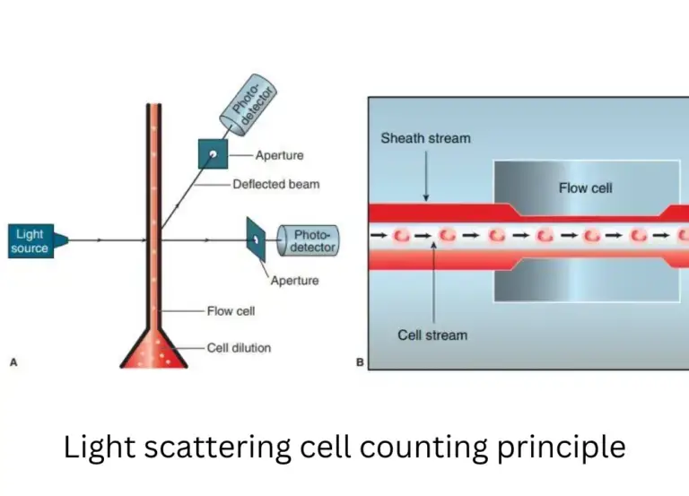
- Light scatter / fluorescence principle: cells are passed through laser, scattered light or emitted fluorescence is detected, signals correlate with size / internal features, cell count is deduced.
- In some instruments hybrid principle is used: impedance + optical or scatter combined, for better discrimination.
- Dilution and proper sample preparation is required so that cells pass singly, avoiding coincidence (two or more cells passing simultaneously) which lead to error.
- Calibration is needed to relate pulse heights or image metrics to actual volumes or diameters, using standards (latex beads etc).
- The principle is such that counting is automated, operator intervention minimal, and reproducibility / speed are improved.


Types of Automated Cell Counters
1. Impedance-Based Cell Counters – This type of counter works on Coulter Principle, where the change in electrical resistance is measured when each cell passes by a small aperture. The cell suspension is flowed by electrolyte, and as the cells interrupt the current, the pulses generated are counted as individual cells. Sometimes it also used for measuring cell volume. These devices are mostly used for RBC/WBC/Platelet counts in hematology. However, accuracy may affected by debris or air bubbles, etc.
2. Optical or Image-Based Cell Counters – In this system, cells are captured by digital imaging using microscope-like optics and advanced software. The captured images are analyzed by algorithms for cell number, size, and viability (using dye staining like trypan blue). The counting is done automatically, though sometimes manual adjustment is required if clumping occurs. Image-based counters are widely used in research labs for mammalian cells, yeast, etc, since they provide visual confirmation of cells.
3. Flow Cytometry-Based Counters – A more advanced type where cells pass single-file by a laser beam, and the light scattered by each cell is measured. Fluorescent dyes can be used to identify cell type, viability, or specific protein expressions. This system provide high accuracy and speed, often used for clinical diagnostics and immunology. However, it’s costlier and need more expertise to operate.
4. Spectrophotometric/Colorimetric Counters – In some instruments, cell concentration is indirectly estimated by measuring optical density (OD) or light absorbance at specific wavelength (like 600nm for bacteria). Although not strictly cell-by-cell counter, it’s automated in many growth monitoring systems. The method is simple and quick but less precise for mixed populations.
5. Electrical/Capacitance Hybrid Counters – Some modern automated counters combine both electrical impedance and optical detection together for better accuracy. The hybrid systems can differentiate live/dead cells more efficiently by combining multiple parameters.
6. Acoustic or Microfluidic-Based Counters – In latest models, microfluidic chips and acoustic waves are used to focus and move cells for detection. These systems are compact and need very less sample, often applied in portable or point-of-care devices.
How to Operate an Automated Cell Counter
- Sample is first prepared (cells detached if adherent, suspended gently) and debris/clumps are removed by pipetting or filtering.
- A small aliquot of cell suspension is taken (for example 10 µL) and mixed with staining dye (like Trypan Blue, or fluorescent dye) if viability measurement is intended
- This stained / mixed sample is allowed to rest briefly so that mixing is uniform, avoid bubbles, let particles settle somewhat.
- The mixture is loaded into counting slide / chamber / cassette carefully, without overloading (avoid too many cells).
- The slide or cassette is inserted into the automated cell counter device (into the slide port or drawer) in correct orientation until full insertion is sensed.
- The instrument then performs autofocus, illumination setting (brightfield / fluorescence) and detection automatically once slide recognized
- Counting is initiated (by pressing “Count” / “Run”) and images or signals are captured, processed by algorithm to identify cells, live / dead, size etc.
- Results (total cell concentration, live cell count, % viability, aggregates, histogram etc) are displayed / saved / printed as needed.
- After measurement, slide / cassette is ejected and disposed or cleaned (if reusable) appropriately (biohazard disposal).
- Device is cleaned / maintained (optical windows, housing) to avoid contamination or residue which may affect later counts
Applications of Automated Cell Counter
- In cell culture labs, monitoring cell growth / proliferation is done by automated cell counters to decide when passaging or seeding new plates.
- Use is made for assessing viability / live vs dead cell ratio during experiments, especially in toxicity or drug testing.
- In bioprocessing / biomanufacturing settings, the counters are used to control cell density in bioreactors (upstream process) to optimize yield.
- For clinical / diagnostic labs, automated counters help in blood cell counts, enumeration of leukocytes, or quality control of cell-based therapies.
- In drug screening / pharmacology, they are applied to measure effect of compounds on cell survival / proliferation (dose response) in assays.
- For microalgae / algal research, image-based counters with fluorescence filters are used to count algae and assess status (chlorophyll fluorescence) in environmental / industrial algal cultures.
- During quality control of cell therapy products, accurate counting and viability measurement are critical (dosing, release criteria) and automated counters are used.
- In high throughput / screening workflows, many samples are processed and automated counters reduce manual labour and variability.
- In vaccine and biologics production, cell concentration and viability must be regularly measured to maintain consistency & product quality.
- For animal / plant cell research, counters are used in experiments with primary cells, stem cells, protoplasts etc to standardize input cell numbers.
Automated cell counter vs Hemocytometer
| Feature | Automated Cell Counter | Hemocytometer |
|---|---|---|
| Accuracy | High | High |
| Speed | Fast | Slow |
| Cost | Expensive | Inexpensive |
| Ease of Use | Easy | Difficult |
| Sample Size | Small | Large |
| Versatility | Limited | Broad |
| Counting Method | Optical | Manual |
- A hemocytometer is manual counting chamber (a thick glass slide with etched grid) that is used with microscope to count cells by eye.
- Automated cell counter is an instrument in which counting is done by sensors / image / algorithms without human counting.
- In manual method, cells are loaded into chamber, grid areas are counted, then average is taken and concentration computed.
- In automated method, sample is loaded into cassette / slide, device focuses, images / signals are captured, software computes count & viability.
- Error / variability is higher in hemocytometer method because user judgment (which particles count as cells, live vs dead) is subjective and differs among users.
- Automated counters reduce user-to-user variability, by using consistent algorithm rules.
- Speed is much greater in automated counter; what may take minutes manually is done in seconds by instrument.
- Range of concentration measurable is wider in automated instruments (low to high) compared to manual limited grid counts.
- Hemocytometer method cost is low (basic equipment) but labor cost / time cost high; automated method has high capital cost and consumables but saves labor & time.
- In certain situations (very low cell count, clumped cells, special cell types) manual method may perform better or allow visual judgment, whereas automated may miscount or misclassify.
- Maintenance / calibration are needed in automated counters; a broken instrument causes downtime. Hemocytometer is simpler, repair / replacement is trivial.
- For labs with high throughput demands, automated counters are more justified; for small occasional counts perhaps hemocytometer suffices.
Advantages of Automated Cell Counter
- Much less human error is introduced when automated cell counter is used, since operator judgment (which particles count, focus selection etc) is eliminated.
- High reproducibility / consistency across runs and users is achieved by automated counters; inter-user variability is greatly reduced.
- Speed is gained: many cells are counted in seconds, throughput is high, time saving is substantial.
- Wider dynamic range is handled: both low and high cell concentrations can be counted more accurately without repeated dilutions.
- Additional parameters (cell size, morphology, viability) can be reported automatically by algorithm / gating / image analysis.
- Data handling and storage is facilitated (export, graphs, software) instead of manual note-taking.
- Fatigue and tedium for personnel is reduced; staff can focus on interpretation rather than counting.
- Cell health is better preserved (cells not held long in stain / suspension) because fast operation reduces exposure time.
- For regulated / GMP / QA environments, compliance features (audit trails, software logs) are possible in automated systems.
- Over long term, labor cost / time cost is reduced, making method more efficient even though instrument cost is higher.
Limitations of Automated Cell Counter
- Accuracy may be compromised when cell aggregates / clumps are present, because the software might count a cluster as one cell instead of many.
- Some algorithms may misclassify debris, dead cell fragments, or non-cellular particles as cells, especially when sample is impure.
- Small cells (or very large cells) falling outside the designed size range / dynamic range of instrument may be underestimated or missed.
- Fluorescence / viability measurement may be inaccurate if staining is uneven or dye penetrance is variable.
- Inter-instrument / inter-model variation is possible: different brands may give different counts for same sample (lack of standardization).
- Consumables (slides, cassettes) may introduce cost / waste, and their defects can lead to error (e.g. scratches, defects).
- Calibration drift or lack of frequent recalibration can cause systematic error over time.
- Instrument failure, clogging, optics misalignment, or contamination may degrade performance or introduce bias.
- In very low cell concentrations, stochastic error and detection limits may cause high relative error.
- Some cell types (irregular shape, very elongated, mixed sizes) may challenge the counting algorithm and yield less accurate results.
Precautions
- Proper mixing of cell suspension must be ensured just before loading, to avoid settling / aggregates which lead to wrong count (mix by pipetting).
- The counting chamber / slide surfaces must be clean and free from dust / smudge so that optical surfaces are not obstructed.
- Bubbles in sample must be avoided because they distort image / signal, and focus is disrupted.
- Sample loading volume must not exceed chamber capacity (do not overfill) to prevent spill into instrument internals.
- The optical surfaces / counting slide surfaces should not be touched by fingers (hold by edges) to avoid smears / fingerprints.
- Viability stains (like Trypan Blue) should be used promptly (within few minutes) after mixing because dye toxicity / diffusion affect result.
- Calibration / validation of instrument must be done periodically to ensure accuracy, and debris / non-cell particles must be excluded via gating settings or thresholds.
- Maintenance & cleaning of internal optics, fluidics and waste lines must be done to prevent contamination, clogging, drift, or carryover.
- Instruments should be operated in appropriate ambient conditions (temperature, vibration, humidity) so that focus, optics, electronics do not drift or fail.
- Slides / consumables must be disposed as biohazardous waste (or cleaned if reusable) following lab safety protocols.
- Samples containing clumps, debris, cell aggregates should be filtered / gently dissociated before counting to reduce error.
- Use of correct slide type / compatible consumables is vital — using wrong slide / chamber may cause wrong reading or instrument damage
FAQ
What is an Automated Cell Counter?
An Automated Cell Counter is a machine that uses imaging or light scattering technology to accurately count cells in a sample.
How does an Automated Cell Counter work?
An Automated Cell Counter uses optical techniques to determine the number of cells in a sample by analyzing light scattering patterns or images of the cells.
What are the benefits of using an Automated Cell Counter?
The benefits of using an Automated Cell Counter include accuracy, speed, ease of use, and small sample size requirements. Automated cell counters are also useful for counting cells in complex suspensions and for use with a wide range of cell types.
How accurate are Automated Cell Counters?
Automated Cell Counters are highly accurate, providing consistent and reproducible results. The accuracy of the count is dependent on the quality of the sample and the performance of the machine.
How fast are Automated Cell Counters?
Automated Cell Counters are typically much faster than manual cell counting methods. The speed of the count depends on the size of the sample, the performance of the machine, and the type of technology used.
What is the sample size requirement for Automated Cell Counters?
The sample size requirement for Automated Cell Counters varies depending on the type of machine, but they typically require a small volume of sample.
What types of samples can be counted using an Automated Cell Counter?
Automated Cell Counters can be used to count a wide range of cell types in complex suspensions, including blood cells, stem cells, and cancer cells.
How does an Automated Cell Counter differentiate between live and dead cells?
Some Automated Cell Counters use fluorescence imaging technology to differentiate between live and dead cells. The machine analyzes the fluorescence of live and dead cells, which allows it to accurately count and differentiate between the two cell populations.
What is the cost of an Automated Cell Counter?
The cost of an Automated Cell Counter varies depending on the type of machine and its features. Automated Cell Counters are typically more expensive than manual cell counting methods, but offer many benefits including accuracy, speed, and ease of use.
What is the maintenance requirement for Automated Cell Counters?
The maintenance requirement for Automated Cell Counters varies depending on the type of machine, but regular cleaning and calibration is recommended to ensure accurate results. Additionally, the machine should be serviced periodically by a qualified technician to ensure optimal performance.
- Green R, Wachsmann-Hogiu S. Development, history, and future of automated cell counters. Clin Lab Med. 2015 Mar;35(1):1-10. doi: 10.1016/j.cll.2014.11.003. Epub 2015 Jan 5. PMID: 25676368.
- https://abu.edu.iq/sites/default/files/lbrary/20058.pdf
- Text Highlighting: Select any text in the post content to highlight it
- Text Annotation: Select text and add comments with annotations
- Comment Management: Edit or delete your own comments
- Highlight Management: Remove your own highlights
How to use: Simply select any text in the post content above, and you'll see annotation options. Login here or create an account to get started.