Zygomycota
- Also known as bread molds they are Zygomycota are a fungus that lives on land with well-developed coenocytic, haploid mycelium.
- Zygomycota or zygote Zygomycota, also known as zygote is a previous division or phylum belonging to the kingdom of Fungi.
- Around 1060 species are recognized.
- Zygomycota’s name refers to the zygosporangia typified by individuals of this group in which spherical resistant spores form during reproduction. Zygos is Greek meaning “joining” also known as “a yoke” that refers to the union of two hyphal strands which produce these spores and mycota is a suffix that refers to a specific fungi group.
- The fruiting bodies of the species are generally small in size. However, their asexually produced sporangia may be more than 5 centimeters tall in certain species.
- In certain conditions, they could in sexual reproduction produce dense-walled resting spores known as Zygospores.
- Certain species, like Rhizopus, Mucor, and Phycomyces are able to grow on a myriad of substrates. A few of them can be human pathogens.
- Numerous species, including Pilobolus are a part of the dung and go through the stomachs of herbivores to gain access to more dung.
- It is an haploid thallus Chitosan and Chitin are important components of the hyphal cell wall.
- The zygomycetes’ asexual reproduction produces nonmotile spores, known as the sporangiospores.
- Sexual spores, also called zygospores are created when two gametangia with morphological similarities of mating forms that are different.
- The fungi listed above are saprophytes, or weak pathogens that cause postharvest molds as well as soft rots. For instance, certain varieties that belong to Mucor are soil dwellers which penetrate the fruits (through wounds or in the calyx) which are sunk to the floor of the orchard. After two months of storage in cold the fruit has completely gone through its decay and fungal mycelium appears in tufts that penetrate the cuticle.
- Within the real Zygomycota The category Trichomycetes includes species that are often closely associated with insects.
- The majority of the entomopathogenic species within Zygomycota belong to one class, called Entomophthorales. More than 200 species of Entomophthorales that are entomopath have been identified.
- They are often the cause of spectacular epizootics, and are identified in all genera, except one (Massospora) through the release of forcefully dispersed primary conidia.
- Numerous species are capable creating different types of secondary conidia from primary conidia. In certain cases, the infection occurs always caused by a secondary conidium.
- A variety of species can also produce resting spores, zygospores that last for a long or Azygospores. These fungi are typically natural pathogens that are obligate however, several species are not easy or even impossible to cultivate using artificial media.

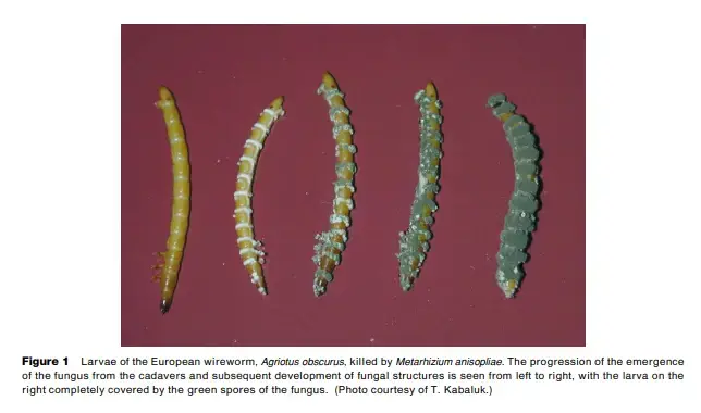
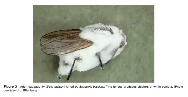
Cell Structure of Zygomycota
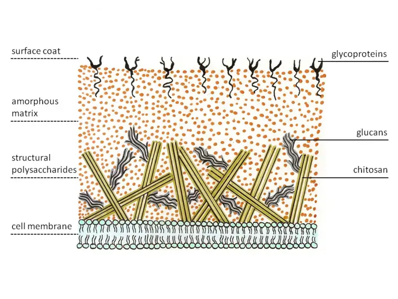
- Cell walls are made up of chitin and chitosan.
- The mature zygospore is thick with walls.
- Zygomycota also has coenocytic mycelium.
- They usually develop in mycellia, or as strings of cells.
- Hyphae are usually devoid of septa or cross-walls They are therefore coeonocytic.
Cell wall structure of Zygomycota
Zygomycetes have a distinct cell wall structure. Chitin is a common structural polysaccharide. Zygomycetes produce chitosan, a homopolymer deacetylated of Chitin. Chitin is made up of b-1,4 bonding N-acetylglucosamine. Fungal hyphae form near the tips. Thus, the specialized vesicles, the chitosomes bring the precursors of chitin as well as its synthesizing enzyme, called chitin synthetase, out to the exterior of the membrane via exocytosis. The enzyme on the membrane catalyzes glycosidic bond formations from the nucleotide sugar substrate, uridine diphospho-N-acetyl-D-glucosamine. The nascent polysaccharide chains are later cleaved by enzyme deacetylase of chitin. This enzyme catalyzes hydrolytic cleavage process of the N-acetamido chitin group. The chitosan-chitosan polymer chain forms micro-fibrils. They are then embedded in an amorphous matrice consisting of glucans, proteins (which are believed to cross-link the fibers of chitosan) Mannoproteins, lipids, and other compounds.
Spores of Zygomycota
Mitospores
In the zygomycetes, Mitospores (sporangiospores)are form in asexual fashion. They form in special structures called mitosporangia (sporangia) which contain a few to a few thousand spores, based upon the type of. Mitosporangia can be carried by specially-designed hyperhae, called mitosporangiophores (sporangiophores). These hyphae that are specialized typically show positive phototropism as well as negative gravitropism, that allow for good dispersal of spores. The sporangia wall is very thin and can be easily destroyed by mechanical forces (e.g. falling raindrops, animals passing by) which leads to the dispersal of ripe mitospores. The spores’ walls contain sporopollenin, in certain species. Sporopollenin is a result of b-carotene. It is resistant to chemical and biological degradation. Zygomycete spores can be further classified according to their longevity:
Chlamydospores
Chlamydospores, asexual spores, differ from the sporangiospores. Chlamydospores’ primary function is to maintain the mycelium , and they release when the mycelium begins to degrade. Chlamydospores do not have a mechanism for dispersal. In zygomycetes, the development of chlamydospores can be described as intercalar. But, it can even be and terminal. According to their role, the chlamydospores have cells with a thick wall and are colored.
Characteristics of Zygomycota
- They are saprobes, or weak parasites that live on plants to parasites that are specialized on animals. Some of them are found in dirt, which makes them coprophilous.
- The thallus is typically composed of well-developed filamentous, branched, and coenocytic mycelium. However, some members have very smaller septate mycelium. In some instances coenocytic mycelium can produce rhizoids , which adhere to hard surfaces thanks to their aid.
- Cell walls are primarily comprised of chitosan and chitin.
- The asexual reproduction occurs typically through non-motile sporangiospores known as aplanospore However, some can reproduce via chlamydospores as well as Oidia formation.
- The reproduction of sexual organs occurs through gametangial copulation. It results in the development of zygospores with thick walls.
- The zygospore develops by generating an sporangiophore germ that eventually bears the germ sporangium.
- These cells, known as motile ones are absent during the entire life cycle.
- Chlamydospore development is a frequent frequency.
- Sexual fusion involves gametangial copulation.
- The economic value of zygomycetes is of significance. Certain of them are utilized to ferment food products while others are used to make acids, enzymes, and other substances. Saprophytic species ruin our food products. Certain zygomycetes are mycorrhizal fungi , and some are pathogens for humans.
- Certain Zygomycetes have been of interest because they have developed unique methods for dispersing spores. “Fungus shotgun” of spore dispersal Pilobolus and the forceful propulsion of the asexual spores of Entomophthorales are the typical examples. The trapp-mechanism used by zoopagales is of great interest.
| Morphologic characteristic | Zygomycetes | Aspergillus spp. | Candida spp. |
|---|---|---|---|
| Hyphal type | Aseptate or nearly aseptate hyphae; may also present as gnarled or “crinkled cellophane” balls in specimens | Septate hyphae | Pseudohyphae |
| Hyphal width | Variable and wide (6–16 μm wide) | Consistently thin (2–3 μm wide) | Consistently thin (2–3 μm wide) |
| Blastoconidia | Absent | Absent | Present |
| Sporulation or conidiation | Absent in tissue | May be present if infected space communicates with air | Not applicable |
| Angioinvasion | Present | Present | May be present |
Use/Significance of Zygomycetes:
- A large portion of Zygomycetes (especially those belonging to the order Mucorales) expand quickly and are usually the first species involved in the decomposition of vegetable matter using the most basic carbohydrates (sugars) effectively leaving polysaccharides that are complex (cellulose, pectin, hemicellulose.) for microorganisms that be able to attack. Due to this, these fungi have commonly been called the “sugar molds”.
- The Rhizopus genus, which includes a variety of species, the fungus that forms bread moulds, are employed in the production of commercial lactic acid. R. Stolonifer for fumaric acid, and R. oryzae to make alcohol. Different species that belong to Mucor along with Actinomucor elegans are used for the production of ‘Sufu’ or Chinese cheese using soybeans. “Tempeh,” a solid food made from soybeans is made using R. the oligosporus.
- The heterothellism phenomenon was first discovered by A.F. Blakeslee, in 1904 in Mucor mucedo as well as M. hiemalis.
- A variety of Zygomycetes contribute to the spoilage of food and textiles as well as leather.
| Species | Product | Uses |
|---|---|---|
| Several Mucor and Rhizopus spp. | Lipases and proteases | Leather, detergent and medical industry (steroid transformation) |
| Rhizopus | Cellulases | Food production (i.e., tempeh) |
| R. oryzae, other Rhizopus spp. | Fumaric acid | Diverse |
| Rhizopus spp. | Lactic acid | Diverse |
| R. delemar | Biotin | Diverse |
| Mortierella romanniana, Mortierella vinacea and Mucor indicus | Linolenic acid | Diverse |
| Mortierella alpina | Arachidonic acid | Diverse |
| Blakeslea trispora | β-carotene | Diverse |
Reproduction of Zygomycota/Life cycle of Zygomycota
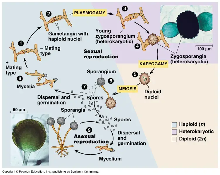
The theory is that zygomycota are able to have zygotic or haplontic life cycle. Zygomycota can reproduce sexually and asexually. Sexual reproduction by zygospores in the course of gametangial fusion is the most defining property of Zygomycota. Sexual reproduction is haploid-dominant and asexual reproduction uses the aplanospores. In Zygomycota sexual reproduction is the result of the fusion of undifferentiated anisogametangia and isogametangia. The mating strains (progametangia) are able to grow towards each to trigger Trisporic acid, a hormone that initiates sexual development. The fusion results in an embryo, which grows into the Zygospore. The method by which Zygomycota reproduce sexually is distinct. In asexual reproduction, spores, also known as sporangiospores, are created in the sporangia. Typically, three sporangia can be produced, however, there are variations in Asexual reproduction. The nuclei are moved to the end of the progametangia and form a septa. Karyogamy and Plasmogamy follow the combination that forms the progametangia. Sexual reproduction ceases when the zygospore forms. Zygospore. Due to the Zgomycota life cycle only the diploid phase occurs within the Zygospore. Zygospores are required to undergo an inactive phase before starting the reproduction cycle once more.
Classification Zygomycota
A third class of fungi called Zygomycota is comprised of fungi that make coenocytic hyphae and reproduce sexually via producing zygospores. Since these fungi were recognized only through their asexual phase (“mitosporic” meaning reproduction via mitosis and not by other means) and therefore, they are called Fungi Imperfecti or imperfect fungi. Many fungi, as well as the majority of the fungal pathogens in humans belong to this category.
In this category are yeast-like fungi that include humans-related pathogens Candida spp. as well as other yeasts. Numerous filamentous fungi that have septate mycelium, which reproduces by creation of conidia are included in this group. In this previous “form group Coelomycetes” are both dematiaceous and hyaline-based and dematiaceous fungi. The most important members of this group include agents of aspergillosis and penicillin producers and the agents of subcutaneous mycosis as well as chromoblastomycosis as well as other fungi.
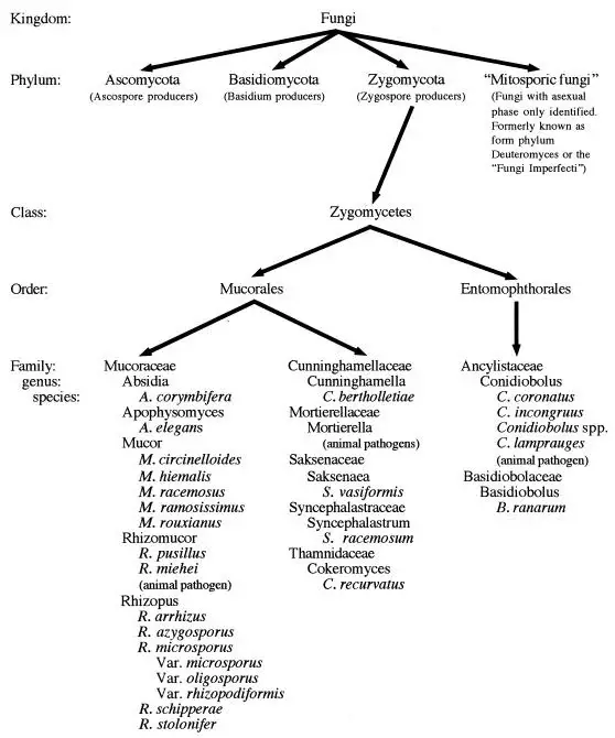
The zygomycetes are classified into a distinct class of phylums, called the Zygomycota. It is made up of organisms that are distinguished by the formation of large ribbon-like aseptate Hyaline Hyaline hyphae (coenocytic hypohae) in addition to sexual reproduction that includes the creation of Zygospores. The phylum is subdivided into two groups, the Trichomycetes which are arthropod-specific symbionts that are obligatory and are free of human pathogens and the Zygomycetes which are the category that contains human pathogens. The class is divided into two orders that comprise the human pathogens that cause Zygomycosis. Mucorales and the Entomophthorales.
Order Mucorales
- Traditionally it is the Mucorales are classified into six families with significance for causing animal or human illness:
- Mucoraceae
- Cunninghamellaceae
- Saksenaea
- Thamnidiaceae
- Syncephalastraceae
- Mortierellaceae.
- In this classification system the majority of human zygomycotic diseases is caused by people belonging to the families Mucoraceae.
- The members of this family comprise the zygomycetes that create asexual sporangiospores that are asack-like structure known as the sporangia.
- Apart from the other six family groups listed in the conventional classification system The family of Absidiaceae is also added on the basis of the existence of an apophysis. the expansion of the terminal part of the sporangiophore in the formation of sporangium.
- The Mucoraceae family Mucoraceae includes nonapophysates, producer of sporangium which may or might not produce rhizoids or stolons and comprises in this family members belonging to the genera Mucor and Rhizomucor.
- A proposed family called Absidiaceae includes zygomycetes that produce apophysate-like sporangiae with desliquescent (dissolving) or persistent sporangial walls. They produce both rhizoids and stolons and produce zygospores with opposing suspensors.
- The most commonly encountered pathogens in this family are found in the genera Rhizopus and Absidia. The majority of recent books on mycology stick to the standard taxonomic scheme with only a few authors embracing the proposed change in classification scheme.
- In the family(ies) Mucoraceae/Absidiaceae, members of the genera Rhizopus, Mucor, Absidia, Rhizomucor, and Apophysomyces have all been implicated in causing human disease.
- In general, Rhizopus species are the most frequently implicated species that cause the condition known as zygomycosis among humans.
- Within the family of Cunninghamellaceae Only just one type, Cunninghamella Bertholletiae has so far been found to be able to infect humans.
- The monotypic Genus Saksenaea includes Saksenaea vasiformis as its sole member.
- Cokeromyces is also monotypic. Cokeromyces recurvatus is an uncommon clinical isolate that can infect the colon of a person and the the genitourinary tract.
- Syncephalastrum racemosum as well as Mortierella wolfii is a pair of zygomycete strains whose classifications as human pathogens is ambiguous. From the limited reports of these organisms that cause human illness, a few are disputed as incorrect identifications of the organism in question and others are not substantiated with full proof of the correct identification.
- S. racemosum has only one case that suggests an insignificant human pathogenicity however Mortierella shouldn’t be considered to be a cause of human illness.
Order Entomophthorales
- The order Entomophthorales includes two families that are home to humans-borne pathogens: Ancylistaceae as well as Basidiobolaceae.
- Like all zygomycetes the Entomophthorales are distinguished by the coenocytic hyphae as well as their reproduction sexually through the producing Zygospores.
- The Entomophthorales are distinguished from Mucorales due to their production of actively expelling sexual sporangioles as well as their distinctively compact and glabrous mycelial form. Both of these features distinguish this group within the category of Zygomycetes.
- While a variety of varieties from Basidiobolus exist in the wild however, all human diseases are now believed to be due to Basidiobolus ranarum.
- Conidiobolus is home to a variety of species that cause illness to mammals. Conidiobolus coronatus is the most important human pathogen.
- C. incongruus has been linked to a variety of dangerous infections that affect humans. C. lamprauges can be not pathogenic to horses.
Methods of Transmission of Zygomycetes
- The primary mode of transmission among zygomycetes is thought to be through the breathing in spores that come from sources in the environment.
- The routes for exposure through the skin are also important in causing infection caused by Zygomycetes.
- The exposure of needle-sticks has been linked in zygomycotic diseases that occur on the site of injections, catheter-insertion places, injection sites used that are used for illicit drug use and tattooing.
- Insect bites and stings are also implicated in transmission of disease in cases of cutaneous or subcutaneous Zygomycosis.
- The formation of wound zygomycosis been observed through a myriad of adhesive products that are used in hospitals.
- Consumption of fermented milk along with bread products that are dried or fermented porridges, as well as alcoholic drinks made from corn can contribute to the development of gastric zygomycosis.
- Spore-contaminated remedies for homeopathic or herbal use are also connected to digestive diseases.
- A number of cases was likely carried out orally via spores contaminated tongue depressors that are used to conduct oral examinations in a clinic for hematology/oncology.
- Consumption of hay with mold and grains are the most likely method to get a bacterial infection in animals.
General Disease Manifestations
The main categories of human disease with the Mucorales are sinusitis/rhinocerebral, pulmonary, cutaneous/subcutaneous, gastrointestinal, and disseminated zygomycosis. Other diseases are seen at a lower rate and include colonization of the vagina or gastrointestinal tract external otitis and allergic diseases.
Rhinocerebral disease
- Rhinocerebral disorder accounts for one-third or half of all cases of zygomycosis.
- The process begins within the sinuses after an influx of fungal spores.
- It is believed at 70% cases of rhinocerebral zygomycosis happen as a result of diabetes ketoacidosis.
- It is a condition that begins with symptoms that are that are consistent with sinusitis. The sinuses are prone to pain, drainage and swelling of the soft tissues are the first signs to be noticed.
- The disease could become progressing, and may spread to neighboring tissues.
- The tissues that are affected turn red, then violaceous and finally , black as blood vessels thrombose and tissues suffer necrosis.
- Extension into the orbital area of the face and then to the orbit is often seen, even at the time of presentation.
- Proptosis, periorbital edema and tearing are the first signs of involvement in the tissues.
- The involvement of the optic nerve is often first spotted by blurring, pain as well as loss of vision within the affected eye.
- A number of nerve palsies in the cranial nerve can be detected.
- The sinuses extend into the mouth is common which causes pain black, necrotic ulcers in the hard palate.
- A discharge of blood from the nose is typically the first sign of the disease having taken over the terbinates, and the brain.
- Patients can experience a change in mental state due to ketoacidosis or central nervous system infiltration.
- If the eye is infected the fungal infection can quickly advance along the optic nerve getting access back into the nervous system central.
- Angioinvasion is a common occurrence and may lead to disseminated disease.
- The most uncommon rhinofacial diseases that have been documented in the literature are sinusitis that is not asymptomatic and uncalcified fungal balls of the sinus.
- The first cases of rhinocerebral Zygomycosis were almost always fatal.
- There’s still a high death rate associated with rhinocerebral diseases however, curative treatments have been implemented with prompt diagnosis, and an aggressive antifungal and surgical treatment. The nature of the disease is the primary element in the survival rate.
Pulmonary disease
- The disease of the lungs is also one of the most frequent manifestations in this particular group of organisms.
- Leukemia, lymphoma, as well as diabetes mellitus account for the majority of patients with primary lung involvement. A variety of disease manifestations are available.
- Upper respiratory diseases can manifest as tracheal involvement or chronic endobronchial zygomycosis.
- In a lot of cases of pulmonary zygomycosis diagnosis is often missed at first when it is then treated as a infection known as a bacterial pneumonia. Chest X-rays can show an image of a lobar but reveal a granulomatous disease later. Infarcts on the lung that are wedge-shaped can be observed, especially in the event of thrombosis in the pulmonary vessels due to fungal angioinvasion.
- The spread of necrosis and the subsequent bleeding into the affected tissues are common.
- In the case of pulmonary disease that is isolated in particular, the mortality rate is less than that of Zygomycosis in general (65 or 80% respectively). This is because of the availability of effective medical and surgical treatment options.
- Localized diseases are most likely curable with surgery, which provides a significant survival benefit for this category of patients. It is essential to detect the disease prior to spreading.
Cutaneous disease
- Cutaneous diseases can develop as a result of an initial infection or due to disseminated disease. The clinical manifestation will differ in both cases.
- The growth of the fungus within an already existing lesion causes an acute inflammatory reaction that manifests as abscess development, pus tissues swelling, necrosis.
- Lesions can be red or indurated, but usually, they develop into black Eschars. The tissue that is necrotic may break off and cause large ulcers.
- Infections can be multimicrobic, and generally are active, even in the presence of medical treatment.
- Sometimes, lesions on the skin produce aerial mycelium that could be visible to the naked eye.
- Primary cutaneous disorders can be quite invasive locally that includes not just the subcutaneous and cutaneous tissues but also muscles, fat and fascial layers below.
- Necrotizing fasciitis could be as a result of subcutaneous or cutaneous Zygomycosis. When it occurs necrotizing fasciitis caused by Mucorales Mucorales is characterized by a significant mortality (80 percent according to one research study).
- In the event of vessel damage, disseminated disease can occur.
- Because of the superficial location for infection, this manifestation is more likely to be identified and properly treated.
- A surgical procedure that is deemed curative may however, cause a significant amount of disfigurement or require the removal of the affected leg. I
- Patients with operable sites for involvement, such as legs are more likely be able to survive than those who have the head or trunk involved.
- In patients with burns the appearance of superficially infiltration of the eschar as well as an invasive condition may be observed.
- The process of superficial colonization could be the first step towards invading disease, and therefore is an important method to recognize and treat patients with this condition.
- While wound- or cutaneous the zygomycosis can be observed in many zygomycetes Apophysomyces elegans Saksenaea vasiformis, Mucor species., Basidiobolus ranarum, and Conidiobolus spp. are the most common. preference for these sites.
- The disseminated disease, as opposed to wound zygomycosis typically manifests as nodular subcutaneous lesions, which can be ulcerated.
Disseminated zygomycosis
- Zygomycosis disseminated can originate from any of the sources of infection.
- Lung involvement is by far the most frequent site of infection in disseminated disease however, it is not the only one. In the host that is infected the zygomycetes swiftly invade blood vessels and can grow rapidly through the hematogenous path.
- There is a high rate of death that is associated with this type of disease that is 96% death reported in one study and 100% in another study.
Gastrointestinal disorders
- Gastrointestinal diseases caused by Zygomycetes is a relatively rare condition.
- Two different syndromes have been identified that involve the gastric tract.
- Gastric colonization is characterized by the appearance of ulcerative lesions. The ulcers that are colonized usually have a smooth surface.
- The symptoms are characterized by the absence of vessel invasion, and a high survival rate.
- Zygomycosis with a zoospora-like invasion more frequently and is characterized by an invasion of fungal organisms into the mucosa and submucosa and vessels.
- The ulcers of the intestinal tract, or gastric, develop, and could rupture, causing peritonitis. The manifestation of this disease is usually fatal, with just two percent of patients getting the illness.
Other Infections
The zygomycetes can cause inflammation in every body part.
- The brain’s involvement even absent of sinus involvement has been proven, with the most notable instances for those who abuse intravenous drugs.
- A renal involvement that is isolated has been reported in a number of cases, and is often linked to intravenous drug usage.
- Cardiac infections can be an indication of disease that has spread through the lungs, or as a result of a secondary to intravenous drug use , or could be an isolated form of cardiac mycosis.
- Many cases of surgery for the postponed Zygomycosis is also a problem that has been observed.
- A pleural involvement isolated after a surgical procedure is also seen.
Treatment of zygomycosis
The treatment of zygomycosis involves several different approaches at once that include surgical intervention, antifungal treatment as well as medical management or correction of the root disease that predisposes the patient to contract the disease.
- Amphotericin B: Amphotericin B is the drug in the majority of instances of zygomycosis caused due to the Mucorales. Amphotericin works through altering the cell walls of fungal cells. This drug binds with Ergosterol, which causes an increase in the permeability of cells. Through permeabilization, ions flow out of the cell, and the membrane becomes depolarized. Lethal effects of amphotericinB occur at doses higher than those that cause the increased permeability.
- Antifungal therapy: Antifungal therapy has yielded different results, depending on the type of organism and the treatment chosen. It is now evident that azoles shouldn’t be employed in the treatment of zygomycosis because of the absence either in vitro and live susceptibility (149 348 434). The first successful antifungal treatment for infections caused by Mucorales Mucorales included the treatment with oral potassium iodide saturated with potassium along with local use of tinctured iodide for skin sites (98, 221 398). This is similar to the treatment method that is effective in treating infections caused by Basidiobolus as well as Conidiobolus spp.
- Hyperbaric oxygen: Hyperbaric Oxygen treatments have been marketed as an effective alternative to the standard medical, surgical, and antifungal treatments, specifically for the treatment of rhinocerebral diseases. Hyperbaric oxygen slows or completely blocks the growth of fungal spores as well as mycelium in the laboratory. The supplementation of hyperbaric treatment to the systemic treatment of drugs could assist in the direct killing of fungal spores or at the very least reduce the rate of growth of fungal spores and allowing the host’s natural immune system to heal.
Preventive Measures
- The most commonly used preventive measures are based on changes and control measures in the surrounding environment that decrease the possibility of exposure to spores that are airborne. A majority of these preventive strategies are targeted towards those who are easily identified as being who are at high risk of developing the disease, i.e., those who are believed to be extremely neutropenic for long durations.
- The chemotherapeutic and transplant wards are typically isolated by The treatment with a Hepafilter of air flow as well as positive pressure in order to avoid the entry of dust into the area.
- The amount of dust should be kept to an absolute minimum in the space which houses these patients.
- Furthermore, floral arrangements or live plants are typically exempt from these wards because they can be home to a variety different fungal agents.
- Patients with a neutropenic level is below 1,000/ml, are advised to wear masks on their heads when leaving the transplant or cancer units, specifically when they leave the wards.
- Monitoring the quality of air especially in times of renovation of buildings and excavations near transplant facilities, is crucial in the control of infection.
- Prevention strategies for patients that aren’t part of the chemotherapy and transplant population need to address the root factors that increase the risk of developing Zygomycosis.
- Controlling diabetes in a proper manner using iron chelators that are not deferoxamine, and limiting the use of buffers containing aluminum for dialysis, as well as aggressive testing of the culture for zygomycosis is among the top preventive measures that can be implemented.
- Maintaining a high degree of suspicion for zygomycosis among those at risk may help in the early detection and implementation of appropriate treatment.
Diagnosis of Zygomycetes
1. Specimens
- Lesion biopsies and tissues of deceased animals and cotyledons and foetal abomasal content and uterine discharges may be taken.
- Tissue samples in 10% formalin, for histopathology.
2. Direct Microscopy
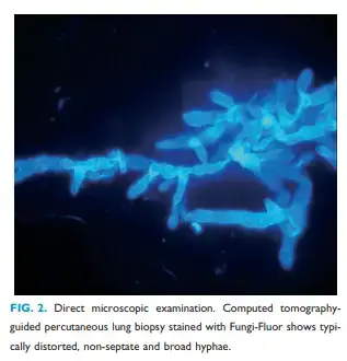
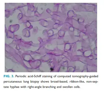
- A histological study of the tissue section
- They are positive for PAS (pink staining).
- staining with silver and methenamine
- KOH prepared in wet form.
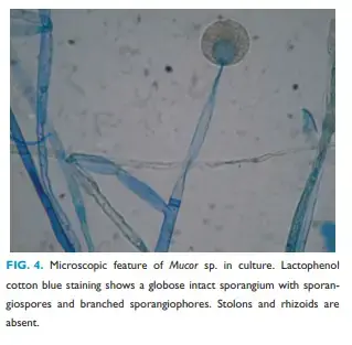
3. Culture
- Sabouraud dextrose agar, without the cycloheximide
- Emmons modification is employed.
- The plates that have been inoculated are then placed in an aerobic environment for as long as 10 days.
- The zygomycetes will thrive on blood Agar plates
4. Serology
The development of serological tests for the diagnosis of zygomycosis through the use of antibodies or antigen detection has been explored. Antibodies to Zygomycetes are detected using enzyme-linked immunosorbent tests (ELISAs) as well as double diffusion. Immunoblot tests have also been utilized to identify Rhizopus arrhizus-related antigens. However, these tests for zygomycosis are not advised without further evaluation by a physician and are not suitable for use in routine at the moment. Recently, Japanese researchers observed that the serum IgE as well as Mucor IgE antibody levels were linked to the invasiveness of mucormycosis orbital.
Colony Characteristics of Zygomycota
| Organism name | Colony morphology |
|---|---|
| Rhizopus spp. | Floccose; rapidly growing |
| Mucor spp. | Floccose; rapidly growing; white, yellow, gray or brown |
| Rhizomucor pusillus | Floccose mycelium 2–3 μm high; colorless to white, turning grey with age |
| Mortierella wolfii | Little aerial mycelium; white, gray, or yellow-gray |
| Cunninghamella bertholletiae | Tall, 0.5–2 cm high; white, yellow, or gray |
| Cokeromyces recurvatus | Low mycelium; tan to gray with concentric zones of color; radial folds form with age |
| Syncephalastrum racemosum | Floccose mycelium 0.5–1.5 cm high; White, green, olive, gray, or almost black |
| Absidia corymbifera | Floccose; first white, turning brown to greenish brown with age |
| Apophysomyces elegans | Floccose, rapidly growing colonies; pale gray to yellowish, turning brown at 37°C |
| Saksenaea vasiformis | Floccose; white colonies |
| Basidiobolus ranarum | Flat furrowed colonies with a waxy texture; yellowish to gray |
| Conidiobolus coronatus | Flat, waxy colonies, becoming powdery, with a short aerial mycelium with age; petri dish lid becomes covered with conidia ejected from older cultures; colonies are white, buff, tan or brown |
| Conidiobolus incongruus | Similar to C. coronatus; dry colonies are produced, becoming more aerial with increased humidity |
| Conidiobolus species | Not known |
| Absidia corymbifera | Floccose; first white, turning brown to greenish brown with age |
References
- C. Lass-Flörl (2009). Zygomycosis: conventional laboratory diagnosis. , 15(Supplement s5), 60–65. doi:10.1111/j.1469-0691.2009.02999.x
- Gould, A.B. (2009). Encyclopedia of Microbiology || Fungi: Plant Pathogenic. , (), 457–477. doi:10.1016/B978-012373944-5.00347-3
- Voigt, K. (2014). Encyclopedia of Food Microbiology || FUNGI | Classification of Zygomycetes. , (), 54–67. doi:10.1016/B978-0-12-384730-0.00136-1
- Volk, Thomas J. (2013). Encyclopedia of Biodiversity || Fungi. , (), 624–640. doi:10.1016/B978-0-12-384719-5.00062-9
- Goettel, M.S. (2005). Comprehensive Molecular Insect Science || Entomopathogenic Fungi and their Role in Regulation of Insect Populations. , (), 361–405. doi:10.1016/B0-44-451924-6/00088-0
- Ribes JA, Vanover-Sams CL, Baker DJ. Zygomycetes in human disease. Clin Microbiol Rev. 2000;13(2):236-301. doi:10.1128/CMR.13.2.236
- https://www.ck12.org/book/ck-12-biology-advanced-concepts/section/12.25/
- http://www.botany.hawaii.edu/faculty/wong/Bot201/Zygomycota/Zygomycota.htm
- https://microbewiki.kenyon.edu/index.php/Zygomycota
- https://courses.lumenlearning.com/wm-biology2/chapter/zygomycota/
- https://www.adelaide.edu.au/mycology/fungal-descriptions-and-antifungal-susceptibility/zygomycota-pin-moulds#:~:text=The%20zygomycota%20are%20usually%20fast,on%20simple%20or%20branched%20sporangiophores.
- https://en.wikipedia.org/wiki/Zygomycota
- https://www.sparknotes.com/biology/microorganisms/fungi/section3/
- http://www.mycolog.com/chapter3b.html
- http://www.ndvsu.org/images/StudyMaterials/Micro/8b_Zygomycetes.pdf
- Text Highlighting: Select any text in the post content to highlight it
- Text Annotation: Select text and add comments with annotations
- Comment Management: Edit or delete your own comments
- Highlight Management: Remove your own highlights
How to use: Simply select any text in the post content above, and you'll see annotation options. Login here or create an account to get started.