

Stage micrometer is a special microscope slide which is used for measuring very small objects under the microscope. It is generally like a normal glass slide in size, about 1 by 3 inches, but it has a fine scale marked on its surface. This scale has known true length.
It is used as a standard scale for calibration of microscope. It helps to calibrate ocular micrometer, eyepiece reticle and also digital imaging system. Without stage micrometer, the divisions of ocular micrometer has no fixed value.
The scale of stage micrometer is etched or engraved on glass or metal surface. Glass stage micrometer is used in transmitted light microscope. Metal stage micrometer is used in reflected light microscope. In some types, the scale is made by chromium coating, so that the marking remain clear and durable.
During calibration, the stage micrometer is placed on the microscope stage in the same focal plane like the specimen. Then its scale is focused and matched with the ocular scale. From this, the value of one ocular division is calculated for a particular objective lens.
Stage micrometer is important because the size seen under microscope changes with different objective lens. So each objective lens is calibrated separately. This gives exact measurement of cells, microorganisms and other microscopic structures in micrometer (µm).
The use of stage micrometer is related with the development of micrometry. Micrometry means measurement of length, area and volume under microscope. Earlier microscope was mainly used for seeing small objects. Later, by using calibrated scale, microscopy became a quantitative study.
The first microscopic measurements were reported by Antonie van Leeuwenhoek in late 1600s. He used fine sand grains as a rough measuring material for estimating the size of human red blood cells. Later, more accurate methods were developed and stage micrometer became a standard reference scale in microscopy.
Principle of Stage micrometer
Stage micrometer is based on the principle of using a known standard scale for calibration of microscope measuring scale. It acts as a transfer scale between the actual measurement and the magnified image seen under microscope.
It has a fine and accurately divided scale marked on a special glass slide. The length of this scale is already known. So it is used to compare with the unknown division of ocular micrometer or digital measuring scale.
During calibration, the stage micrometer is placed on the microscope stage. It is focused in the same focal plane where the specimen is normally observed. So the stage micrometer scale and specimen get same magnification by the objective lens.
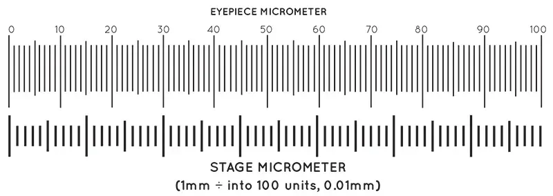
The known divisions of stage micrometer are then matched with the divisions of ocular micrometer. From this comparison, the value of one ocular division is calculated for that particular objective lens.
This calculated value is then used for measuring microscopic object. When the specimen is observed, its size in ocular divisions is multiplied with the calibrated value. Thus the apparent size of the magnified specimen is converted into actual size in micrometer (µm).
Parts of Stage micrometer
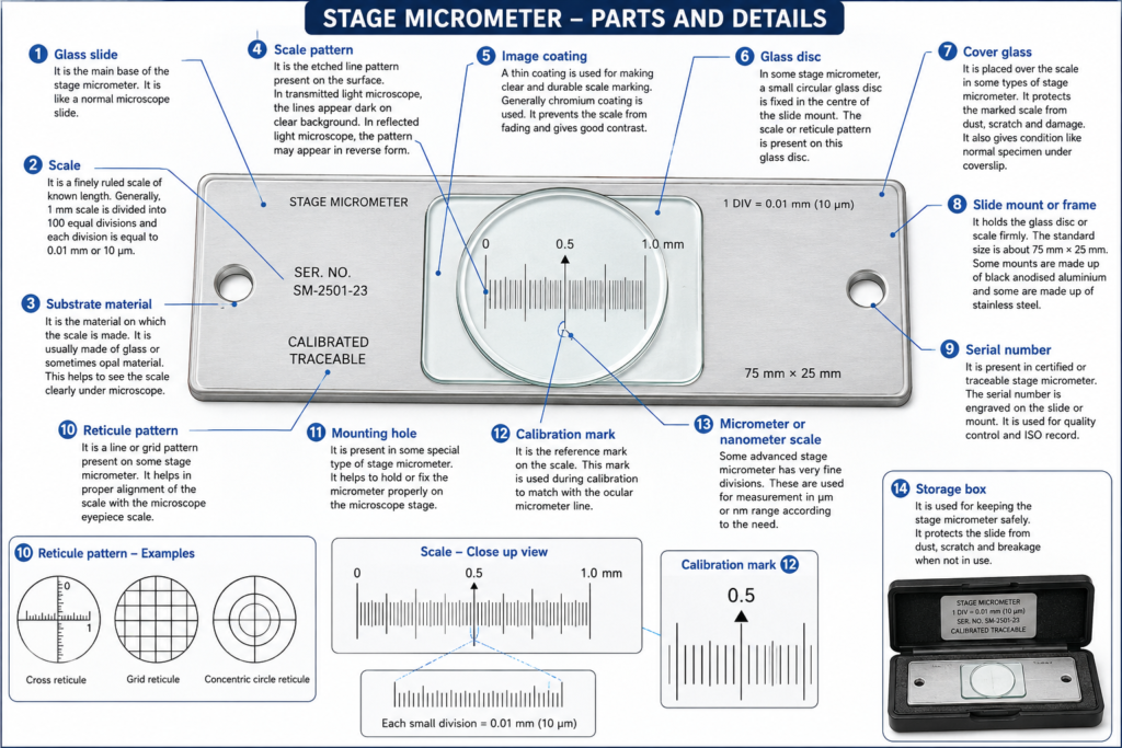
- Glass slide – It is the main base of the stage micrometer. It is like a normal microscope slide. The fine measuring scale is present on this slide.
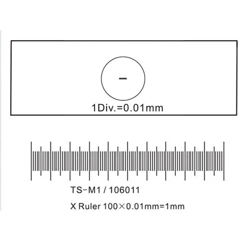
- Scale – It is the most important part of stage micrometer. It is a finely ruled scale of known length. Generally, 1 mm scale is divided into 100 equal divisions and each division is equal to 0.01 mm or 10 µm.
- Substrate material – It is the material on which the scale is made. It is usually made of glass or sometimes opal material. This helps to see the scale clearly under microscope.
- Scale pattern – It is the etched line pattern present on the surface. In transmitted light microscope, the lines appear dark on clear background. In reflected light microscope, the pattern may appear in reverse form.
- Image coating – A thin coating is used for making clear and durable scale marking. Generally chromium coating is used. It prevents the scale from fading and gives good contrast.
- Glass disc – In some stage micrometer, a small circular glass disc is fixed in the centre of the slide mount. The scale or reticule pattern is present on this glass disc.
- Cover glass – It is placed over the scale in some types of stage micrometer. It protects the marked scale from dust, scratch and damage. It also gives condition like normal specimen under coverslip.
- Slide mount or frame – It holds the glass disc or scale firmly. The standard size is about 75 mm × 25 mm. Some mounts are made up of black anodised aluminium and some are made up of stainless steel.
- Serial number – It is present in certified or traceable stage micrometer. The serial number is engraved on the slide or mount. It is used for quality control and ISO record.
- Reticule pattern – It is a line or grid pattern present on some stage micrometer. It helps in proper alignment of the scale with the microscope eyepiece scale.
- Mounting hole – It is present in some special type of stage micrometer. It helps to hold or fix the micrometer properly on the microscope stage.
- Calibration mark – It is the reference mark on the scale. This mark is used during calibration to match with the ocular micrometer line.
- Micrometer or nanometer scale – Some advanced stage micrometer has very fine divisions. These are used for measurement in µm or nm range according to the need.
- Storage box – It is used for keeping the stage micrometer safely. It protects the slide from dust, scratch and breakage when not in use.
Types of Stage Micrometer
A. On the basis of illumination
- Transmitted light type – It is used in compound microscope and biological microscope. The scale is made on clear glass disc. Light passes through the slide from below. It usually has cover glass for protecting the scale.
- Reflected light type – It is used in metallurgical and industrial microscope. In this type, light falls from the upper side on the micrometer. It does not have cover glass. The scale gives good contrast in reflected light.
B. On the basis of scale pattern
- Linear scale type – It is the common type of stage micrometer. It has a straight ruled scale with equal divisions. It is used for normal calibration of ocular micrometer.
- Crossed line type – It has two scales crossing each other at right angle. It is used for calibration of both X-axis and Y-axis. This is useful when measurement is needed in two directions.
- Grid pattern type – It has many small squares or rectangles. It is used for counting cells, measuring particles and estimating size of small microscopic objects.
- Dual scale type – It has two measuring scales on same slide. Metric scale and inch scale may be present side by side. It is used when different unit is required.
- High resolution type – It has very fine divisions like 0.002 mm or 2 µm. It is used for high magnification and oil immersion objective calibration.
C. On the basis of special use
- Finder slide type – It has many labelled positions on the slide. It is used to locate again the same cell, tissue part or small object under microscope.
- Advanced imaging target type – It has special line, star or grid pattern. It is used to check resolving power, distortion and field of view of microscope or imaging system.
How to Use Stage micrometer
- The stage micrometer slide is first placed on the microscope stage. It is fixed properly with stage clip so that it does not move during focusing.
- The scale of stage micrometer is focused under low power objective. Coarse adjustment knob is used first and then fine adjustment knob is used to make the scale clear.
- The ocular micrometer scale in the eyepiece should also be seen clearly. If it is not clear, the eyepiece is adjusted until the scale becomes sharp.
- The eyepiece is rotated slowly. The lines of ocular micrometer are made parallel with the lines of stage micrometer.
- The mechanical stage is moved carefully. The zero line of ocular micrometer is placed exactly over the zero line of stage micrometer.
- After the zero lines are matched, the scales are observed towards the right side. A second point is searched where one line of ocular micrometer again matches with one line of stage micrometer.
- The divisions of stage micrometer are counted from zero line to the second matching point. This gives the known actual distance on stage micrometer.
- The divisions of ocular micrometer are also counted for the same distance. This shows how many ocular divisions are equal to that known stage distance.
- The value of one ocular division is calculated by dividing the stage micrometer distance by the number of ocular divisions. Value of 1 ocular division = Stage micrometer distance / Number of ocular divisions
- If the value is in mm, it is converted into µm by multiplying with 1000. This gives the actual value of one ocular division in micrometer.
- The same process is repeated for each objective lens. It is done separately for low power, high power and oil immersion objective, because magnification changes the value.
- The calculated value for every objective lens is written in the notebook or calibration chart. These values are used later for measuring cells, microorganisms and other microscopic objects.
Calibration of Eyepiece Graticule with stage micrometer Video
Precaution of Stage micrometer
- Stage micrometer should be handled very carefully. It should not be treated like ordinary glass slide, because any damage on the scale can make it useless for calibration.
- The scale surface should not be touched with finger. Fingerprint, oil and dust may cover the fine lines and give wrong reading.
- The micrometer should not be rubbed strongly during cleaning. Strong rubbing may scratch the etched scale or remove the marking.
- Only clean and soft lens tissue should be used for cleaning. Dry and damage-free cleaning method is better for removing dust from the scale.
- The stage micrometer should not be placed directly on hard laboratory bench. If it is kept outside the microscope, it should be placed on clean lint-free cotton wiper.
- The micrometer should be protected from dirt, stain and biological sample. Any contamination on the scale may make the divisions unclear under microscope.
- After use, it should be kept in its original storage box. The box protects it from dust, scratch and breakage.
- Uncovered stage micrometer should be used with more care. The scale of this type is exposed and can be damaged easily, especially in reflected light micrometer.
- It should be stored in dry and clean place. High humidity may cause fungal growth on glass and rusting in metallic mount.
- Before calibration, the stage micrometer and microscope should be kept in same room temperature for some time. This helps to avoid error due to thermal expansion.
- The slide should be fixed properly on the microscope stage. It should not be moved roughly, because the scale may shift and alignment may become wrong.
- The objective lens should not touch the surface of micrometer. This can scratch the scale and also damage the lens.
Applications of Stage micrometer
- Stage micrometer is used for calibration of ocular micrometer. It gives the actual value of one ocular division for each objective lens.
- It is used for calibration of digital microscope and imaging software. The known scale of stage micrometer helps to convert image measurement into real measurement.
- It is used for measuring the size of cells, tissues and microorganisms. Bacteria, erythrocytes, spermatozoa and other small structures can be measured by using calibrated microscope.
- It is used in medical and biological research work. It helps to find the exact dimension of microscopic specimen under different magnification.
- It is used in metallurgy for measuring grain size of metals and alloys. It is also used for observing microstructure and thickness of coating.
- It is used in forensic laboratory for measuring trace evidences. Fibres, fingerprints and other small particles can be examined and measured.
- It is used in electronics and semiconductor work. It helps to check PCB, solder joint and small features present on semiconductor wafer.
- It is used in pharmaceutical industry for particle size measurement. It is also used for tablet inspection and checking of active pharmaceutical particles.
- It is used for particle analysis of dust, pigments, abrasives, fertilizers, seeds and soil grains. Their size and number can be compared under microscope.
- It is used in quality control work. It helps to check surface cracks, small defects and other microscopic changes in manufactured materials.
- It is used as a calibration standard for some measuring instruments. It can help in checking instruments like Vickers, Rockwell and Brinell hardness tester.
- It is used in teaching laboratory. Students use it as a reference scale for learning microscope calibration and measurement of microscopic objects.
Advantages of Stage micrometer
- Stage micrometer gives a known standard scale for microscope calibration. It helps to change the eyepiece division into actual measurement.
- It gives more accurate measurement of microscopic objects. The size of cells, microorganisms and other small structure can be measured in micrometer (µm).
- It helps to reduce error in microscope measurement. As the scale is placed in same focal plane of specimen, the optical error becomes less.
- It gives same and reliable result when used properly. The measurement can be repeated again with same objective lens and same calibration value.
- It makes microscopic measurement simple and fast. After calibration, the specimen can be measured directly by using ocular micrometer value.
- It is useful for different objective lens. Each objective lens can be calibrated separately by stage micrometer.
- It helps in digital microscope calibration also. The known scale is used to set the correct value for camera and image analysis software.
- It is important in quality control work. Certified stage micrometer gives traceable calibration value which is needed in medical, pharmaceutical and industrial laboratory.
- It helps to compare the size of different samples. The measured value becomes more standard and not only based on visual observation.
- It is small, simple and easy to use. It can be kept on microscope stage like normal slide and used whenever calibration is needed.
Limitations of Stage micrometer
- Stage micrometer can be damaged easily if it is not handled properly. Scratches, dust and fingerprint may cover the fine scale lines and make it unsuitable for calibration.
- The exposed scale type is more sensitive to damage. In reflected light micrometer, cover glass may not be present, so the scale can be scratched very easily.
- It needs careful alignment with ocular micrometer. If the lines are not matched properly, the calculated value may become wrong.
- Human error may occur during counting of divisions. At high magnification, the lines may appear thick or close and counting becomes difficult.
- It is difficult to use at very high magnification. Only few divisions of stage micrometer can be seen in the field, so accurate comparison becomes less easy.
- The accuracy also depends on the resolving power of microscope. If the microscope cannot show clear separate lines, the measurement will not be reliable.
- Low quality stage micrometer may give less accurate result. Some cheap micrometers have thick, rough or unclear lines which are not good for precise measurement.
- It cannot measure specimen directly by itself. It is mainly used for calibration, and actual measurement needs ocular micrometer or digital measuring system.
- It must be calibrated separately for each objective lens. One calibration value cannot be used for all magnifications.
- Without proper certificate, its true scale value cannot be fully guaranteed. Certified stage micrometer is needed in quality control, research and standard laboratory work.
References
- ResearchGate. (n.d.). (PDF) An alternative stage micrometer for use at light microscope.
- Office of Justice Programs. (1999). 218830.pdf – Forensic science communications.
- PACE Technologies. (n.d.). ASTM E112 – Standard test methods for determining average grain size.
- Echemi. (n.d.). Acetone vs alcohol: Key differences, uses, and safety compared.
- Biocompare. (n.d.). Automated microscopy for faster and more reliable results.
- Mueez, A., & Vyas, S. (2026). Bridging foundation models and ASTM metallurgical standards for automated grain size estimation from microscopy images. arXiv.
- cyanoScope. (n.d.). Calibrating a microscope for use in with AmScope software [Video]. YouTube.
- Eduqas. (n.d.). Calibration of microscope.
- Centers for Disease Control and Prevention (CDC). (n.d.). Calibration of the ocular micrometer.
- Centers for Disease Control and Prevention (CDC). (n.d.). Calibration of the ocular micrometer.
- Chemtronics. (n.d.). Choosing the best swab for aggressive solvents.
- The W.O. Hickok Manufacturing Company. (n.d.). Do you know these practical tips for micrometer measurement?
- Thermo Fisher Scientific. (n.d.). Forensic microscopy.
- Bruker. (n.d.). Forensics.
- Ibañez H., P. C. (n.d.). Grain size ASTM E 112: What metallurgist must know about. INGINTEGRAL.
- Light in Glass. (n.d.). Glass substrates for photomask and photoetching.
- Evident Scientific. (n.d.). Grain size analysis in metals and alloys.
- INGPLUS. (n.d.). Grain size analysis – ASTM E112 & E1382.
- Thorlabs. (n.d.). Grid distortion test targets.
- Almicroinstruments. (n.d.). How to use digital microscope with measurement software.
- Graticules Optics. (n.d.). How to use a stage micrometer.
- Edmund Optics. (n.d.). Image analysis & stage micrometers test targets.
- Fellers, T. J., & Davidson, M. W. (n.d.). Linear measurements (micrometry). Nikon’s MicroscopyU.
- Microscope World. (n.d.). Measuring with a microscope eyepiece reticle & stage micrometer.
- Unknown Author. (n.d.). Microanalysis and examination of trace evidence.
- World Precision Instruments. (n.d.). Micrometer maintenance tips: Ensure precision & longevity.
- Travers Tool Co. (n.d.). Micrometer maintenance: Ensure precision and longevity for machinists.
- Keyence. (n.d.). Micrometers | Measurement system types and characteristics.
- Danaher Life Sciences. (n.d.). Microscope imaging software: Seamless workflow, AI integration.
- Microscope World. (n.d.). Microscope stage micrometers.
- Microscope World. (n.d.). NIST certified stage micrometers.
- Unknown Author. (n.d.). Ocular and stage micrometers.
- Richard J. Bagan, Inc. (n.d.). Proper techniques for micrometer usage.
- Thorlabs. (n.d.). Resolution test targets.
- SSM College of Pharmacy and Research Centre. (n.d.). SSM College of Pharmacy and Research Centre, VIAJAYAPUR-586101, Department of Pharmacognosy.
- SPI Supplies. (n.d.). Stage micrometer FAQs.
- Science Services. (n.d.). Stage micrometers.
- Nikon Instruments Inc. (n.d.). Slide scanning | Clinical research | Solutions.
- Electron Microscopy Sciences. (n.d.). Stage graticules.
- Thorlabs. (n.d.). Stage micrometers.
- Edmund Optics. (n.d.). Stage micrometers | Precision microscopy calibration tools.
- Graticules Optics. (n.d.). Stage micrometers, calibration scales / grids, UKAS & NPL calibration.
- Graticules Optics. (n.d.). Stage micrometers, calibration standards & grids.
- Pyser-SGI Limited. (2013). Stage micrometers & calibration standards.
- Micro to Nano. (n.d.). Stage micrometers and graticules for light microscopy.
- MEETOPTICS Academy. (n.d.). Substrate.
- Shin-Etsu MicroSi. (n.d.). Synthetic quartz glass substrates.
- Maguire, C. (2013). TES09 – Polarized light microscopy for fiber examination. District of Columbia Department of Forensic Sciences.
- Unknown Author. (n.d.). The definitive guide to stage micrometers: Principles, metrology, and multidisciplinary applications in optical calibration.
- Thorlabs. (n.d.). Thorlabs · Calibration targets.
- Thorlabs. (n.d.). Thorlabs · Stage micrometers.
- Structure Probe, Inc. (SPI Supplies). (n.d.). Traceable stage micrometers.
- Groshon, N. (2017). Webinar calibrating your microscope. The McCrone Group.
- CNSI Microfluidics Lab. (2023). solvent_clean_sop [Innovation Workshop Wiki].