What is Rhizopus?
- Nestled within the world of fungi, the genus Rhizopus stands as a remarkable example of adaptability and ecological diversity. These saprophytic fungi showcase an impressive range, thriving on a plethora of organic substrates, from the remnants of “mature fruits and vegetables” to bread, leather, and even tobacco.
- While their versatility spans various niches, some members of this genus have a more sinister side, as opportunistic pathogens that can lead to a severe and often fatal condition known as mucormycosis. Within the expansive realm of fungi, Rhizopus, boasting a lineage of at least eight discernible species, presents a captivating tale of existence and interaction.
- Microscopically, the filamentous nature of Rhizopus species comes to life, with branching hyphae forming a sprawling network. These hyphae, distinctive for their lack of cross-walls (coenocytic), serve as the foundation for both asexual and sexual reproduction.
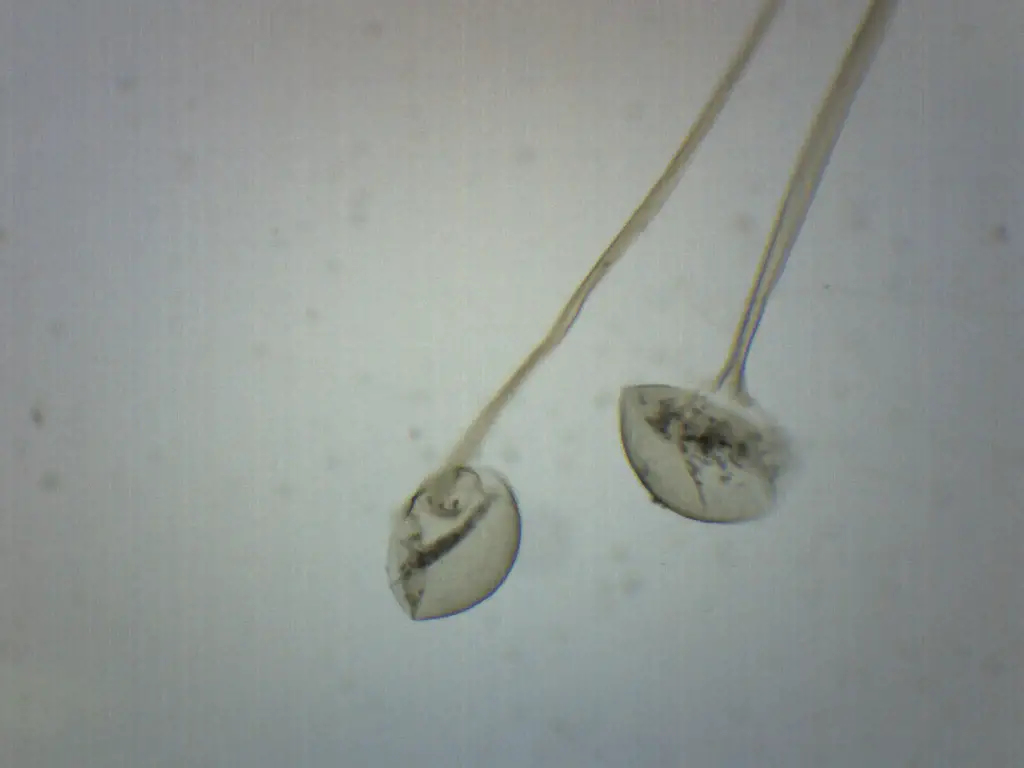
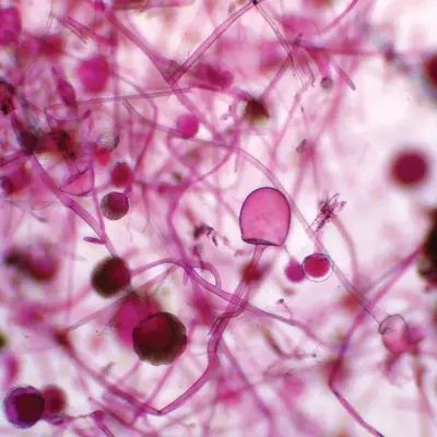
- Asexual reproduction unfolds within spherical structures called sporangia, which contain sporangiospores. Towering above the sporangia is a notable structure—the sporangiophore—resembling a stalk that supports the sporangium. Root-like rhizoids add to the distinctiveness of this organism’s appearance, as sporangiophores emerge among these structures.
- On the other hand, sexual reproduction involves the fusion of compatible mycelia, culminating in the formation of dark zygospores. The offspring that arise from germinating zygospores possess genetic uniqueness, setting the stage for an ongoing cycle of diversity.
- Rhizopus species have managed to insinuate themselves into various human activities and industries. Rhizopus oligosporus, for instance, takes center stage in the creation of tempeh, a beloved fermented soybean product.
- The fungus lends its transformative touch to soybeans, enhancing their nutritional profile and culinary appeal. Meanwhile, Rhizopus oryzae plays a pivotal role in parts of Asia and Africa, contributing to the production of alcoholic beverages.
- In the realm of agriculture, Rhizopus stolonifer, often referred to as black bread mold, takes on a dual role—inducing fruit rot on strawberries, tomatoes, and sweet potatoes while also being harnessed for the commercial synthesis of fumaric acid and cortisone. However, its influence isn’t limited to the plant kingdom, as it can provoke soft rot in sweet potatoes and Narcissus.
- Rhizopus goes beyond its direct applications to influence ecosystems and processes. This genus contributes to nutrient development, its growth within soil fostering the fermentation of plant matter. This fermentation curtails the proliferation of toxigenic fungi, promoting healthier soil and vegetation.
- In a fascinating turn, specific Rhizopus strains exhibit unexpected health benefits. Rhizopus microsporus-fermented soybean tempe emerges as a potential ally in the battle against colon carcinogenesis.
- It achieves this by enhancing factors such as mucins, immunoglobulin A, and organic acids, thereby offering protection against malignancies. Moreover, this remarkable fungus lends a hand in safeguarding piglets from Escherichia coli infections by hindering adhesion to intestinal membranes.
- The genetic tapestry of Rhizopus reveals intriguing insights as well. Comparative studies shed light on the species’ structural flexibility, setting it apart from its genus counterparts. Genome analyses unveil a topology length three times that of its closest relatives, underscoring its unique genomic makeup.
- In essence, Rhizopus captivates us with its ability to shape environments, influence health, and find its place in the intricate dance of life. From the fermentative magic of tempeh to its involvement in the production of crucial compounds, Rhizopus remains an emblem of adaptation and scientific intrigue within the fungal realm.
Mold or Rhizopus Under the Microscope
Preparing specimen
Studying the intricate world of mold and fungi under the microscope offers a fascinating glimpse into the hidden realms of life. While one can readily discover mold on decaying organic matter in nature, cultivating and preparing specimens for microscopic observation provides a controlled and insightful approach. If you’re intrigued by the prospect of delving into the microscopic structures of molds like Rhizopus, Neurospora, Aspergillus, and Penicillium, here is a step-by-step guide to preparing your specimen:
Caution: Before embarking on this journey, it’s essential to note that this procedure involves handling mold, which can be an allergen. Individuals with respiratory issues or allergies are advised to exercise caution or consult a medical professional before attempting this experiment.
Materials Needed:
- Soft bread without preservatives
- Fruits like oranges or potatoes
- Plastic paper bag (moist)
- Dark and warm location
Procedure:
- Selecting the Substrate: Begin by choosing a suitable substrate to encourage mold growth. Soft bread without preservatives is an excellent option, as it fosters quicker mold development compared to bread with preservatives. Alternatively, you can opt for fruits like oranges or potatoes.
- Exposing to Open Air: Allow the chosen food (bread, fruit, etc.) to sit in open air for approximately 30 minutes to an hour. This initial exposure allows natural spores present in the environment to settle on the surface of the food.
- Moist Bag Enclosure: Place the food item into a plastic paper bag to create a controlled environment for mold growth. Ensure the bag is slightly moist to provide the necessary humidity for mold development. Seal the bag securely to retain moisture.
- Dark and Warm Environment: Find a suitable location that is both dark and warm to facilitate mold growth. Ideally, this location should mimic the conditions in which molds thrive. A cupboard, closet, or a corner of a room away from direct light can serve as an appropriate setting.
- Incubation Period: Allow the specimen to incubate for about 5 days. During this period, the mold should proliferate and become visible to the naked eye. Monitor the progress periodically without disturbing the environment.
- Microscopic Examination: After the incubation period, carefully retrieve the specimen from the bag. If mold growth is evident to the naked eye, the specimen is ready for microscopic examination. Handle the specimen with care to avoid inhaling spores or making direct contact with bare hands.
Safety Measures:
- Wear gloves and a mask to minimize the risk of inhalation or direct contact with mold spores.
- Work in a well-ventilated area or consider using a fume hood if available.
- Properly dispose of the specimen and any materials used in a responsible manner, following recommended guidelines for mold disposal.
Requirements for Observation Under Microscope
- Glass Slide: A clear glass slide serves as the foundation for your mold specimen.
- Cover Slip: A thin, transparent cover slip is placed over the specimen to create a protective barrier and enhance clarity.
- Mounting Medium: Water, Shear’s mounting medium, Melzer’s solution, or Lacto-fuchsin are options for creating a suitable environment for the mold specimen on the slide.
- Gloves: Wearing gloves ensures proper hygiene and minimizes contamination during the process.
- Microscope: A microscope equipped with various magnification levels is essential for viewing the mold specimen.
- Dropper: A dropper aids in precise placement of liquid substances on the slide.
- Methylene Blue Solution: This solution can be utilized to improve visibility and highlight specific structures.
Procedure:
- Preparing the Specimen:
- Place a glass slide on a clean, flat surface.
- Using a dropper, carefully dispense a small drop of water (or chosen mounting medium) at the center of the glass slide. This will serve as the foundation for your mold specimen.
- Collecting Mold Sample:
- Gently scrape a tiny amount of mold from the chosen source, such as the bread, using a toothpick.
- Introduce the collected mold onto the drop of water on the glass slide.
- Covering the Specimen:
- Hold a cover slip at a slight angle and gently lower it onto the specimen. This technique helps remove air bubbles and ensures a smooth, even surface.
- Microscopic Observation:
- With the prepared slide in hand, place it onto the microscope stage.
- Begin with the lowest magnification (low power) and gradually increase the magnification (high power) to explore different levels of detail.
Enhancing Visibility:
- To augment visibility and bring out finer structures, a drop of methylene blue solution can be added to the specimen on the slide before covering it with the cover slip.
About molds
Fungi, a diverse group of organisms, inhabit a multitude of environments, ranging from the beneficial to the potentially harmful. While mushrooms exemplify edible fungi, molds offer an intriguing avenue for study, even yielding antibiotics with transformative medical applications. Understanding the intricate world of molds unveils captivating structures and characteristics, shedding light on their varied forms and functions.
Hyphal Networks: The Canvas of Growth
Molds, often encountered as unwelcome guests on food, arise from complex networks of hyphae. These thread-like structures intertwine to form intricate mold colonies, initiated by a solitary mold spore. The hyphal appearance can range from soft and fuzzy to vividly colorful, a visual testament to the diversity within the fungal realm. Employing the power of microscopy, one can discern and classify mold types by scrutinizing the unique configurations of their hyphae.
Rhizopus: Starch’s Sublime Consumer
Among the pantheon of molds, Rhizopus takes center stage as a common inhabitant of bread. Its dietary preferences lean toward starch and sugar, heralding its role as a bread mold protagonist. The initial stage may manifest as delicate white hair-like structures, ultimately maturing into distinctive black spots. Through the lens of a microscope, Rhizopus reveals itself as short strands crowned with oval-shaped heads, akin to balloons on strings. These heads encapsulate the mold’s spores, a vital facet of its reproductive strategy.
Aspergillus: The Grain Guardian
Another notable inhabitant of the fungal kingdom is Aspergillus, frequently encountered on food items, particularly grains. Cloaked in bluish-green hues, with a delicate white ring encircling each colony, Aspergillus boasts an unmistakable appearance. Some variants exhibit a noir demeanor. Delving into the microscopic realm, Aspergillus unveils its intricate structure—a thin, branch-like formation, crowned with blooming flower-like heads that release spherical spores. This microscopic canvas portrays the essence of Aspergillus’ presence.
Penicillium: Antibiotics Unveiled
Penicillium, celebrated as the source of the potent antibiotic Penicillin, showcases a spectrum of colors—blue-green or gray, often adorned with a fuzzy white perimeter. Akin to Aspergillus, Penicillium’s macroscopic visage can be deceivingly familiar. Microscopy, however, unravels its distinctive attributes—a head with slender segments branching from the main strand, culminating in the delicate elegance of small spores. This remarkable mold serves as a testament to nature’s capacity for both artistry and life-saving potential.
Evaluating the Microcosm: Clean-Up and Closure
Upon the culmination of these microscopic explorations, a responsible approach to clean-up is paramount. Discarding the bread and any related materials, including gloves and aprons, in a heavy-duty plastic bag ensures containment. The transient nature of the microscopic slides prompts their proper disposal as well. Cleaning the workspace thoroughly, whether through bleach wipes or soap and water, concludes the experiment, leaving behind a pristine environment for future endeavors.
In unraveling the mysteries of molds, we encounter a realm where growth, adaptation, and the pursuit of knowledge intertwine. The microscope serves as a portal, revealing a cosmos of structures and interactions, while responsible practices ensure a harmonious fusion of exploration and stewardship.
Rhizopus Under Microscope Pictures


Rhizopus Under Microscope Video
FAQ
What is Rhizopus?
Rhizopus is a type of filamentous fungus commonly known as bread mold. It is often found on decaying organic matter and is easily identifiable by its rapid growth and distinct appearance.
Why observe Rhizopus under the microscope?
Microscopic observation of Rhizopus allows for a detailed exploration of its structures, such as hyphae and spores, providing insights into its morphology and reproductive mechanisms.
What equipment do I need for observing Rhizopus under the microscope?
You’ll need a microscope with various magnification levels, glass slides, cover slips, a mounting medium (water or suitable solution), and optionally, stains like methylene blue for enhanced visibility.
How do I prepare a Rhizopus specimen for microscopy?
Place a drop of water or mounting solution on a glass slide, gently scrape a bit of Rhizopus from your sample onto the drop, cover with a cover slip, and ensure minimal air bubbles.
What can I expect to see under the microscope?
You’ll witness the intricate hyphal structures of Rhizopus, which resemble branching threads, as well as the distinctive spore-bearing structures.
What magnification should I use?
Start with lower magnifications (e.g., 10x or 40x) to get an overview of the specimen. Then, switch to higher magnifications (e.g., 100x) for more detailed examination.
How can I distinguish different parts of Rhizopus under the microscope?
The main body consists of branching hyphae, while the reproductive structures include sporangia (spore-containing structures) atop sporangiophores.
What do Rhizopus spores look like under the microscope?
Rhizopus spores are typically contained within sporangia. When mature, the sporangia rupture, releasing spores that may appear spherical or oval-shaped, depending on the species.
Are there any precautions I should take when handling Rhizopus for microscopy?
Wear gloves to avoid direct contact, as some people might be sensitive to mold. Work in a well-ventilated area to minimize inhalation of spores.
How can I differentiate Rhizopus from other molds under the microscope?
Each mold species has unique characteristics. Rhizopus often displays short strands with balloon-like heads (sporangia) at the tips. Comparing your observations with reference images or descriptions can aid in accurate identification.
References
- https://www.microbiologyresearch.org/content/journal/micro/10.1099/00221287-37-2-205
- https://askfilo.com/user-question-answers-science/to-examine-rhizopus-or-common-mold-under-the-microscope-it-32383437373839
- https://petroarcintl.com/custom-slide-selections/rhizopus-nigricans-asexual-sporangia-400x/
- https://worldundermicroscope.wordpress.com/2013/01/17/rhizopus-bread-mold-under-microscope/
- https://www.carolina.com/fungi-microscope-slides/rhizopus-sporangia-wm-microscope-slide/297770.pr
- https://www.microscopemaster.com/mold-under-the-microscope.html
- https://www.eiscolabs.com/products/bs18126
- https://www.luc.edu/biology/111lab/9-thefungi/photos/rhizopuszygosporangia40x/
- https://learning-center.homesciencetools.com/article/microscope-mold-project/
- https://ask.learncbse.in/t/examine-rhizopus-or-common-mould-under-microscope/18006