- Pseudomonas aeruginosa is a Gram-negative bacterium that belongs to the genus Pseudomonas. It is commonly found in various moist environments and exhibits extensive metabolic diversity, allowing it to thrive in different ecological niches. While it can be a pathogen responsible for various hospital-acquired infections, its infections are particularly severe among individuals with compromised immune systems.
- To study and grow Pseudomonas aeruginosa in microbial culture, several media are used, including Pseudomonas isolation agar, LB Broth, King A, and MOPS. When growing the bacterium in Pseudomonas isolation agar, clean and sterile plates are used, and the agar is poured into the plates and allowed to dry before inoculation. Inoculation involves streaking the specimen over the surface of the agar using a sterile loop to avoid contamination.
- After incubation for one day at 37 degrees C in an aerobic environment, Pseudomonas aeruginosa appears as blue-green colonies. Other Pseudomonas or non-fermenting bacteria present will not have a blue-green color. The pigments produced by Pseudomonas aeruginosa can vary depending on the medium or strain of bacteria and include pyocyanin (blue-green), pyoverdine (yellow-green), and pyorubin (red-brown).
- Pseudomonas aeruginosa is a non-fermentor, and it is associated with acid production in culture rather than gases commonly seen with fermenting bacteria. Studies have shown that it can grow slowly in an anaerobic environment at about 42 degrees C in the presence of nitrate.
- Apart from Pseudomonas isolation agar, the bacterium can also be grown in MacConkey agar, where it forms flat and smooth colonies of about 2 to 3mm in diameter with regular margins. These colonies have an alligator skin-like appearance when viewed from above and are colorless in MacConkey agar since the bacterium does not ferment lactose present in this medium.
- In Centrimide agar, Pseudomonas aeruginosa colonies appear greenish-blue and are medium-sized with irregular growth. In Nutrient agar, the bacterium is associated with several odors ranging from sweet to earthy smells.
- Overall, Pseudomonas aeruginosa is a versatile bacterium that can adapt to various environments and poses a significant threat, especially in healthcare settings, making its study and identification crucial for understanding and managing infections caused by this pathogen.
Requirements for Gram stain
- Sample – Pseudomonas aeruginosa culture: The first requirement is the bacterial culture itself. Pseudomonas aeruginosa, which was grown in a suitable medium, will serve as the sample for the Gram stain. The bacterial culture should be obtained from a pure culture to ensure accurate results.
- Glass slides: Clean and sterile glass slides are essential for preparing bacterial smears. These slides provide a flat surface for holding the bacterial material during the staining process.
- Gram stain reagents: The Gram stain involves a series of reagents that facilitate the staining process. The key Gram stain reagents include:a. Crystal violet (primary stain): This stain imparts a purple color to bacterial cells. b. Iodine solution (mordant): It enhances the binding of crystal violet to the bacterial cells, forming a complex that is difficult to wash out. c. Ethanol or alcohol (decolorizing agent): It is used to wash out the crystal violet-iodine complex from certain bacterial cells. d. Safranin (counterstain): This stain imparts a pink color to bacterial cells that do not retain the crystal violet-iodine complex.
- Wire loop: A sterile wire loop or an inoculation loop is used to transfer a small amount of the bacterial culture to the glass slide for making the bacterial smear.
- Water: Distilled or deionized water is used to wash the slide during the staining process. It helps in removing excess stain and reagents without disturbing the bacterial smear.
- Burner: A Bunsen burner or a suitable heat source is used to heat-fix the bacterial smear on the glass slide. Heat-fixing helps to attach the bacterial cells firmly to the slide, preventing them from washing off during the staining process.
Procedure
- Prepare the Sample: If the bacterial sample is obtained from a culture plate, add a small drop of water onto a clean glass slide. Aseptically transfer a small amount of bacteria from the culture plate onto the drop of water using a sterile wire loop.
- Smear Preparation: Using the wire loop or a glass stir stick/rod, spread the bacterial sample evenly on the glass slide to create a thin smear. Ensure that the smear is made at the middle of the slide.
- Air Dry the Smear: Allow the smear to air dry completely. If the sample was taken from a broth, ensure that all the liquid has evaporated before proceeding.
- Heat Fixing: Once the smear has dried, heat fix the slide by passing it over the flame of a Bunsen burner or other heat source. Pass the slide over the flame about three times, ensuring not to overheat the sample.
- Crystal Violet Staining: Flood the slide with crystal violet stain and let it stand for approximately 1 minute. The crystal violet is the primary stain that imparts a purple color to all the bacterial cells.
- Rinse with Water: Gently rinse the slide with water to remove excess crystal violet stain.
- Gram’s Iodine Treatment: Apply Gram’s iodine solution to the slide and let it stand for about 1 minute. Gram’s iodine acts as a mordant, forming a complex with crystal violet, making it difficult to wash out of certain bacterial cells.
- Rinse with Water: Gently rinse the slide with water to remove excess iodine solution.
- Decolorization: Add a few drops of 95 percent alcohol (or ethanol) onto the slide. This step is crucial as it determines the Gram reaction. Allow the alcohol to act for a few seconds to remove the crystal violet-iodine complex from certain bacterial cells. Rinse the slide immediately with water.
- Counterstaining: Flood the slide with safranin (the counterstain) to impart a pink color to the bacterial cells that did not retain the crystal violet-iodine complex.
- Rinse with Water: Gently rinse the slide with water to remove excess safranin.
- Mounting and Observation: Add a drop of immersion oil onto the stained smear, and then place a coverslip over it. Observe the slide under a microscope using oil immersion lenses.
- It is recommended to prepare several slides (approximately 3 slides) from the same sample to ensure consistency and accuracy in the staining results.
Observation of Pseudomonas Aeruginosa Under Microscope
When observing Pseudomonas aeruginosa under a microscope, several distinctive characteristics can be noted:
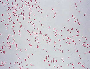
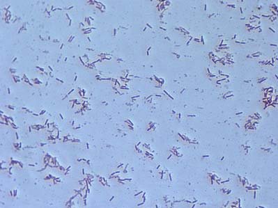
- Shape and Color: Pseudomonas aeruginosa appears as reddish/pink rods when stained with the Gram stain. This coloration indicates that they are Gram-negative bacteria, as they do not retain the primary stain (crystal violet) during the staining process.
- Size: Under high magnification, Pseudomonas aeruginosa cells typically range from 0.5 to 0.8 micrometers in diameter and 1.5 to 3.0 micrometers in length. They have a rod-like shape, which is a common characteristic of many bacterial species.
- Motility: Pseudomonas aeruginosa is motile, and its motility is facilitated by flagella. Most strains of Pseudomonas aeruginosa have a single polar flagellum, which is a long, whip-like structure that extends from one end of the bacterial cell. This flagellum allows the bacterium to move in liquid environments.
- Multiple Flagella: Some strains of Pseudomonas aeruginosa have been found to possess two to three polar flagella, which can enhance their motility and movement capabilities.
- Pili: Pseudomonas aeruginosa also possesses pili on its surface. Pili are hair-like structures that protrude from the bacterial cell and serve various functions, including adhesion to surfaces and facilitating a form of motility known as twitching motility. Twitching motility involves the extension and retraction of pili to enable the bacterium to crawl along surfaces.
In summary, under the microscope, Pseudomonas aeruginosa can be identified as Gram-negative rods with a reddish/pink coloration. They have a rod-like shape, are motile with a polar flagellum (sometimes more than one), and possess pili for adhesion and twitching motility. These characteristics are essential for distinguishing Pseudomonas aeruginosa from other bacterial species and play a crucial role in its pathogenesis and survival in various environments.
Pseudomonas Aeruginosa Under Microscope Pictures and Video


FAQ
What is the appearance of Pseudomonas aeruginosa under the microscope after Gram staining?
Pseudomonas aeruginosa appears as reddish/pink rods under the microscope after Gram staining, indicating that it is a Gram-negative bacterium.
How do you differentiate Pseudomonas aeruginosa from Gram-positive bacteria under the microscope?
Pseudomonas aeruginosa can be distinguished from Gram-positive bacteria by its pink/red coloration, as it does not retain the crystal violet stain during the Gram staining process.
What are the typical dimensions of Pseudomonas aeruginosa cells when viewed under high magnification?
Under high magnification, Pseudomonas aeruginosa cells are approximately 0.5 to 0.8 micrometers in diameter and 1.5 to 3.0 micrometers in length, displaying a rod-like shape.
What type of motility does Pseudomonas aeruginosa exhibit under the microscope?
Pseudomonas aeruginosa is motile and utilizes flagella for movement. Most strains have a single polar flagellum, while some may possess two to three polar flagella.
How does Pseudomonas aeruginosa use pili?
Pseudomonas aeruginosa possesses pili on its surface, which serve several functions. They aid in adhesion to surfaces and are involved in twitching motility, a form of movement where the bacterium crawls along surfaces.
What color does Pseudomonas aeruginosa appear after the Gram staining process?
Pseudomonas aeruginosa appears pink/red after Gram staining, indicating its Gram-negative nature.
Why is Gram staining important for identifying Pseudomonas aeruginosa?
Gram staining is crucial for identifying Pseudomonas aeruginosa as it helps differentiate between Gram-positive and Gram-negative bacteria based on their cell wall characteristics, aiding in accurate bacterial identification.
Can Pseudomonas aeruginosa exhibit different flagellar arrangements?
Yes, Pseudomonas aeruginosa can have variations in its flagellar arrangements. While most strains possess a single polar flagellum, some may have multiple polar flagella, enhancing their motility.
How is the Gram staining procedure used to identify Pseudomonas aeruginosa in clinical settings?
In clinical settings, Gram staining is one of the initial steps in identifying the causative agent of an infection. The characteristic appearance of Pseudomonas aeruginosa under the microscope after Gram staining can help in its preliminary identification.
Are there any other distinguishing features of Pseudomonas aeruginosa visible under the microscope?
Apart from its reddish/pink rod shape and motility, Pseudomonas aeruginosa can be identified by its single polar flagellum or, in some strains, multiple polar flagella. These features aid in distinguishing it from other bacterial species during microscopic examination.
- Text Highlighting: Select any text in the post content to highlight it
- Text Annotation: Select text and add comments with annotations
- Comment Management: Edit or delete your own comments
- Highlight Management: Remove your own highlights
How to use: Simply select any text in the post content above, and you'll see annotation options. Login here or create an account to get started.